Surgical Approach to the Ulnar Nerve: Anatomy, Repair, and Transposition
Key Takeaway
The surgical approach to the ulnar nerve requires precise anatomical knowledge from the axilla to the wrist. This comprehensive guide details the step-by-step exposure of the ulnar nerve, techniques for managing nerve gaps including anterior transposition and interfascicular grafting, and evidence-based postoperative rehabilitation protocols. Mastery of these techniques is essential for optimizing motor and sensory recovery following complex ulnar nerve injuries.
INTRODUCTION AND SURGICAL PRINCIPLES
The ulnar nerve is a critical terminal branch of the medial cord of the brachial plexus (C8, T1), responsible for the fine motor control of the hand and sensory innervation to its ulnar aspect. Surgical intervention upon the ulnar nerve is frequently indicated for traumatic lacerations, severe traction injuries, compressive neuropathies (such as cubital tunnel syndrome or Guyon’s canal syndrome), and tumor resections.
Achieving a successful outcome in ulnar nerve surgery requires an exhaustive understanding of its topographical anatomy, its relationship to adjacent neurovascular structures, and the biomechanical principles governing nerve excursion. The primary goal of any nerve repair (neurorrhaphy) is a tension-free coaptation. When segmental tissue loss occurs, the surgeon must employ advanced techniques—ranging from extensive mobilization and anterior transposition to interfascicular nerve grafting—to bridge the defect without compromising the microvascular supply (vasa nervorum).
SURGICAL ANATOMY AND EXTENSILE EXPOSURE
The approach to the ulnar nerve can be tailored to the specific zone of injury, but an extensile approach allows for complete visualization from the axilla to the distal palmar crease.
Exposure in the Axilla and Proximal Arm
In the axilla, the ulnar nerve is exposed utilizing a standard distal brachial plexus incision.
* Incision: Begin the incision over the tendon of the pectoralis major. Curve it gently into the natural folds of the axilla to prevent postoperative scar contracture, and continue distally along the medial aspect of the upper arm.
* Deep Dissection: In the axilla and proximal upper arm, the ulnar nerve lies immediately medial to the brachial artery and is typically situated deep to the brachial vein.
🔪 Surgical Pitfall: Nerve Identification
The medial antebrachial cutaneous (MABC) nerve runs in close proximity to the ulnar nerve in the proximal arm and can easily be misidentified. The MABC is generally smaller, more superficial, and courses with the basilic vein. Precise identification using a nerve stimulator prior to any transection or extensive mobilization is mandatory.
Exposure in the Mid-to-Distal Arm
As the dissection proceeds distally to the middle third of the upper arm, the anatomical relationships shift significantly.
* Course: The ulnar nerve diverges from the main neurovascular bundle. It gradually courses posteriorly, piercing the medial intermuscular septum (often passing through the arcade of Struthers, located approximately 8 cm proximal to the medial epicondyle).
* Incision Modification: At a point 6 to 8 cm proximal to the elbow, curve the incision posteriorly, passing slightly behind the medial epicondyle to avoid crossing the flexion crease of the elbow at a right angle.
* Triceps Relationship: The nerve travels superficial to the medial head of the triceps muscle before entering the ulnar groove (cubital tunnel) behind the medial humeral epicondyle.
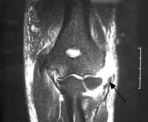
Exposure at the Elbow (Cubital Tunnel)
In the region of the ulnar groove, the nerve is constrained by Osborne’s ligament (the cubital tunnel retinaculum).
* Branching: Within the groove itself, the ulnar nerve gives off no major motor branches to the forearm or hand. However, it does provide critical articular branches to the elbow joint and one or two proximal motor branches to the flexor carpi ulnaris (FCU).
* Distal Mobilization: To trace the nerve into the forearm, the surgeon must release the two heads of the FCU (the humeral and ulnar heads) connected by Osborne’s fascia. This can be achieved by carefully splitting the FCU fascia or, in complex reconstructive cases, by performing a medial epicondylectomy.
Exposure in the Forearm and Wrist
- Incision: Continue the incision distally along the ulnar border of the volar forearm, extending to the proximal flexor crease of the wrist.
- Course: Distal to the cubital tunnel, the nerve gives off its main muscular branches to the medial half of the flexor digitorum profundus (FDP) and additional branches to the FCU. It then courses distally, resting on the FDP muscle belly, positioned on the radial side of the FCU muscle belly.
- Neurovascular Convergence: At the junction of the proximal and middle thirds of the forearm, the ulnar artery approaches the nerve from its lateral (radial) side. The artery and nerve travel together as a neurovascular bundle into the hand via Guyon's canal.
- Dorsal Cutaneous Branch (DCB): The DCB originates 5 to 8 cm proximal to the pisiform. It winds dorsally, deep to the FCU tendon, to provide sensation to the dorsum of the ulnar hand. Extreme care must be taken not to avulse this branch during distal mobilization.
MANAGEMENT OF NERVE GAPS: MOBILIZATION AND TRANSPOSITION
The ulnar nerve possesses a unique anatomical advantage: a gap in its continuity can often be closed more easily than in any other major peripheral nerve. This is primarily due to the ability to transpose the nerve anterior to the axis of elbow rotation, effectively shortening its required path.
Biomechanics of Gap Closure
Historically, Bunnell and Zachary reported that massive gaps of up to 13 to 15 cm could be closed through a combination of extensive mobilization, anterior transposition, and extreme flexion of the wrist and elbow. However, modern biomechanical and cadaveric studies have refined these parameters:
* Modern Limits: Trumble and McCallister demonstrated that anterior transposition reliably overcomes a 4-cm gap at the elbow and a 2-cm gap in the proximal forearm.
* Distal Forearm Limitations: In cadaveric studies, ulnar nerve transposition at the elbow has no effect on closing nerve gaps in the distal forearm or wrist.
* Joint Positioning: In the proximal forearm, wrist and elbow flexion of greater than 45 degrees is required to reduce a nerve gap of more than 11 mm following transposition.
Techniques for Anterior Transposition
If transposition is elected to achieve a tension-free primary neurorrhaphy, the nerve must be transposed only after painstaking intraneural dissection (epifascicular neurolysis) of the motor branches to the FDP and FCU, allowing the main trunk to move anteriorly without tethering.
- Subcutaneous Transposition:
- The nerve is placed anterior to the medial epicondyle, resting on the fascia of the flexor-pronator mass.
- To prevent the nerve from subluxating back into the ulnar groove, a thick layer of subcutaneous fat is mobilized and sutured to the medial fascia, creating a soft-tissue sling.
- Submuscular Transposition:
- The nerve is placed deep to the flexor-pronator muscle group.
- This is achieved by either detaching the flexor-pronator origin at its tendinous insertion on the medial epicondyle (and later repairing it) or by performing a medial epicondyle osteotomy with subsequent rigid fixation.
- The nerve is transposed anteriorly, coming to lie adjacent to the median nerve.
🚨 Surgical Warning: The Medial Intermuscular Septum
Regardless of the transposition technique utilized, the medial intermuscular septum must be radically excised proximal to the elbow. Failure to resect the septum will result in acute kinking, tethering, and secondary ischemic neuropathy of the ulnar nerve when the elbow is extended.
Interfascicular Nerve Grafting (The Modern Gold Standard)
While extreme joint flexion and extensive mobilization can close large gaps, these techniques place the repaired nerve under chronic tension upon joint extension, leading to intraneural ischemia and scarring. Furthermore, prolonged joint flexion causes severe flexion contractures.
* Current Recommendation: As an alternative to awkward positioning and extensive mobilization, interfascicular nerve grafting (using the sural nerve or MABC) is the preferred modern technique for gaps exceeding 2 to 3 cm that cannot be closed with simple transposition and mild joint flexion.
CRITICAL LIMITS OF DELAY FOR NEURORRHAPHY
Time is the most critical variable in peripheral nerve reconstruction. Prolonged denervation leads to irreversible motor endplate degradation and muscle atrophy. The "critical limit of delay" defines the timeframe beyond which surgical repair is futile.
- High Lesions (Axilla/Proximal Arm):
- Useful Motor Recovery: Should not be expected if suture is delayed beyond 9 months.
- Sensory Recovery: Rarely occurs after 9 months.
- Absolute Limit: Motor and sensory return cannot be expected after a delay of 29 months in lesions above the FCU branches.
- Low Lesions (Distal Forearm/Wrist):
- Useful Motor Recovery: Should not be expected if suture is delayed beyond 15 months.
- Sensory Recovery: Has been reported to occur up to 31 months after injury.
- Absolute Limit: Motor return cannot be expected after a delay of 18 months in lesions below the branches of the FDP.
POSTOPERATIVE PROTOCOL AND REHABILITATION
Postoperative immobilization is dictated by the degree of tension on the repair and the techniques used to close the gap.
Immobilization Phase
- Transposition with Elbow/Wrist Flexion: If the nerve was transposed and the elbow/wrist flexed to achieve coaptation, a well-padded, custom-molded posterior plaster splint is applied from the axilla to the metacarpophalangeal (MCP) joints.
- Wrist Flexion Only: If the lesion is in the distal forearm and the gap was closed by flexing the wrist alone, a posterior splint from just distal to the elbow to the MCP joints is sufficient.
- Duration: Sutures are typically removed at 7 to 10 days. The splint is maintained for a total of 4 weeks. During this period, the patient is strictly instructed to perform active range of motion of the fingers to keep the MCP and interphalangeal joints supple and prevent intrinsic contractures.
Mobilization Phase
- After 4 weeks, the static splint is removed.
- The elbow and wrist are gradually extended over a period of 2 to 3 weeks using an adjustable hinged brace. The rate of extension depends entirely on the intraoperative assessment of tension at the suture line.
- Once full extension is achieved, aggressive physical therapy is initiated to restore joint kinematics, maximize tendon glide, and begin sensory re-education. Splints are rarely required once the limb can be fully extended.
CLINICAL OUTCOMES AND PROGNOSIS
When evaluating the results of ulnar nerve neurorrhaphy, it is universally accepted that motor recovery is more critical than sensory recovery, given the profound functional deficit associated with an ulnar claw hand and loss of pinch grip.
Motor Recovery
- General Outcomes: Approximately 50% of patients will demonstrate a return of function in the long flexors (FDP, FCU) and some useful, albeit weak, function in the hypothenar and interossei muscles.
- Intrinsic Function: True, independent function of the interossei is notoriously difficult to achieve, occurring in only about 5% of patients. Independent finger motion is seen in roughly 16% of cases.
- Favorable Circumstances: Under optimal conditions (clean, sharp transection, immediate repair, young patient), up to 78% of patients may regain "useful" motor recovery.
- Grafting Outcomes: Modern interfascicular nerve grafting yields a return of motor power of M3 (Medical Research Council scale) or better in 79.5% of cases.
- Anatomical Level: The poorest motor return is universally reported following repairs of the ulnar nerve high in the axilla, due to the immense distance regenerating axons must travel to reach the intrinsic muscles of the hand.
Sensory Recovery
- With Overresponse: Approximately 50% of patients will regain sensitivity to touch and pain within the autonomous zone of the ulnar nerve, but this is often accompanied by a persistent, uncomfortable overresponse (hyperesthesia/allodynia).
- Without Overresponse: Only about 30% of patients regain touch and pain sensation without overresponse. Under highly favorable circumstances, this ideal sensory return may be achieved in up to half of the patient population.
You Might Also Like