Comprehensive Management of Adult Spinal Disorders: Stenosis, Deformity, and Tumors
Key Takeaway
This comprehensive chapter outlines the evaluation and operative management of complex adult spinal disorders. It provides an evidence-based approach to lumbar spinal stenosis, degenerative and isthmic spondylolisthesis, adult idiopathic scoliosis, and inflammatory arthritides such as rheumatoid arthritis and ankylosing spondylitis. Furthermore, it details the pathoanatomy, biomechanics, and surgical techniques required for the safe decompression, stabilization, and resection of primary and metastatic spinal tumors.
Introduction to Adult Spinal Pathologies
The management of adult spinal disorders requires a profound understanding of spinal biomechanics, spinopelvic alignment, and the natural history of degenerative, inflammatory, and neoplastic conditions. This comprehensive masterclass details the evidence-based evaluation and surgical management of complex spinal pathologies, ranging from degenerative stenosis and adult deformity to advanced reconstructive techniques for spinal oncology.
Lumbar Spinal Stenosis
Lumbar spinal stenosis (LSS) represents a progressive narrowing of the spinal canal, lateral recesses, or neural foramina, resulting in compression of the cauda equina or exiting nerve roots.
Anatomy and Pathophysiology
The pathoanatomy of LSS is typically a cascade of degenerative changes. Intervertebral disc desiccation and loss of height lead to altered load-sharing, placing increased stress on the facet joints. This results in facet hypertrophy, osteophyte formation, and buckling or hypertrophy of the ligamentum flavum. The combination of these factors transforms the normal neural canal into a constricted, trefoil shape.
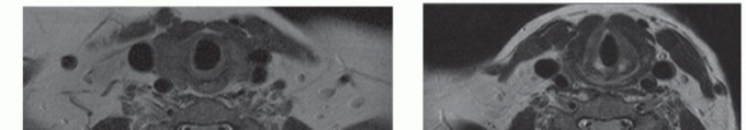
Clinical Evaluation and Diagnostic Imaging
Patients classically present with neurogenic claudication—radiating leg pain, numbness, or weakness exacerbated by standing or walking and relieved by sitting or lumbar flexion (the "shopping cart sign").
* Magnetic Resonance Imaging (MRI): The gold standard for evaluating soft tissue, disc pathology, and neural compression.
* Computed Tomography (CT) Myelography: Reserved for patients with contraindications to MRI (e.g., incompatible pacemakers) or those with severe scoliosis where MRI artifact obscures neural elements.
Operative Treatment: Decompression
Surgical intervention is indicated for patients with progressive neurologic deficits or refractory neurogenic claudication that fails conservative management (e.g., physical therapy, epidural steroid injections).
Principles of Spinal Stenosis Surgery:
The primary goal is adequate neural decompression while preserving iatrogenic stability.
1. Positioning: The patient is placed prone on a radiolucent frame (e.g., Jackson table) with the abdomen free to reduce venous engorgement and minimize epidural bleeding.
2. Approach: A standard midline posterior approach is utilized. Subperiosteal dissection exposes the spinous processes and lamina.
3. Decompression: A high-speed burr and Kerrison rongeurs are used to perform a laminectomy or bilateral laminotomies. The ligamentum flavum is carefully resected to expose the dura.
4. Foraminotomy: The lateral recess and neural foramina are decompressed by undercutting the medial facet.
Surgical Warning: When performing a medial facetectomy, the surgeon must preserve at least 50% of the pars interarticularis and the lateral aspect of the facet joint to prevent iatrogenic postoperative instability.
Degenerative Spondylolisthesis and Scoliosis
Degenerative spondylolisthesis is the forward translation of one vertebra over another with an intact neural arch, most commonly occurring at L4-L5.
Anatomy and Biomechanics
The primary biomechanical driver is the sagittal orientation of the facet joints combined with disc degeneration. As the disc space collapses, the sagittally oriented facets fail to resist anterior shear forces, leading to subluxation.
Operative Treatment: Decompression and Fusion
While decompression alone may be considered in elderly patients with stable, non-mobile slips, the gold standard for symptomatic degenerative spondylolisthesis is decompression combined with instrumented fusion.
Posterior and Transforaminal Lumbar Interbody Fusion (PLIF/TLIF):
Interbody fusion restores disc height, provides indirect foraminal decompression, and maximizes fusion rates by placing bone graft under compression.
* TLIF Technique: A unilateral facetectomy is performed to access the disc space, minimizing dural retraction compared to a PLIF. The disc is thoroughly prepped, cartilaginous endplates are removed, and an interbody cage packed with autograft/allograft is inserted.
* 360-Degree Fusion: In cases of severe instability or revision surgery, an Anterior Lumbar Interbody Fusion (ALIF) combined with posterior pedicle screw instrumentation provides robust biomechanical stability.
Clinical Pearl: During a TLIF, over-distraction of the disc space using the interbody cage can lead to contralateral nerve root stretch or compression. Always assess the contralateral foramen if significant height restoration is achieved.
Adult Idiopathic Scoliosis
Adult idiopathic scoliosis presents a unique challenge, often combining the structural deformity of adolescent idiopathic scoliosis with superimposed degenerative changes, leading to pain, radiculopathy, and sagittal imbalance.
Sagittal Balance and Spinopelvic Parameters
Modern deformity surgery is dictated by the restoration of global sagittal balance. Key parameters include:
* Pelvic Incidence (PI): A fixed morphologic parameter.
* Lumbar Lordosis (LL): Must be matched to the PI (ideally PI - LL < 10°).
* Sagittal Vertical Axis (SVA): The distance from the C7 plumb line to the posterior superior corner of S1 (normal is < 5 cm).
Operative Treatment
Surgical intervention aims to halt curve progression, decompress neural elements, and restore spinopelvic alignment.
* Posterior Instrumentation and Fusion: Utilizing pedicle screw constructs and rod derotation techniques. Osteotomies (e.g., Smith-Petersen or Pedicle Subtraction) may be required for rigid deformities.
* Combined Anterior and Posterior Fusions: Severe, rigid curves may require an anterior release and interbody fusion (ALIF or Lateral Lumbar Interbody Fusion - LLIF) followed by posterior instrumentation. This maximizes lordosis restoration and provides a massive surface area for arthrodesis.
Adult Isthmic Spondylolisthesis
Unlike degenerative spondylolisthesis, isthmic spondylolisthesis involves a defect in the pars interarticularis (spondylolysis), most frequently at L5-S1.
Biomechanics and Natural History
The L5-S1 junction is subjected to immense shear forces due to the sacral slope. A pars defect uncouples the anterior vertebral body from the posterior stabilizing elements. Over time, the L5 body slips anteriorly, potentially causing L5 radiculopathy as the nerve root is compressed in the foramen by the fibrocartilaginous pars defect (Gill body).
Operative Treatment
Symptomatic high-grade slips or those with progressive neurologic deficit require surgical stabilization.
* Anterior Lumbar Interbody Fusion (ALIF): ALIF is highly effective at L5-S1. It places the graft in compression, restores the lumbosacral angle, and provides indirect decompression of the L5 nerve root by restoring foraminal height.
* Posterior Instrumentation: Pedicle screws are placed to stabilize the segment. Reduction of the slip is controversial; partial reduction is often preferred to restore sagittal balance while minimizing the risk of neurologic injury.
Pitfall: Aggressive instrumental reduction of a high-grade L5-S1 isthmic spondylolisthesis carries a high risk of L5 nerve root stretch injury. In situ fusion or partial reduction is often safer and yields excellent clinical outcomes.
Rheumatoid Arthritis of the Spine
Rheumatoid arthritis (RA) is a systemic inflammatory disease that frequently targets the cervical spine, leading to ligamentous laxity, pannus formation, and profound instability.
Cervical Instability Patterns
- Atlantoaxial Subluxation (AAS): The most common pattern, caused by destruction of the transverse ligament. Evaluated via the Anterior Atlantodens Interval (ADI). An ADI > 3 mm is abnormal; > 9 mm indicates high risk of neurologic injury.
- Basilar Invagination (Cranial Settling): Upward migration of the odontoid process into the foramen magnum, risking brainstem compression.
- Subaxial Subluxation: Stair-step deformity of the lower cervical spine.
Operative Treatment
- C1-C2 Fusion: Indicated for progressive AAS. Techniques include Harms construct (C1 lateral mass screws and C2 pedicle/pars screws) or transarticular screws.
- Occipitocervical Fusion: Required for basilar invagination or when the C1 lateral masses are incompetent. The construct extends from the occipital squama down to the subaxial spine.
Ankylosing Spondylitis
Ankylosing spondylitis (AS) is a seronegative spondyloarthropathy characterized by progressive ossification of the spinal ligaments and intervertebral discs, resulting in a rigid, brittle "bamboo spine."
Pathoanatomy and Surgical Risks
The ankylosed spine acts as a long bone. Even minor trauma can cause highly unstable, transdiscal, or transvertebral fractures. These patients are at extreme risk for epidural hematomas and neurologic deterioration.
Osteotomy of the Spine for Deformity Correction
Patients with AS often develop a severe, rigid cervicothoracic or thoracolumbar kyphosis, severely impairing forward gaze and quality of life.
* Smith-Petersen Osteotomy (SPO): A posterior column shortening technique through the facet joints. Yields approximately 10° of lordosis per level. Requires a mobile anterior column (often not present in AS unless the anterior ossification is fractured).
* Pedicle Subtraction Osteotomy (PSO): A three-column, closing-wedge osteotomy performed through the vertebral body. It can provide 30° to 40° of lordosis at a single level.
* Eggshell Osteotomy: A technique involving decancellation of the vertebral body, allowing the spine to be hinged backward, closing the posterior defect.
Surgical Warning: During a PSO in an ankylosed spine, the surgeon must meticulously control the hinge. Premature closure or asymmetric closure can lead to catastrophic sagittal or coronal translation, resulting in spinal cord transection.
Tumors of the Spine
Spinal oncology requires a multidisciplinary approach. Tumors are broadly categorized into benign, primary malignant, and metastatic lesions.
Benign Tumors
- Posterior Element Tumors: Osteoid osteoma and osteoblastoma typically present with painful scoliosis and nocturnal pain relieved by NSAIDs. Surgical excision or radiofrequency ablation is curative.
- Vertebral Body Tumors: Hemangiomas are usually asymptomatic but can occasionally cause vertebral collapse or epidural extension, requiring embolization and decompression.
Primary Malignant Tumors
Primary malignancies of the spine are rare and require aggressive surgical management based on the Enneking or Weinstein-Boriani-Biagini (WBB) staging systems.
* Chordoma: Arises from notochordal remnants, most commonly in the sacrum or clivus. They are radioresistant. The gold standard is en bloc wide marginal resection.
* Osteosarcoma and Ewing Sarcoma: Require neoadjuvant chemotherapy followed by aggressive en bloc resection.
Metastatic Tumors
The spine is the most common site for skeletal metastases (Breast, Lung, Thyroid, Renal, Prostate).
* Pathoanatomy: Metastases typically seed the highly vascularized vertebral body via Batson's venous plexus, leading to structural collapse and epidural spinal cord compression (ESCC).
* Operative Treatment: The decision to operate is guided by the SINS (Spinal Instability Neoplastic Score) and the NOMS (Neurologic, Oncologic, Mechanical, Systemic) framework.
* Separation Surgery: For radioresistant tumors (e.g., renal cell, melanoma) causing cord compression, a posterior decompression is performed to create a safe margin between the tumor and the dura. This is followed by posterior stabilization and postoperative stereotactic radiosurgery (SRS) to ablate the remaining tumor.
* Palliative Decompression and Stabilization: Indicated for intractable pain, progressive neurologic deficit, and mechanical instability in patients with a life expectancy greater than 3 to 6 months.
Postoperative Management in Spinal Oncology
Postoperative protocols must balance the need for early mobilization to prevent deep vein thrombosis and pulmonary complications with the necessity of protecting complex reconstructive constructs. Bracing is often utilized, and coordination with radiation oncology is critical to time postoperative radiotherapy without compromising wound healing.
You Might Also Like