Proximal Humerus Fractures: Epidemiology, Advanced Anatomy & Biomechanics
Key Takeaway
Proximal humerus fractures are complex injuries with bimodal epidemiology. Management is individualized, guided by Neer or AO/OTA classification. Crucial surgical anatomy includes the humeral head's blood supply, tuberosities, and vital neurovascular structures like the axillary nerve. Understanding deforming biomechanical forces is essential for effective anatomical reduction and fixation.
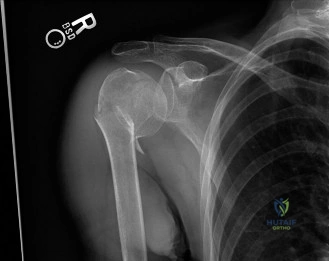
An 82-year-old female presents to the Emergency Department following a mechanical fall. She is clutching her right arm in pain. Examination reveals bruising tracking down the arm. Radiographs demonstrate a displaced 3-part proximal humerus fracture. Please describe the blood supply to the humeral head and explain its clinical significance in the management of this fracture.

Candidate: The blood supply is mainly from the anterior circumflex humeral artery. The ascending branch, the arcuate artery, enters the head through the bicipital groove. If this is disrupted, especially in 3- or 4-part fractures, the head can die, leading to avascular necrosis (AVN).
Candidates often oversimplify the anatomy by focusing only on the anterior circumflex artery. Failing to mention the posteromedial soft tissue hinge (containing the arcuate artery) or the role of the posterior circumflex humeral artery demonstrates a lack of surgical depth regarding why some fractures are more "at-risk" than others.
The dominant blood supply is via the arcuate artery, a branch of the anterior circumflex humeral artery (ACHA). Crucially, this vessel enters the humeral head through the posteromedial hinge. The risk of AVN is predicated on the disruption of this hinge. Therefore, I classify fractures not just by fragment number, but by whether this posteromedial metaphyseal blood supply is compromised. If this hinge is disrupted in a 3- or 4-part fracture, the risk of AVN increases significantly, which influences my decision toward primary arthroplasty versus salvage attempts.
Look at these radiographs. How would you classify this injury, and what are the key elements you look for when deciding between non-operative management and ORIF?
Candidate: Using the Neer classification, this is a displaced fracture. To decide on treatment, I would consider the patient's age, bone quality, and functional demand. If it's a 2-part fracture, I might consider ORIF. If it's a 4-part, I would think about an arthroplasty.
Failing to define "displaced" using the Neer criteria (1cm/45 degrees) is a major oversight. Additionally, failing to mention the ProXimal trial or the importance of the tuberosity healing makes the candidate look like they are relying on outdated "dogma" rather than evidence-based medicine.
I would use the Neer classification, noting that displacement is defined as >1cm or >45 degrees of angulation. However, decision-making is multifactorial: 1) Patient factors: Physiological age, comorbidities, and baseline functional demand. 2) Fracture factors: Degree of comminution, articular involvement, and integrity of the medial calcar. 3) Evidence: I am cognizant of the ProXimal trial, which showed no significant long-term functional difference between operative and non-operative management for many displaced fractures. Therefore, in an elderly low-demand patient, I have a low threshold for non-operative management, whereas for a younger, high-demand patient with a displaced greater tuberosity fracture, I would lean toward ORIF to restore the rotator cuff footprint.
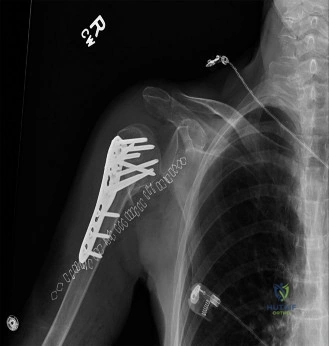
During your ORIF using a deltopectoral approach, you are struggling to achieve reduction. You are worried about the axillary nerve. Where exactly is it, and what maneuvers should you avoid to prevent iatrogenic injury?
Candidate: The axillary nerve is about 5cm below the acromion. I should be careful when retracting the deltoid so I don't stretch it.
Being vague about the "safe zone." Simply saying "5cm" is inadequate. The examiner wants to see that you appreciate that the nerve is at risk not just from direct incision, but from aggressive lateral retraction of the deltoid and when placing screws into the posterior or inferior glenoid/humeral head.
The axillary nerve courses around the surgical neck, approximately 5-7cm distal to the lateral acromion. During the deltopectoral approach, I must avoid aggressive lateral retraction of the deltoid, as this puts the nerve under tension against the humeral neck. Furthermore, I am cautious when inserting locking screws into the inferior and posterior aspects of the plate; I use a 'down-and-in' technique under direct visualization or fluoroscopic guidance to ensure the nerve is not in the path of the drill or screw tip, particularly in the 'danger zone' of the posterior-inferior quadrant.
