Solving Persistent Hip Pain: Arthroscopic Osteoid Osteoma Lesion Excision
Key Takeaway
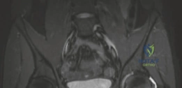
Looking for accurate information on Solving Persistent Hip Pain: Arthroscopic Osteoid Osteoma Lesion Excision? An osteoid osteoma lesion often presents as persistent hip pain, worse with activity and at night, typically in young adults. This 22-year-old male's case highlights its identification via CT and MRI, showing a characteristic nidus with surrounding sclerosis after initial misdiagnosis. Treatment options include radiofrequency ablation or surgical excision.
A 19-year-old male presents with a 10-month history of worsening left hip pain. The pain is worse at night and improves significantly with ibuprofen. He has developed an antalgic gait and reduced internal rotation. Initial AP and lateral radiographs are non-specific. Given this presentation, what is your primary differential diagnosis, and how would you proceed with imaging?
Candidate: Given the age, nocturnal pain, and dramatic response to NSAIDs, the classic diagnosis is an osteoid osteoma. Since it is in the hip, I am concerned it is intra-articular and causing reactive synovitis. I would order a thin-slice CT scan to identify the nidus and an MRI to evaluate for reactive synovitis or joint pathology.
Failure to mention the "intra-articular" nature of the lesion. A poor candidate treats this as a generic bone tumor, ignoring that intra-articular osteoid osteomas of the hip are "great masqueraders" that cause severe synovitis, often leading to a delayed diagnosis of 8-24 months because they mimic septic arthritis or inflammatory arthropathy.
Identify the clinical triad: young male, nocturnal pain, NSAID responsiveness. Explicitly label the differential as "intra-articular osteoid osteoma." Emphasize that the hip capsule lack of periosteum prevents typical sclerosis, making it harder to spot. Recommend thin-slice CT as the gold standard for nidus localization and MRI to assess the inflammatory synovial milieu (synovitis) and rule out other causes of hip pain like FAI or labral tears.
You have confirmed a 1.2cm nidus located on the superior femoral neck, within 5mm of the articular cartilage. What are your considerations regarding surgical planning and the risk of avascular necrosis (AVN)?

Candidate: This lesion is intra-articular and near the articular cartilage, which makes RFA relatively contraindicated due to the risk of chondrolysis and thermal injury. I would opt for arthroscopic excision. Regarding AVN, the main risk is damage to the ascending retinacular branches of the medial femoral circumflex artery (MFCA). I must be meticulous during the synovectomy along the posterosuperior femoral neck to protect these vessels.
Suggesting an open approach or just stating "I will be careful." A failing candidate misses the specific anatomical landmarks (Weitbrecht's retinacula) and fails to address the "tension vs. compression side" biomechanical risk of an iatrogenic fracture.
Structure the answer: 1. Contraindication: RFA (thermal necrosis risk to cartilage/vessels). 2. Approach: Arthroscopic excision (minimally invasive). 3. Vascular preservation: Avoid aggressive thermal usage near the posterosuperior aspect where the MFCA retinacular vessels travel. 4. Biomechanics: Acknowledge the lesion is on the "tension side" (superior neck) and suggest prophylactic cannulated screw fixation if the resection exceeds 30% of the neck diameter.
During the procedure, you have successfully localized the nidus under fluoroscopy. You have removed the lesion. How do you decide if the patient requires internal fixation before closing?
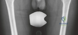
Candidate: I would evaluate the size and location of the cortical defect. If the defect is greater than 30% of the femoral neck diameter, or if it is located on the superior "tension side," the risk of a pathological fracture is high. In these cases, I would perform prophylactic fixation using 2 or 3 cannulated screws percutaneously.
Providing a vague answer like "I'll use a screw if it looks big." The examiner demands the "30% rule" and the distinction between the tension side (superior) vs. compression side (inferior/calcar) of the femoral neck.
A rigorous answer: 1. Assess the defect size relative to the femoral neck (30% threshold). 2. Assess location (Tension vs. Compression side). 3. Mention that a large/tension-sided defect acts as a stress riser. 4. State clearly that prophylactic fixation is the standard of care for these high-risk defects to prevent catastrophic femoral neck fracture.
