Lambotte’s Principles and the AO-ASIF Philosophy of Surgical Fracture Management
Key Takeaway
Lambotte’s historical principles form the foundation of modern AO-ASIF fracture management guidelines. These dictate anatomical reduction, stable internal fixation, preservation of vascularity, and early active mobilization. Successful surgical intervention requires meticulous preoperative planning, respect for soft tissue biology, and precise execution of provisional and definitive stabilization. Understanding the biomechanical demands and inherent risks of operative osteosynthesis is critical for orthopedic surgeons to optimize patient outcomes and prevent nonunion or infection.
INTRODUCTION: THE EVOLUTION OF OSTEOSYNTHESIS
The surgical management of fractures is governed by a delicate balance between mechanical stabilization and biological preservation. While the conceptual desire to stabilize broken bones spans centuries, it was Albin Lambotte—often heralded as the "Father of Modern Osteosynthesis"—who formalized the principles of surgical fracture treatment. Lambotte’s foundational tenets remain as applicable today as they were during their inception. Building upon this historical bedrock, the Arbeitsgemeinschaft für Osteosynthesefragen (AO-ASIF) refined these concepts into four universal treatment guidelines that dictate contemporary orthopedic trauma surgery.
The ultimate goal of operative fracture management is the rapid, complete restoration of limb function. This is achieved not merely by mechanical carpentry, but by a profound understanding of bone biology, soft tissue envelopes, and the biomechanical demands of the injured extremity.
THE FOUR AO-ASIF PRINCIPLES OF FRACTURE MANAGEMENT
The AO-ASIF formulated four distinct guidelines derived from Lambotte’s original principles. These pillars form the cognitive framework for every orthopedic trauma procedure.
1. Anatomical Reduction
The necessity for anatomical reduction varies depending on the anatomical location of the fracture.
* Intra-articular Fractures: Absolute anatomical reduction of the articular surface is mandatory. Even a 1-2 mm step-off can alter joint contact pressures exponentially, leading to early post-traumatic osteoarthritis.
* Diaphyseal Fractures: The paradigm has shifted from absolute anatomical reduction to "functional reduction." The primary goals are the restoration of length, alignment (coronal and sagittal), and rotation. Attempting to anatomically reduce every comminuted diaphyseal fragment often requires excessive soft tissue stripping, which devitalizes the bone and leads to nonunion.
2. Stable Internal Fixation
Fixation must fulfill the local biomechanical demands of the fracture pattern and the patient's functional requirements.
* Absolute Stability: Achieved through interfragmentary compression (e.g., lag screws, compression plates). This eliminates micro-motion at the fracture site, dictating primary bone healing via cutting cones without callus formation. It is indicated for simple articular and diaphyseal fractures.
* Relative Stability: Achieved through splinting techniques (e.g., intramedullary nails, bridge plating, external fixators). This allows controlled micro-motion, stimulating secondary bone healing via endochondral ossification and robust callus formation. It is the treatment of choice for highly comminuted diaphyseal and metaphyseal fractures.
3. Preservation of Blood Supply
The most mechanically sound construct will fail if the biology is compromised. The preservation of the vascularity to the bone fragments and surrounding soft tissues is paramount. Modern techniques emphasize Minimally Invasive Plate Osteosynthesis (MIPO) and closed intramedullary nailing to protect the periosteal and endosteal blood supply.
4. Early, Active Mobilization
Prolonged immobilization leads to "fracture disease," characterized by joint stiffness, muscle atrophy, osteopenia, and complex regional pain syndrome (CRPS). The surgical construct must be robust enough to permit active, pain-free mobilization of adjacent muscles and joints immediately postoperatively.
Clinical Pearl: The modern orthopedic surgeon must act as a "biological watchmaker." The mechanical construct must be perfectly tailored to the biological capacity of the host tissue. Do not sacrifice biology on the altar of mechanical perfection.
CONTRAINDICATIONS AND HIGH-RISK SCENARIOS
While surgical stabilization is the gold standard for many fractures, there are distinct clinical scenarios where the probability of surgical failure is unacceptably high. In these instances, alternative strategies or non-operative management must be employed.
1. Severe Osteoporosis
Osteoporotic bone lacks the trabecular density required for standard screw purchase, leading to a high risk of hardware pull-out and construct failure.
* Management Strategy: Utilization of fixed-angle locking plates, intramedullary load-sharing devices, or polymethylmethacrylate (PMMA) cement augmentation to enhance screw purchase.
2. Compromised Soft Tissue Envelope
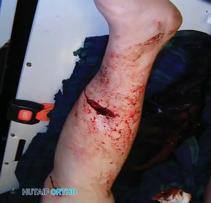
Soft tissues overlying the fracture or the planned surgical approach may be of exceptionally poor quality due to severe contusions, fracture blisters, scarring, burns, active dermatitis, or prior radiation.
* Management Strategy: Immediate internal fixation through compromised tissue inevitably leads to wound dehiscence and deep infection. The standard of care is Damage Control Orthopedics (DCO) utilizing a spanning external fixator until the soft tissues declare themselves and swelling subsides (the "wrinkle sign").
3. Active Infection or Osteomyelitis
Placing internal hardware into an actively infected field provides a nidus for biofilm formation, making eradication of the infection nearly impossible.
* Management Strategy: External fixation combined with aggressive surgical debridement and targeted systemic antibiotics. Occasionally, antibiotic-coated intramedullary nails are utilized by highly experienced trauma surgeons as a salvage procedure, but this is not routinely recommended for the general practitioner.
4. Irreconstructible Comminution
Severe intra-articular fractures where impaction has completely destroyed the articular cartilage (e.g., severe pilon or tibial plateau fractures) may preclude successful anatomical reconstruction.
* Management Strategy: Primary arthrodesis or delayed arthroplasty may be required. Spanning external fixation is often used to maintain length and alignment while the soft tissues heal.
5. Medical Contraindications
Patients with severe systemic comorbidities (ASA Class IV or V) who cannot tolerate the physiological stress of anesthesia or blood loss are poor candidates for prolonged reconstructive procedures.
6. Stable, Undisplaced Fractures
Fractures that are undisplaced or stably impacted in an acceptable position generally do not require surgical exposure.
* Exception: Prophylactic fixation is indicated in specific scenarios, such as valgus-impacted femoral neck fractures, where the risk of secondary displacement and subsequent avascular necrosis (AVN) is unacceptably high.
7. Inadequate Resources
Surgical intervention should be aborted or transferred if there is inadequate equipment, fluoroscopy, or surgeon experience to manage the specific fracture pattern and its potential complications.
DISADVANTAGES AND INHERENT RISKS OF SURGICAL INTERVENTION
Surgical treatment inherently adds a "second hit" of trauma to an already injured extremity. The surgeon must ensure that the benefits of stabilization outweigh the biological costs of the surgical approach.
The Biological Cost of Dissection
Any surgical dissection produces scar tissue and can cause contracture of the musculotendinous units required for functional rehabilitation. Furthermore, aggressive periosteal stripping destroys the extrinsic blood supply to the bone, precipitating delayed union or nonunion.
Neurovascular Risks
Surgical approaches carry a constant risk of iatrogenic injury to major neurovascular structures. Approaches must strictly follow established internervous and intermuscular planes to prevent denervation of the musculature.
Systemic and Blood-Borne Risks
Operative intervention carries the risk of significant blood loss, necessitating transfusions that carry inherent risks of immunological reactions and transmission of blood-borne pathogens (e.g., Hepatitis, HIV). The American Academy of Orthopaedic Surgeons (AAOS) mandates strict protocols to minimize HIV transmission, heavily favoring closed reduction techniques (such as closed intramedullary nailing) over open exposures whenever feasible to minimize blood loss and contamination.
Surgical Warning: The decision to open a fracture site must be justified by the inability to achieve an acceptable reduction via closed or percutaneous methods. Every scalpel stroke alters the natural biology of fracture healing.
STEP-BY-STEP SURGICAL EXECUTION
Successful operative fracture management requires meticulous adherence to a structured surgical workflow.
1. Preoperative Planning
The operation is won or lost before the patient enters the theater.
* Templating: Utilize orthogonal radiographs and CT scans to understand the fracture personality. Digital templating is mandatory to select the appropriate implant size, screw lengths, and trajectory.
* Equipment: Ensure all necessary implants, reduction forceps, radiolucent tables, and fluoroscopy units are available and functioning.
2. Patient Positioning and Fluoroscopy
Positioning must allow for unhindered fluoroscopic imaging in both the anteroposterior (AP) and lateral planes without moving the injured extremity.
* Traction tables or radiolucent flat tables should be utilized based on the fracture location.
* The extremity must be prepped and draped to allow for full range of motion during the procedure to assess joint stability and implant impingement.
3. Surgical Approaches
When open reduction is mandated, the approach must be atraumatic.
* Utilize true internervous planes (e.g., the Henry approach to the radius between the median and radial nerve innervations).
* Employ full-thickness fasciocutaneous flaps. Avoid undermining the subcutaneous tissues, which compromises dermal vascularity.
* Retract soft tissues gently using blunt retractors; avoid excessive tension that causes tissue ischemia.
4. Reduction Techniques
- Direct Reduction: Used for articular fractures. Involves direct visualization of the fracture site, clearing of hematoma, and precise manipulation of fragments using pointed reduction forceps or dental picks.
- Indirect Reduction: Used for diaphyseal fractures. Relies on ligamentotaxis, traction, or the use of the implant itself (e.g., a plate or nail) to restore length and alignment without exposing the fracture site.
5. Provisional Stabilization
Once acceptable reduction is achieved, it must be provisionally held to allow for radiographic confirmation and definitive implant placement.
* Technique: Kirschner wires (K-wires) or independent lag screws are most commonly used.
* Pitfall Avoidance: Provisional fixation must be placed strategically so that it does not obstruct the planned trajectory of the definitive plate, intramedullary nail, or locking screws. Failure to plan K-wire placement often results in the loss of reduction when the wires must be prematurely removed to accommodate the definitive implant.
6. Definitive Stabilization
The definitive construct must provide the mechanical stability dictated by the preoperative plan.
* Construct Selection: The mechanical construct (nail, plate, or external fixator) must possess sufficient fatigue life to withstand physiological loads until bony union occurs.
* Load Sharing vs. Load Bearing: Whenever possible, constructs should be load-sharing (e.g., intramedullary nails) rather than load-bearing. Load-sharing devices transmit physiological stresses across the fracture site, stimulating Wolff’s Law and promoting robust callus formation. Rigid, load-bearing constructs shield the bone from stress, which can lead to osteopenia and hardware failure if union is delayed.
POSTOPERATIVE PROTOCOLS AND REHABILITATION
The surgical procedure is only the first phase of fracture management. Postoperative care is dictated by the stability of the fixation and the biological status of the patient.
Immediate Postoperative Phase (Days 0-14)
- Edema Control: Elevation of the extremity above the level of the heart is critical to minimize swelling and prevent wound dehiscence.
- Early Mobilization: Passive and active-assisted range of motion of adjacent joints should begin immediately to prevent arthrofibrosis and promote venous return, mitigating the risk of deep vein thrombosis (DVT).
- Wound Care: Dressings should remain undisturbed until the first postoperative check unless there is excessive strike-through or signs of infection.
Weight-Bearing Protocols
Weight-bearing status is entirely dependent on the fracture pattern and the chosen fixation method.
* Diaphyseal Fractures (IM Nailing): Often permit immediate weight-bearing as tolerated, as the load-sharing nature of the nail biomechanically supports axial loads.
* Articular Fractures (Plate Osteosynthesis): Typically require a period of restricted weight-bearing (e.g., toe-touch or non-weight-bearing for 6-12 weeks) to prevent subsidence of the articular fragments while primary bone healing occurs.
Long-Term Monitoring
Patients must be followed with serial clinical examinations and orthogonal radiographs to monitor for signs of union (bridging callus on three out of four cortices) or complications such as hardware failure, loss of reduction, or infection.
By strictly adhering to Lambotte’s historical principles and the modern AO-ASIF guidelines, the orthopedic surgeon can navigate the complex biomechanical and biological demands of fracture management, ultimately restoring the patient to their pre-injury level of function.
You Might Also Like