THA Wear Explained: Decoding the Critical Modes of Wear
Key Takeaway
Here are the crucial details you must know about THA Wear Explained: Decoding the Critical Modes of Wear. The **modes of wear** in joint replacement surgery are categorized into four types. Mode 1 involves wear debris from intended motion between primary bearing surfaces. Mode 2 describes a primary bearing surface rubbing an unintended secondary surface. Mode 3 includes two primary bearing surfaces with interposed third-body particles. Mode 4 refers to two non-bearing surfaces rubbing together, such as back-sided liner wear or fretting.
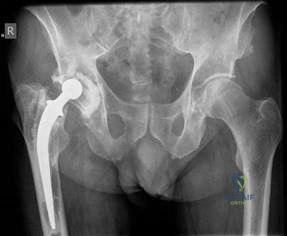
This is an anteroposterior (AP) radiograph of a 72-year-old male who had a cemented THA performed 17 years ago. Please describe the radiographic findings and provide your management thoughts.
Candidate: The radiograph shows a cemented THA with evidence of failure. There is clear eccentric wear of the polyethylene, resulting in superior migration of the femoral head. I see lucencies in all three DeLee and Charnley acetabular zones, suggesting component loosening. Furthermore, there is a continuous radiolucency at the femoral cement–bone interface. I would assess this patient clinically for pain, examine the hip for function, and order further imaging, likely CT, to quantify bone loss before planning revision surgery.
Candidates often jump straight to "The patient needs a revision." Failing to describe the specific zones of loosening (DeLee/Charnley) or neglecting to mention the "effective joint space" concept makes the candidate look like they are guessing rather than evaluating periprosthetic biology.
A systematic description is key: start with the implant type, then assess the acetabulum (Zones 1, 2, 3), then the femoral side (cement-bone interface). Explicitly link these findings to the "Effective Joint Space" hypothesis. Mention the biological process (macrophage-mediated osteolysis via RANK/RANKL) to demonstrate depth of knowledge regarding why the bone is failing, rather than just stating "it's loose."
You mentioned wear as a cause for the loosening. Can you define the four modes of wear in joint replacement?
Candidate: Mode 1 is expected articulation between the bearing surfaces. Mode 2 is a primary bearing surface articulating against a non-intended surface (e.g., metal shell). Mode 3 involves third-body particles (like cement/bone debris) between surfaces. Mode 4 is non-bearing surface wear, such as back-sided wear or Morse taper fretting.
Mixing up Mode 3 (third-body) and Mode 4 (non-bearing) is a classic error. Failing to provide clinical examples for each mode demonstrates a lack of practical understanding of how these mechanisms present in revision theatre.
Structure the answer clearly as Mode 1 through 4. For each, provide the definition and a concrete, high-yield clinical example (e.g., Mode 4: Morse taper fretting at the trunnion). Finish by noting that while they occur simultaneously, Mode 1 is the dominant contributor in well-functioning implants.
Explain the concept of 'Effective Joint Space' and how it relates to periprosthetic osteolysis.
Candidate: Coined by Schmalzreid, the "effective joint space" includes all periprosthetic regions accessible to synovial fluid and wear debris. Particles migrate through these spaces—such as screw holes or areas of non-ingrown porous coating—and trigger an inflammatory cascade at the bone-implant interface involving macrophages and cytokine release.
Forgetting to credit Schmalzreid, et al. (1992) is a missed opportunity for a "brownie point." Additionally, failing to mention the actual mechanism—that this space allows debris to reach the bone-cement interface—makes the answer superficial.
Define the term, name the authors (classic paper!), and explain the clinical implication: access of debris leads to the biological response. Conclude by briefly explaining how modern design (e.g., circumferential coating, sealed screw holes) aims to minimize this space to prevent osteolysis.
