Structured Hip Oral: Master This Exam Question & Case
Key Takeaway
This article provides essential research regarding Structured Hip Oral: Master This Exam Question & Case. A structured hip oral is an orthopedic assessment where an examiner presents complex hip cases, like a loose hemiarthroplasty with suspected infection, to evaluate a candidate’s diagnostic and management skills. It involves interpreting radiographs, identifying pathologies, and discussing investigation strategies, including the sensitivity and specificity of inflammatory markers for prosthetic joint infection.
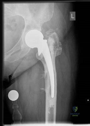
This is a radiograph of a 77-year-old woman who sustained a displaced intracapsular fractured neck of femur 3 years earlier, managed with a cemented Thompson’s hemiarthroplasty. She presents with increasing left hip pain and difficulty mobilizing. How do you interpret these initial findings?
Candidate: "This is an AP radiograph of a cemented Thompson’s hemiarthroplasty. There is evidence of component subsidence below the lesser trochanter, with significant radiolucencies in Gruen zones 1, 4, 5, and 7. There is also a faint rim of soft tissue calcification laterally. The implant orientation appears neutral. I require an immediate comparison with post-operative films and an orthogonal lateral view to assess the extent of loosening and cortical bone integrity."
Failing to mention the specific Gruen zones or ignoring the soft tissue changes. Candidates often look only at the metal-bone interface and miss the systemic implication of the "calcification" (which may suggest particle disease or chronic infection).
A systematic radiological assessment: 1) Identification (View/Prosthesis type), 2) Zone-by-zone analysis (Gruen zones 1,4,5,7), 3) Implant-bone interface integrity, 4) Soft tissue evaluation, 5) Comparative analysis (comparing current films to immediate post-op), and 6) Requesting necessary orthogonal views.
Here is the lateral radiograph. Given the progression of symptoms, you suspect infection. How do you investigate, and how reliable are inflammatory markers?
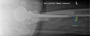
Candidate: "I would order CRP and ESR. However, I recognize they have limited sensitivity and specificity for prosthetic joint infection (PJI). Per the Berbari et al. systematic review (2010), serum markers are adjuncts. I would also perform a thorough history—specifically checking for pain unrelated to movement, prior wound discharge, or systemic sepsis—and consider joint aspiration if the pre-test probability is high."
Relying solely on lab values. A common failure is to ignore the "clinical suspicion" aspect—if a patient has a sinus or systemic symptoms, the lab markers are secondary to the clinical presentation.
Structure the answer into: 1) Clinical assessment (History/Exam), 2) Serum markers (CRP/ESR limitations), 3) Advanced investigation (Aspiration criteria per Spangehl et al.), and 4) Acknowledging the role of microbiology/MDT input. Quote the sensitivity/specificity limitations clearly.
Your aspiration is negative, yet the patient has severe, debilitating pain and wants a solution. She is physiologically frail with significant comorbidities. How do you categorize the infection, and what is your surgical plan?
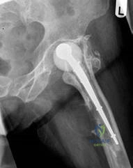
Candidate: "I would categorize the infection using the Tsukayama classification. Given her pain, I would advocate for a two-stage revision. This allows for rigorous debridement, assessment of soft tissue response to antibiotic therapy, and the use of an antibiotic-loaded spacer (e.g., PROSTALAC) to maintain length and function between stages. A one-stage procedure is generally reserved for healthy patients with a low-virulence, known organism and sufficient bone stock."
Failing to address the patient's frailty. A poor candidate suggests a radical, long operation without discussing the physiological cost, leading to "surgical hubris."
Demonstrate maturity: Categorize the infection (Tsukayama/McPherson), justify the two-stage approach over one-stage (Safety/Versatility/Clinical clearance), and highlight the importance of shared decision-making given the patient's co-morbid history.
