Operative Principles and Biomechanics of Tendon Transfer in the Upper Extremity
Key Takeaway
Tendon transfer surgery restores lost motor function by re-routing a functional, expendable muscle-tendon unit to substitute for a paralyzed one. Success relies on strict adherence to biomechanical principles, including adequate muscle strength, appropriate excursion amplitude, synergistic action, and a straight line of pull. Preoperative correction of fixed contractures and meticulous intraoperative tensioning are critical to achieving optimal functional outcomes and preventing secondary joint deformities.
INTRODUCTION TO TENDON TRANSFER SURGERY
Tendon transfer surgery remains a cornerstone of reconstructive orthopaedics, offering a dynamic solution for the restoration of hand and upper extremity function lost to traumatic, congenital, infectious, or vascular etiologies. The procedure involves the detachment of a functioning, expendable muscle-tendon unit at its insertion and its surgical relocation to a new attachment site, thereby substituting for a paralyzed or structurally deficient muscle.
To achieve predictable, high-quality outcomes, the orthopaedic surgeon must possess a profound understanding of upper extremity biomechanics, muscle physiology, and spatial anatomy. Haphazard transfers inevitably lead to functional failure, exacerbation of muscle imbalance, and the development of secondary joint deformities.
Clinical Pearl: A tendon transfer does not create new power; it merely redistributes existing power. The fundamental goal is to maximize the efficiency of this redistribution while minimizing the donor site deficit.
PREOPERATIVE PLANNING: THE "BALANCE SHEET" APPROACH
Regardless of the underlying pathology, the affected extremity must be systematically evaluated. The surgeon must construct a "balance sheet" detailing:
1. Function Lost: The specific motor deficits requiring reconstruction.
2. Function Retained: The intact muscles available for potential transfer.
3. Function Possible: The realistic functional goals achievable through reconstruction.
It is highly recommended to list the required functions in one column and the available, expendable donor muscles in an opposite column. Matching these columns allows for the logical and accurate design of a surgical blueprint.
Prerequisites for Tendon Transfer
Before any tendon transfer is executed, several absolute prerequisites must be met:
* Supple Joints: A transferred muscle cannot overcome a fixed joint contracture. Maximum passive range of motion must be achieved preoperatively through therapy, serial casting, or surgical release.
* Adequate Soft Tissue Bed: Tendons must glide through healthy, well-vascularized subcutaneous tissue. Scarred, irradiated, or grafted skin beds severely impede excursion.
* Stable Skeletal Architecture: Fractures must be healed, and unstable joints must be stabilized (dynamically or via arthrodesis) to provide a solid foundation for the transfer.
EVALUATING MUSCLES FOR TENDON TRANSFER
The selection of an appropriate donor muscle is governed by strict biomechanical criteria. The two most critical factors are expendability and strength. Restoring one major function (e.g., finger extension) is absolutely contraindicated if it is done at the expense of another major function (e.g., finger flexion).
Muscle Strength and the "One Grade Loss" Rule
The strength of a potential donor muscle is evaluated clinically using the Medical Research Council (MRC) grading system:
* 0 (Zero): No palpable or visible contraction.
* 1 (Trace): Palpable contraction only; no joint motion.
* 2 (Poor): Moves the joint through a range of motion with gravity eliminated.
* 3 (Fair): Moves the joint through a range of motion against gravity.
* 4 (Good): Moves the joint against gravity and moderate resistance.
* 5 (Normal): Normal strength against full resistance.
Surgical Warning: A muscle universally loses at least one MRC grade of strength following transfer. This occurs due to the trauma of mobilization, altered biomechanical vectors, and the formation of peritendinous adhesions. Therefore, only muscles graded 4 (Good) or 5 (Normal) are suitable for transfer.
Amplitude of Excursion
The amplitude of excursion (the distance a tendon glides when the muscle contracts from maximum stretch to maximum shortening) must be sufficient to provide satisfactory function at the recipient site. While the donor tendon's excursion may not perfectly match the tendon it replaces, it must be functionally adequate.
For example, the brachioradialis (BR) is a highly expendable muscle, but it possesses a naturally short excursion (approximately 3 cm). It is entirely unsuitable for restoring full finger flexion (which requires ~7 cm of excursion). However, it can be an excellent donor for the flexor pollicis longus (FPL), as even limited flexion of the thumb interphalangeal (IP) joint is highly functional.
Technical Modification: The excursion of the brachioradialis can be surgically increased by dissecting its tendon proximally and meticulously freeing all of its fascial attachments to the radius and surrounding septa.
Synergy and Cortical Re-education
Rehabilitation is significantly accelerated when the transferred muscle is synergistic with the lost function. Synergistic muscles normally contract together to facilitate a specific movement. For instance, wrist flexors naturally contract during finger extension to stabilize the wrist. Therefore, transferring a wrist flexor (e.g., Flexor Carpi Radialis) to restore finger extension (e.g., Extensor Digitorum Communis) utilizes pre-existing cortical motor patterns, making postoperative re-education intuitive.
INTRAOPERATIVE TECHNICAL CONSIDERATIONS
Assessing Muscle Viability
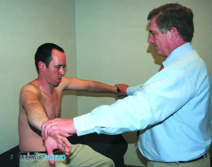
While preoperative clinical evaluation is paramount, the intraoperative appearance of the muscle provides the final confirmation of its suitability.
* Healthy Muscle: Appears dark pink or red, indicating robust vascularity, satisfactory nutrition, and the presence of normal, contractile muscle fibers.
* Unsuitable Muscle: Appears pale pink, fibrotic, and is smaller than normal (atrophic). Its amplitude of excursion will be demonstrably less than normal when tested intraoperatively.
Muscles that fail to contract briskly in response to a direct mechanical stimulus (pinch) or electrical stimulus (low-voltage electrocautery or nerve stimulator) are nonfunctional and must be abandoned as active donors.
Neurovascular Preservation
During the proximal mobilization of a donor muscle, meticulous care must be taken to avoid stretching, compressing, or transecting the neurovascular bundle. The primary neurovascular pedicle typically enters the muscle belly at its proximal third. Overzealous proximal dissection to gain excursion can inadvertently denervate the muscle, rendering the transfer useless.
Line of Pull and Pulleys
The biomechanical efficiency of a transferred muscle is directly proportional to its line of pull. The straighter the muscle vector from its origin to its new insertion, the more efficient its action.
* Avoid Acute Angles: A muscle and its tendon should never be routed around an acute angle.
* Pulleys: If a change in direction is absolutely necessary, a fascial or retinacular pulley must be utilized. However, surgeons must recognize that pulleys inherently diminish the mechanical efficiency of the muscle due to friction and vector alteration.
Tendon Routing and the Gliding Bed
A transferred tendon cannot be expected to glide if it is routed across raw, decorticated bone, passed through restrictive fascial windows, or buried within dense scar tissue.
* With few exceptions, transferred tendons should be routed subcutaneously through healthy adipose tissue, which provides the optimal low-friction gliding environment.
* When passing a tendon through the interosseous membrane, the fascial window must be made generously large to prevent postoperative tethering as scar tissue forms.
Tensioning and Fixation
Setting the correct tension is arguably the most challenging and critical step of the procedure.
* Contracted Muscles: A muscle that has been detached from its insertion for a prolonged period will have developed a physiologic contracture. Consequently, its tendon must be anchored under slightly more tension than usual, as the muscle belly will gradually stretch and regain its resting length and excursion postoperatively.
* Splitting Tendons: When a single donor tendon is split to power multiple recipient tendons (e.g., transferring to the four slips of the Extensor Digitorum Communis), the muscle will act primarily on the slip anchored under the greatest tension. The surgeon must meticulously equalize the tension across all slips during fixation to ensure a balanced, synchronous cascade of the digits.
* Insertion Site: The more distal to a joint a tendon is anchored, the greater the moment arm and the more power the muscle can exert across that joint. However, this increased power comes at a cost: a distal insertion requires a greater amplitude of tendon excursion to achieve the same arc of joint motion.
RESTORATION OF THUMB OPPOSITION (OPPONENSPLASTY)
The thumb is responsible for approximately 40% to 50% of hand function, and opposition is its most critical movement. Opposition is not a single plane of motion but a complex, coordinated, multi-planar function involving:
1. Abduction of the thumb away from the palm.
2. Flexion of the first metacarpal at the carpometacarpal (CMC) joint.
3. Internal rotation (pronation) of the first metacarpal.
4. Radial deviation of the proximal phalanx.
5. Motion of the thumb pad toward the fingers.
While opposition requires the coordinated action of multiple intrinsic and extrinsic muscles, the Abductor Pollicis Brevis (APB) is the single most important muscle in this complex movement. It internally rotates and abducts the thumb away from the index metacarpal, internally rotates and abducts the proximal phalanx, and assists the Extensor Pollicis Longus (EPL) in extending the thumb IP joint.
Clinical Pearl: Because the APB is the primary driver of opposition, the most biomechanically sound opponensplasty techniques route the transferred tendon to insert directly into the tendon of the APB.
Correction of Pre-existing Thumb Deformity
To restore thumb function properly, secondary deformities must be corrected before or during the opponensplasty. In the setting of median nerve palsy (loss of intrinsic opposition), patients frequently develop substitution patterns.
The EPL Substitution Pattern:
Patients may habitually use the Extensor Pollicis Longus to adduct the thumb against the index finger as a substitute for true opposition. In this pattern, the flexed tip of the thumb is brought against the base of the index proximal phalanx by the pull of the EPL toward Lister’s tubercle. Pinch occurs at the base of the finger rather than the tip. To pick up an object, the patient must adopt an awkward posture: pronating the wrist, elevating the elbow, and abducting the shoulder.
Over time, this substitution pattern leads to the EPL tendon migrating into the first web space, resulting in a fixed adduction and external rotation contracture of the thumb.
Surgical Release of the First Web Space:
Any fixed adduction contracture must be eradicated before tendon transfer.
1. Soft Tissue Release: This is usually accomplished by dividing the deep fascia in the web space between the first and second metacarpals, releasing the fascia over the first dorsal interosseous muscle, and performing a subperiosteal stripping of the adductor pollicis origin from the ulnar side of the first metacarpal.
2. Skin Coverage: If the skin web is severely contracted, a broad Z-plasty or a local transposition flap is required to deepen the web space.
3. Skeletal Management: If the deformity is rigid and cannot be corrected by soft tissue release alone, a rotational osteotomy of the first metacarpal or an arthrodesis of the first CMC joint may be indicated.
Note on CMC Arthrodesis: A tendon transfer for opposition may still be highly functional even after a CMC arthrodesis, provided the more proximal joints (scaphotrapezial) allow some compensatory motion. Alternatively, if mobility is prioritized over absolute stability, excision of the trapezium (trapeziectomy) can release the soft tissues sufficiently to make arthrodesis unnecessary.
Dynamic Stabilization and Arthrodesis of the Thumb
For an opponensplasty to function effectively, the thumb must have dynamic stability in flexion and extension. If the FPL, EPL, or Abductor Pollicis Longus (APL) are paralyzed, concurrent tendon transfers to these units are mandatory.
If available donor muscles are insufficient to dynamically stabilize the thumb, or if the joints are rendered unstable by capsuloligamentous laxity, selective arthrodesis is required:
* Metacarpophalangeal (MCP) Joint Arthrodesis: Indicated for severe MCP hyperextension instability (often seen in Charcot-Marie-Tooth disease or severe median/ulnar palsy). The joint should be fused in 15 degrees of flexion and slight internal rotation (pronation) to facilitate pinch. Arthrodesis is also indicated if a previous opponensplasty was anchored incorrectly, resulting in iatrogenic hyperflexion or hyperextension of the MCP joint.
* Interphalangeal (IP) Joint Arthrodesis: Indicated when terminal pinch stability is lost and cannot be reconstructed dynamically.
POSTOPERATIVE PROTOCOL AND REHABILITATION
The success of a tendon transfer is equally dependent on meticulous surgical technique and rigorous postoperative rehabilitation.
- Immobilization Phase: The extremity is typically immobilized in a custom orthosis or cast for 3 to 4 weeks. The joints are positioned to relieve all tension on the transfer (e.g., wrist extended and fingers extended for an EDC transfer).
- Early Protected Motion: At 3 to 4 weeks, the cast is removed, and a removable thermoplastic splint is fabricated. Active, gravity-eliminated motion is initiated. Passive stretching of the transfer is strictly avoided to prevent elongation of the healing tendon repair.
- Cortical Re-education: The patient is instructed to perform the original action of the donor muscle to activate the new function. For synergistic transfers, this occurs rapidly. For non-synergistic transfers, biofeedback and intensive occupational therapy are required to remap the motor cortex.
- Strengthening: Progressive resistance exercises are delayed until 8 to 10 weeks postoperatively, once the tendon-to-tendon or tendon-to-bone healing is biologically robust.
By adhering strictly to these biomechanical principles, respecting tissue handling, and executing precise tensioning, the orthopaedic surgeon can reliably restore profound functional independence to the upper extremity.
📚 Medical References
- tendon transfer for residual dynamic supination deformity in treated clubfeet, J Pediatr Orthop B 9:207, 2000.
- Ferreira RC, Costo MT, Frizzo GG, de Fonseca Filho FF: Correction of neglected clubfoot using the Ilizarov external fi xator, Foot Ankle Int 27:266, 2006.
- Fisher RL, Shaffer SR: An evaluation of calcaneal osteotomy in congenital clubfoot and other disorders, Clin Orthop Relat Res 70:141, 1970.
- Flynn JM, Donohoe M, Mackenzie WG: An independent assessment of two clubfoot-classifi cation systems, J Pediatr Orthop 18:323, 1998.
- Freedman JA, Watts H, Otsuka NY: The Ilizarov method for the treatment of resistant clubfoot: is it an effective solution? J Pediatr Orthop 26:432, 2006.
- Galdino MJ Jr, Siff SJ, Butler JE, et al:
You Might Also Like