Pectoralis Major Muscle Injuries: Causes, Symptoms & Treatment
Key Takeaway
Looking for accurate information on Pectoralis Major Muscle Injuries: Causes, Symptoms & Treatment? A pectoralis major muscle tear or rupture typically affects weight-lifters during bench press, occurring with the arm extended and externally rotated under maximal contraction. Patients report pain, swelling, ecchymosis, weakness, and often an audible pop. Examination may reveal loss of the axillary fold and a cord-like fascia mistaken for an intact tendon, with MRI confirming the diagnosis.
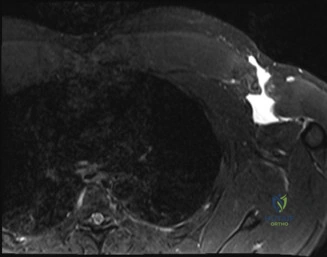
A 32-year-old competitive powerlifter presents to the clinic complaining of acute right-sided chest and axillary pain following a heavy "one-rep max" bench press session two days ago. He describes a "tearing sensation." On examination, you note significant ecchymosis extending from the chest wall into the proximal arm and a palpable defect at the anterior axillary fold. He has profound weakness in shoulder adduction and internal rotation. Based on the clinical presentation and the provided image, what is your primary diagnosis and immediate management plan?
Candidate: The patient has an acute pectoralis major rupture. Given the history of a bench press injury and the physical findings of a palpable defect and ecchymosis, it is likely a complete humeral avulsion. I would order an MRI to confirm the diagnosis and assess the degree of retraction. As he is a competitive athlete, I would recommend surgical repair within the acute window of 3-6 weeks.
Failing to mention the specific anatomical location (humeral insertion vs. musculotendinous) or neglecting the importance of timing. A poor candidate might simply suggest "surgery" without discussing the importance of the acute window (3-6 weeks) or the impact of retraction on the surgical approach.
The patient has a classic presentation of a complete pectoralis major tendon rupture, most likely at the humeral insertion (60-80% of cases). My immediate management includes: 1. Clinical confirmation: Assess for neurological deficits and inspect the deltopectoral interval. 2. Imaging: MRI is the gold standard to categorize the injury (humeral vs. musculotendinous) and assess the degree of muscle retraction and atrophy. 3. Counseling: Given his high functional demand as a powerlifter, surgical repair is indicated. I would advise that acute repair within the 3-6 week window is optimal to avoid the technical challenges of chronic retraction, scarring, and fatty degeneration of the muscle belly. I would also discuss the risks of stiffness, re-rupture, and the potential need for graft reconstruction if the tear is neglected.
The MRI confirms a complete humeral avulsion of the sternocostal head of the pectoralis major. The clavicular head remains intact. Explain the surgical anatomy and why this injury is common in this specific patient population.
Candidate: The pectoralis major has two heads: the clavicular and sternocostal. The insertion is unique; it is bilaminar and twisted, with the sternocostal fibers inserting more posteriorly and inferiorly than the clavicular head. In the bench press, the arm is in abduction and external rotation, which places the sternocostal head under maximum tension. A forceful eccentric contraction in this position causes the tendon to avulse from the bicipital groove.
Forgetting to mention the "twisted" nature of the insertion. The candidate must explicitly state that the sternocostal fibers are the ones that untwist and become tight during the bench press maneuver, which is why they are the most commonly injured fibers.
The insertion is bilaminar and twisted. The sternocostal head fibers insert more inferiorly and posteriorly on the lateral lip of the bicipital groove, effectively running 'under' the clavicular head. During the bottom phase of a bench press (abduction and external rotation), this configuration becomes untwisted and maximally tensioned. The sternocostal head, having longer fibers and a more posterior attachment, undergoes the highest eccentric load, predisposing it to avulsion at the humeral footprint. Knowledge of this anatomy is crucial for placing anchors appropriately during the anatomical reattachment of the bilaminar footprint.
During your repair, you encounter significant scar tissue and the muscle belly has retracted significantly. How does your surgical strategy change, and what are the potential complications of this repair?
Candidate: I would perform an extensive mobilization of the muscle belly, releasing adhesions from the pectoralis minor and chest wall, while carefully protecting the medial and lateral pectoral nerves. If I cannot achieve a tension-free repair, I would consider an allograft augmentation, such as an Achilles tendon graft, to bridge the defect. Potential complications include stiffness, re-rupture, cosmetic deformity, and damage to the pectoral nerves.
Omitting the protection of neurovascular structures. The nerves pass deep to the pectoralis major; aggressive blind mobilization can result in denervation of the muscle, leading to further atrophy and poor functional outcome.
My priority is restoring muscle length through meticulous release of adhesions. I must identify and preserve the medial and lateral pectoral nerves which enter the deep surface of the muscle. 1. Strategy: If the tendon is insufficient, I utilize a robust Krackow locking stitch and transition to allograft reconstruction (e.g., Achilles or semitendinosus) if the gap is too large to tension without over-constraining the shoulder. 2. Fixation: Use double-row or suture bridge construct to maximize footprint healing. 3. Complications: I would counsel the patient on the risk of post-operative stiffness (10-20%), persistent cosmetic deformity, and the risk of re-rupture if the post-operative rehabilitation protocol—which forbids active adduction and internal rotation—is not strictly followed.
