Corticosteroid-Induced Avascular Necrosis (AVN) of the Humeral Head: Etiology, Pathophysiology, and Clinical Insights

Key Takeaway
Avascular necrosis (AVN) of the humeral head, or ONFH, is bone cell death due to interrupted blood supply. Corticosteroid use is a significant non-traumatic risk factor. Pathophysiology involves fat embolization, hypercoagulability, and increased intraosseous pressure. Diagnosis uses systems like Ficat, Steinberg, and ARCO, with treatment guided by these stages.
A 42-year-old patient with systemic lupus erythematosus (SLE) presents with a 6-month history of progressive left shoulder pain. Plain radiographs are unremarkable. You suspect osteonecrosis of the humeral head (ONFH). What is the preferred initial imaging modality to confirm the diagnosis, and what specific findings would you look for?
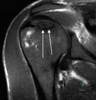
Candidate: I would order an MRI of the shoulder. It is the gold standard for early detection. I'd look for a low signal intensity band on T1-weighted images and potentially the "double-line sign" on T2-weighted images.
Candidates often stop at "MRI is the test." A high-scoring candidate must explain why (sensitivity) and specifically mention the "double-line sign" (hypervascular granulation tissue at the interface of necrotic and viable bone), demonstrating an understanding of the underlying pathophysiology.
MRI is the gold standard due to its superior sensitivity for Stage I disease. On T1-weighted images, I look for a well-demarcated band of low signal intensity. On T2-weighted images, I look for the 'double-line sign'—an inner bright line of hypervascular granulation tissue and an outer dark line of reactive sclerotic bone. This confirms early, pre-radiographic Stage I disease, which is critical for potentially joint-preserving interventions like core decompression.
Examine this radiographic image. Given the clinical presentation of chronic corticosteroid use and the current findings, what is your staging and proposed management?
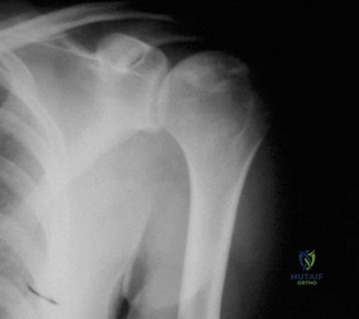
Candidate: This radiograph shows subchondral lucency consistent with the "crescent sign." This is Stage III disease. Since there is structural collapse, core decompression is no longer indicated; I would discuss arthroplasty options.
Failing to mention the Cruess staging by name or omitting the clinical correlation (mechanical failure). A poor candidate might suggest core decompression, showing a lack of understanding that the window for joint preservation has closed once the crescent sign appears.
This is Cruess Stage III disease, indicated by the pathognomonic 'crescent sign' representing subchondral bone collapse. Management should shift from joint-preserving techniques to arthroplasty. I would further perform a CT scan to evaluate the glenoid morphology and humeral head collapse geometry. Given the patient's age and etiology, if the glenoid is pristine, I would discuss the merits of hemiarthroplasty versus anatomic TSA with the patient, noting the trend toward TSA to prevent secondary glenoid erosion.
Discuss the vascular supply of the humeral head and its clinical significance in the context of surgical approach for AVN.
Candidate: The blood supply comes from the anterior and posterior humeral circumflex arteries. The ascending branch of the anterior humeral circumflex artery is the main one. During surgery, I'd use the deltopectoral approach to avoid damaging these vessels.
Relying on outdated concepts. The "arcuate artery of Laing" is no longer considered the primary supply. A failing candidate ignores the major contribution of the PHCA.
While historically the ascending branch of the Anterior Humeral Circumflex Artery (AHCA) was thought to be dominant, modern studies demonstrate that the Posterior Humeral Circumflex Artery (PHCA) provides up to 64% of the blood supply to the humeral head, particularly the superior and posterior quadrants. The PHCA traverses the quadrangular space. During a deltopectoral approach, it is vital to respect this posterior-medial vascular network. Understanding this anatomy emphasizes why the superior-central portion of the head—which is biomechanically loaded—becomes ischemic in corticosteroid-induced AVN.
