Hybrid Primary Total Hip: A Solution for Complex Joint Pain
Key Takeaway
Looking for accurate information on Hybrid Primary Total Hip: A Solution for Complex Joint Pain? A primary total hip arthroplasty treats severe hip osteoarthritis. A "hybrid" approach typically involves an uncemented acetabular component and a cemented femoral component. This method aims to provide optimal fixation and long-term stability, relieving pain and improving mobility for patients like the 86-year-old woman described, who suffered from debilitating right hip pain.
A 74-year-old patient presents with end-stage hip osteoarthritis. Pre-operative radiographs demonstrate a Dorr Type C femoral canal. You are planning a primary total hip arthroplasty. Please interpret the femoral morphology and discuss your choice of fixation.
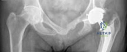
Candidate: The patient has a Dorr Type C femur, which means the cortical bone is thin and the canal is wide. I would probably choose a cemented femoral stem because press-fit uncemented stems might not have enough purchase in this type of bone, leading to a risk of fracture or subsidence. I would use an uncemented acetabular cup as the bone quality there is usually better.
Candidates often fail to classify the femur systematically or provide the biomechanical rationale for the "hybrid" choice. A poor answer focuses solely on the femur and ignores the acetabulum, or fails to mention the risk of "stress shielding" associated with modern uncemented stems in this specific population.
A perfect answer defines the Dorr Type C femur as a "stovepipe" morphology. It explains that the lack of metaphyseal flare and attenuated cortices makes press-fit fixation unreliable. The candidate should justify the hybrid construct: using a cemented stem to provide immediate, circumferential load transfer and a uniform cement mantle, which minimizes the risk of intraoperative periprosthetic fracture and early subsidence. Simultaneously, they justify the uncemented acetabular cup based on preserved acetabular bone stock and the efficacy of biological ingrowth in that region.
During the femoral preparation, you are using a cement gun to deliver the PMMA. What are the three critical steps required to ensure optimal cement-bone interdigitation for a cemented femoral stem?
Candidate: You need to clean the canal, use a plug, and put the cement in from the bottom up.
This is too colloquial. Failing to mention "pulsatile lavage," "distal cement restrictor," and "drying the canal" shows a lack of technical rigor expected at the FRCS level.
The candidate must articulate: 1. Canal Cleansing: Use of pulsatile lavage to remove fat, marrow, and debris to ensure the cement-bone interface is not compromised. 2. Distal Pressurization: Placement of an intramedullary cement restrictor (plug) to allow for retrograde filling and hydraulic pressurization of the cement into the cancellous interstices. 3. Canal Drying: Meticulous drying with swabs or compressed air to ensure moisture does not interfere with cement polymerization and bonding.
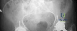
Look at this acetabular preparation image. What is the clinical goal of this reaming process, and how do you define the "safe zone" for acetabular component placement?
Candidate: You ream to get down to bleeding bone for the cup to stick to. The safe zone is about 40 degrees of inclination and 20 degrees of anteversion.
Failing to mention the biological goal of osseointegration or the risk of neurovascular injury during screw placement. Also, using "about 40 degrees" is vague; candidates should use the standard "Lewinnek's Safe Zone."
The candidate should state: 1. Goal of Reaming: To expose bleeding subchondral bone, facilitating biological osseointegration into the porous coating of the cup, while creating a press-fit (typically 1-2mm undersized). 2. Safe Zone: Reference the Lewinnek Safe Zone (40° ± 10° inclination, 15° ± 10° anteversion) to optimize stability and prevent impingement. 3. Neurovascular awareness: Crucially, mention avoiding screw placement in the anteroinferior quadrant of the acetabulum to prevent injury to the external iliac vessels and obturator nerve.
