Operative Management of Biceps Brachii and Posterior Tibial Tendon Displacement
Key Takeaway
Tendon displacement, particularly involving the long head of the biceps brachii and the posterior tibial tendon, presents unique biomechanical challenges. Surgical management requires meticulous approach selection, ranging from open subpectoral tenodesis to arthroscopic release, often dictated by concurrent rotator cuff pathology. This guide details the indications, step-by-step operative techniques, and evidence-based rehabilitation protocols necessary to restore optimal kinematics and prevent recurrent subluxation in high-demand patients.
Introduction to Tendon Displacement Pathoanatomy
Tendon displacement—encompassing subluxation and frank dislocation—represents a complex biomechanical failure of the retinacular and ligamentous pulley systems that stabilize musculotendinous units across joints. While tendon ruptures and ischemic myositis (such as acute or chronic exertional compartment syndrome) often dominate the discourse on soft-tissue trauma, the subtle yet debilitating nature of tendon instability requires an equally rigorous diagnostic and surgical approach.
This masterclass focuses on two distinct but biomechanically critical manifestations of tendon instability: the displacement of the long head of the biceps brachii (LHB) and the dislocation of the posterior tibial tendon (PTT). Both conditions demand a profound understanding of local anatomy, precise surgical execution, and tailored postoperative rehabilitation to restore native joint kinematics and prevent recurrent pathology.
Part I: Displacement of the Long Head of the Biceps Brachii
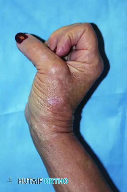
Displacement of the long head of the biceps tendon from the bicipital groove is rarely an isolated event; it is intimately associated with pathology of the rotator cuff, specifically the subscapularis and the supraspinatus tendons. The stability of the LHB is maintained by the "biceps pulley," a capsuloligamentous complex comprising the superior glenohumeral ligament (SGHL), the coracohumeral ligament (CHL), and the distal attachment of the subscapularis tendon.
When this pulley system fails, the LHB subluxates or dislocates medially. Historically, the transverse humeral ligament was considered the primary stabilizer; however, modern biomechanical studies have relegated its importance, emphasizing the SGHL and CHL. Consequently, surgical management has evolved from simple ligamentous repair to definitive tenodesis or tenotomy.
Indications for Surgery
Surgical intervention is indicated in patients with symptomatic, MRI-confirmed LHB displacement who have failed conservative management (NSAIDs, targeted physical therapy, corticosteroid injections). The choice of procedure depends heavily on the presence of concurrent rotator cuff pathology.
Clinical Pearl: Medial dislocation of the biceps tendon is pathognomonic for a subscapularis tear. Always evaluate the integrity of the subscapularis when LHB instability is identified on advanced imaging or during diagnostic arthroscopy.
Surgical Technique: Isolated Biceps Displacement (Anterior Approach)
In the rare instance where LHB displacement occurs without concomitant rotator cuff pathology, an anterior approach through the deltopectoral interval is utilized.
- Positioning and Anesthesia: The patient is placed in the beach-chair position under general anesthesia, often supplemented with an interscalene regional block.
- Incision and Dissection: A standard anterior incision is made over the deltopectoral groove. The cephalic vein is identified and retracted laterally with the deltoid to preserve its venous drainage.
- Exposure of the Bicipital Groove: The deltopectoral interval is developed. The long head of the biceps tendon is identified as it exits the joint capsule.
- Ligamentous Release: An incision is made through the transverse humeral ligament, extending distally through the proximal third of the pectoralis major tendon to fully expose the bicipital groove.
- Decision Making (Repair vs. Tenodesis):
- Historical Repair: Rarely, if the pulley system is acutely avulsed but structurally robust, the biceps tendon can be reduced into the groove, and the transverse humeral ligament/pulley complex repaired using heavy non-absorbable interrupted sutures.
- Definitive Tenodesis: More commonly, due to chronic attenuation of the pulley and tendon degeneration, a tenodesis is performed. The tendon is externalized, the diseased proximal portion is resected, and the healthy tendon is secured into the bicipital groove or subpectoral region using a bio-tenodesis screw or suture anchors.
Surgical Technique: Biceps Displacement with Rotator Cuff Pathology (Anterosuperior Approach)
When LHB subluxation is accompanied by rotator cuff tears (supraspinatus/subscapularis), an anterosuperior approach provides optimal exposure for both tenodesis and cuff repair.
- Incision: Begin the incision just lateral to the acromioclavicular (AC) joint, extending it distally in line with the anterior deltoid fibers.
- Deltoid Dissection: Perform wide subcutaneous dissection to expose the deltoid at its insertion. Split the deltoid beginning just lateral to the AC joint, continuing exactly 5 cm distally into the deltoid raphe.
Surgical Warning: Do not extend the deltoid split beyond 5 cm from the acromion. Extending the split further risks iatrogenic transection of the axillary nerve. Furthermore, meticulously preserve the acromial branch of the thoracoacromial artery during proximal dissection to ensure adequate vascularity to the anterior deltoid.
- Acromioplasty: Carefully dissect the deltoid off the anterior acromion using electrocautery. Perform an anterior acromioplasty to decompress the subacromial space, and excise the coracoacromial ligament.
- Tendon Management: Flex the shoulder to relax the musculature. Identify the long head of the biceps tendon. Perform a tenodesis to the proximal humerus using your preferred fixation method (e.g., interference screw or cortical button).
- Intra-articular Excision: Excise the redundant intra-articular portion of the LHB tendon to prevent mechanical catching within the glenohumeral joint.
- Rotator Cuff Repair: Proceed with the mobilization, footprint preparation, and repair of the rotator cuff tear using standard suture anchor techniques.
- Closure: Meticulously repair the deltoid split and its acromial origin through transosseous drill holes if detached. Close the subcutaneous tissue and skin in layers.
Arthroscopic Alternative
With advancements in minimally invasive techniques, the proximal biceps tendon attachment can be released arthroscopically.
* A standard posterior viewing portal and an anterior working portal are established.
* The LHB is identified, and an arthroscopic electrocautery device or biting punch is used to release the tendon at its superior labral origin.
* The tendon is allowed to retract distally into the bicipital groove.
* A subsequent mini-open subpectoral tenodesis is then performed to secure the tendon, removing it entirely from the inflammatory environment of the bicipital groove.
Postoperative Rehabilitation (Biceps Tenodesis)
- Phase I (0-2 Weeks): The patient is placed in a shoulder immobilizer. Passive range of motion (PROM) for the shoulder is initiated, but active elbow flexion and supination are strictly prohibited to protect the tenodesis site.
- Phase II (2-4 Weeks): The immobilizer is transitioned to a standard sling. Active-assisted range of motion (AAROM) begins.
- Phase III (4+ Weeks): Active use and progressive strengthening exercises are initiated.
- Note: If a concurrent rotator cuff repair was performed, the rehabilitation protocol must be dictated by the size and tension of the cuff repair, often delaying active motion for 4 to 6 weeks.
Part II: Posterior Tibial Tendon (PTT) Dislocation
Posterior tibial tendon dislocation is an exceedingly rare clinical entity, often misdiagnosed as a medial ankle sprain. It typically occurs secondary to forced dorsiflexion and inversion or eversion of the ankle, which forcefully avulses or ruptures the flexor retinaculum, allowing the PTT to subluxate anteriorly over the medial malleolus.
Pathoanatomy and Clinical Presentation
The PTT is stabilized behind the medial malleolus by the retromalleolar groove and the overlying flexor retinaculum. In patients with a shallow retromalleolar groove, the threshold for retinacular failure is significantly lowered.
Patients typically present with medial ankle pain, swelling, and a palpable snapping sensation over the medial malleolus during active dorsiflexion and inversion. Ouzounian and Myerson’s landmark review of recurrent subluxation of the PTT highlighted that successful outcomes depend on addressing both the soft-tissue incompetence and the underlying bony morphology.
Surgical Technique: Retinacular Repair and Groove Deepening
Operative intervention is the gold standard for recurrent PTT dislocation, as conservative management universally fails to restore the mechanical block required to prevent subluxation.
- Positioning: The patient is placed supine with a bump under the contralateral hip to externally rotate the operative leg, providing excellent access to the medial ankle. A thigh tourniquet is applied.
- Incision: A curvilinear incision is made posterior to the medial malleolus, following the course of the PTT.
- Exposure: The flexor retinaculum is identified. In chronic cases, it is often attenuated or healed in an elongated, incompetent state. The retinaculum is incised longitudinally, leaving a sufficient cuff for later repair.
- Tendon Inspection: The PTT is inspected for longitudinal split tears or severe tendinosis. Any degenerative tissue is debrided, and split tears are tubularized using a running 5-0 non-absorbable suture.
- Groove Deepening:
- A periosteal flap is elevated from the posterior aspect of the medial malleolus.
- Using a high-speed burr, the retromalleolar groove is deepened by 3 to 5 millimeters.
- Alternative: A cortical trapdoor can be elevated, the underlying cancellous bone impacted or burred, and the cortical roof tamped down to deepen the groove while preserving the smooth fibrocartilaginous gliding surface.
- Relocation and Repair: The PTT is relocated into the newly deepened groove. The flexor retinaculum is repaired tightly over the tendon using heavy non-absorbable sutures. If the retinaculum is deficient, a strip of periosteum or a local fascial flap can be utilized to augment the repair.
- Closure: The subcutaneous tissues and skin are closed meticulously to prevent wound breakdown in this watershed area.
Postoperative Rehabilitation (PTT Relocation)
- Weeks 0-2: The patient is placed in a non-weight-bearing short leg cast with the ankle in slight plantarflexion and inversion to remove tension from the repaired retinaculum.
- Weeks 2-6: The patient is transitioned to a controlled ankle motion (CAM) boot. Weight-bearing is gradually advanced. Gentle active range of motion is initiated out of the boot, strictly avoiding forced dorsiflexion and eversion.
- Weeks 6-12: Formal physical therapy commences, focusing on PTT strengthening, proprioception, and eccentric loading. Return to pre-injury activity is typically achieved by 4 to 6 months, with studies showing marked improvement and high rates of return to sport.
Part III: Differential Diagnosis and Associated Pathologies
When evaluating patients for tendon displacement, the orthopedic surgeon must maintain a broad differential diagnosis. The lower extremity, in particular, is susceptible to overlapping pathologies that can mimic or complicate tendon instability.
Chronic Exertional Compartment Syndrome (CECS)
Medial leg pain, often attributed to PTT pathology or medial tibial stress syndrome, may actually represent Chronic Exertional Compartment Syndrome (CECS) of the deep posterior compartment. As extensively documented by Rorabeck, Mubarak, and Whitesides, CECS is characterized by exercise-induced pain secondary to elevated intracompartmental pressures that compromise microvascular perfusion.
Diagnostic Pitfall: Do not mistake the ischemic pain of deep posterior compartment syndrome for PTT tendinopathy. If a patient presents with bilateral, exercise-induced medial leg pain that resolves with rest, intracompartmental pressure testing (using a slit-catheter or solid-state transducer) is mandatory before any tendon exploration is considered.
If CECS is confirmed (resting pressure > 15 mm Hg, 1-minute post-exercise pressure > 30 mm Hg, or 5-minute post-exercise pressure > 20 mm Hg), the treatment is a surgical fasciotomy. Modern techniques, including endoscopically assisted single-portal or two-portal minimal incision fasciotomies, have shown excellent long-term outcomes in returning athletes to high-level competition.
Acute Compartment Syndrome and Muscle Ruptures
In the setting of acute trauma, tendon dislocations can be accompanied by severe muscle strains or ruptures (e.g., medial head of the gastrocnemius or pectoralis major). The resulting hematoma and edema can precipitate acute compartment syndrome. The surgeon must remain vigilant; an unrecognized compartment syndrome can lead to irreversible ischemic myositis and Volkmann’s ischemic contracture. Immediate four-compartment fasciotomy of the leg is the definitive treatment if acute compartment syndrome is suspected, prioritizing limb salvage over isolated tendon repair.
Conclusion
The operative management of tendon displacement requires a nuanced appreciation of joint biomechanics and soft-tissue envelopes. Whether addressing the complex pulley system of the long head of the biceps in the shoulder or reconstructing the retromalleolar groove for the posterior tibial tendon in the ankle, the principles remain the same: restore the anatomical restraint, address concurrent intra-articular or tendinopathic pathology, and protect the repair through a phased, biologically sound rehabilitation protocol. Mastery of these techniques ensures that the orthopedic surgeon can reliably return patients to their highest level of function.
You Might Also Like