Holstein–Lewis Fracture: Comprehensive Clinical Management & Radial Nerve Injury
Key Takeaway
A Holstein–Lewis fracture is a specific spiral or oblique fracture of the distal one-third of the humerus, uniquely characterized by its high association (10-20% incidence) with radial nerve injury. The nerve is vulnerable as it pierces the lateral intermuscular septum, often entrapped by displaced fracture fragments, necessitating careful neurovascular assessment and tailored management.
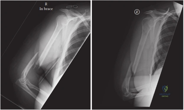
You are in the A&E department. A 32-year-old male presents following a fall from a bicycle. He has a closed injury to his right arm. Clinical examination reveals an obvious deformity of the distal third of the humerus. Crucially, the patient has an inability to extend his wrist and digits. Look at the radiograph provided below.
What is the specific diagnosis, and how would you manage the radial nerve palsy in this specific scenario?
Candidate: The patient has a Holstein-Lewis fracture of the distal humerus with a radial nerve palsy. Because there is a complete palsy, I would take the patient to surgery for ORIF and exploration of the radial nerve.
A failing candidate simply says "operate" without justifying the decision-making process. They often fail to mention the specific mechanism of radial nerve risk in Holstein-Lewis fractures (impingement at the lateral intermuscular septum) and don't distinguish between initial vs. post-reduction nerve palsy.
The candidate should state: "This is a Holstein-Lewis fracture—a spiral fracture of the distal third of the humerus. It is associated with a high risk of radial nerve palsy (10-20%) due to entrapment as the nerve pierces the lateral intermuscular septum. In the presence of a primary complete radial nerve palsy, the consensus is to proceed to early surgical exploration and internal fixation (ORIF). This allows for anatomical reduction and direct visualization of the nerve to rule out entrapment or transection. If the palsy were to develop post-reduction, urgent/immediate exploration would be mandatory to rule out iatrogenic injury."
Let's talk about the surgical approach. You have decided to proceed with ORIF for this patient. Which surgical approach do you favor, and how do you systematically identify the radial nerve?
Candidate: I would perform a posterior approach, either splitting or sparing the triceps. I would identify the nerve proximally in the spiral groove where it is safer, and then trace it distally towards the site of injury.
Candidates often suggest an anterolateral approach without acknowledging that the posterior approach is superior for direct visualization of the nerve in the distal third. Failing to mention the ulnar nerve at the medial elbow during a posterior approach is a significant safety omission.
The candidate must define: "I prefer a posterior approach as it offers the most reliable visualization of the radial nerve. 1. Ulnar Nerve: I identify and protect the ulnar nerve medially. 2. Triceps: Reflect or split the triceps. 3. Proximodistal approach: Identify the radial nerve proximal to the fracture site in the spiral groove, where it is often uninvolved in the hematoma/fragmentary zone. 4. Release: Trace it distally through the lateral intermuscular septum to ensure it is not entrapped by the displaced proximal fragment. 5. Protection: Use vessel loops to gently retract the nerve during the reduction and instrumentation phases."
During the procedure, you find the radial nerve is intact but contused. You have reduced the fracture. What is your preferred construct for stable fixation, and why?
Candidate: I would use dual plating. I'd place a posterior plate and a lateral plate, usually at 90 degrees to each other, to provide stable fixation for early rehabilitation.
Failing to mention interfragmentary compression (lag screws) before applying the plate. The spiral/oblique nature of a Holstein-Lewis fracture means that compression is the most important step to achieve stability.
The candidate must explain: "Given the spiral/oblique nature of the fracture, I would achieve interfragmentary compression first using independent lag screws across the fracture line. Then, I would apply dual plating (orthogonal or parallel) using locking compression plates. This construct provides the rotational stability required for early range-of-motion, which is essential to prevent arthrofibrosis and stiffness in the elbow. I would ensure at least 6-8 cortices of purchase on either side of the fracture."
