Bunionette Deformity: Epidemiology, Surgical Anatomy, Biomechanics & Management
Key Takeaway
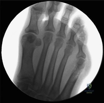
A bunionette (Tailor's Bunion) is a painful lateral forefoot prominence of the fifth metatarsal. It's categorized into three types: Type I (enlarged lateral condyle), Type II (lateral shaft bowing), and Type III (increased 4-5 IMA). Lateral condylar resection is a primary treatment for Type I deformities.
You are seeing a 45-year-old female complaining of chronic lateral-sided foot pain. She describes "difficulty with shoe wear" and a painful lump on the side of her foot. She has tried wide shoes and orthotics for 6 months with minimal improvement. Please describe the clinical classification of this condition and what your initial assessment would entail.
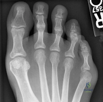
Candidate: This patient likely has a "Tailor's Bunion." I would classify it using the Kilmartin classification: Type I is lateral condylar hypertrophy, Type II is lateral bowing of the 5th metatarsal shaft, and Type III is increased 4-5 intermetatarsal angle. My assessment would include a clinical exam for skin changes and neurovascular status, followed by weight-bearing radiographs of the foot.
Candidates often jump straight to "I would offer surgery" or "I would resect the prominence" without performing a formal clinical exam or mentioning the importance of distinguishing between the three types. Failing to emphasize the 4-5 IMA measurement on radiographs is a major oversight.
A high-scoring answer defines the pathology as a Bunionette (Tailor's Bunion) and immediately invokes the Kilmartin classification. The candidate must systematically detail the physical exam (identifying the location of tenderness, neurovascular assessment of the lateral dorsal cutaneous nerve, and joint stability) and specify that standard weight-bearing AP, lateral, and oblique radiographs are required to measure the 4-5 IMA and assess for shaft bowing.
You have reviewed these weight-bearing radiographs. Based on the findings, what is your surgical decision-making process?
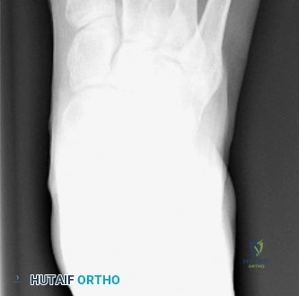
Candidate: If the patient has a Type I deformity with normal 4-5 IMA and no significant shaft bowing, I would consider a lateral condylar resection. If it were Type II or III, I would need an osteotomy to correct the shaft or metatarsal splay, as simple condylar resection would lead to a high rate of recurrence.
Proposing lateral condylar resection for all bunionettes. Failing to recognize that Type II and III deformities require osteotomy, not just soft tissue/condylar excision.
The candidate must explicitly state that the surgical choice is driven by the deformity type. "For a Type I deformity, lateral condylar resection is indicated because the pathology is localized to the articular prominence. For Type II or III, an osteotomy—such as a distal chevron or proximal closing wedge—is required to address the metatarsal alignment. I would emphasize that condylar resection for Type II/III is a known cause of surgical failure."
During your surgical approach for a lateral condylar resection, what is the most critical structure to protect, and how do you ensure its safety? Provide the intraoperative steps shown in the images.
Candidate: The most critical structure is the lateral dorsal cutaneous nerve, a branch of the sural nerve. I ensure safety through meticulous longitudinal dissection, careful retraction, and staying superficial. During the resection, I use an oscillating saw to remove the prominence while sparing the lateral collateral ligament and the articular cartilage.
Ignoring the nerve. Failing to mention the specific danger of over-resection, which compromises joint stability.
Identify the lateral dorsal cutaneous nerve immediately. The response should outline: 1. Careful blunt dissection to identify the nerve. 2. Use of a dorsolateral longitudinal incision. 3. Subperiosteal elevation. 4. Precise bone removal (3-5mm) with a saw while protecting the articular cartilage margin. 5. Smooth the bone with a rasp. 6. Ensure no impingement with passive ROM. 7. Meticulous closure without nerve entrapment.
