Comprehensive Diagnosis of Plantar Plate Rupture in Athletes: A Challenging Football Injury Case Study
Key Takeaway
Plantar plate rupture in athletes is diagnosed through a meticulous clinical exam revealing dorsal hammertoe deformity, interdigital splaying, and MTP joint instability (positive vertical stress test). Crucial imaging includes weight-bearing radiographs showing dorsal subluxation and definitive high-resolution MRI, often after initial misdiagnosis as a forefoot sprain.
A 22-year-old semi-professional athlete presents with persistent pain and instability in the second toe two weeks after a hyperextension injury during a football match. Physical examination shows a dorsal hammertoe deformity of the second digit. What is your clinical diagnosis, and how would you verify it?
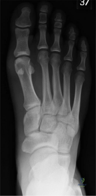
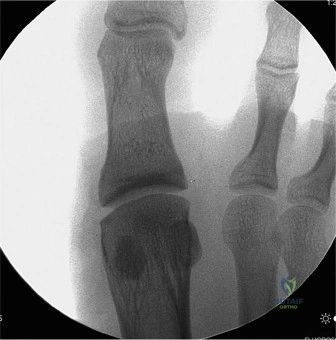
Candidate: I suspect a second metatarsophalangeal (MTP) joint plantar plate rupture. I would examine for a positive 'Lachman' equivalent test of the MTP joint, which would show sagittal plane instability. I would then order weight-bearing radiographs to look for dorsal subluxation and consider an MRI to confirm the soft-tissue injury.
Candidates often rely on non-weight-bearing films and state they are "normal," failing to appreciate that the dynamic subluxation is only visible on weight-bearing views or physical stress. They may also confuse this with a Morton's neuroma due to the location, missing the critical diagnosis of structural joint instability.
A structured response: 1. **Clinical Diagnosis:** Likely a complete rupture of the second MTP plantar plate. 2. **Examination:** Specifically mention the "Vertical Stress Test" (Lachman) and note the presence of a "crossover" or hammertoe deformity. 3. **Imaging:** Emphasize weight-bearing lateral radiographs (to assess subluxation) and high-resolution MRI to grade the injury (Type I-IV) and evaluate for collateral ligament involvement. Mentioning the exclusion of Freiberg Infraction and Morton's neuroma shows high clinical maturity.
The MRI confirms a complete distal avulsion of the plantar plate with collateral ligament attenuation. You have decided to proceed with a surgical repair via a dorsal approach with a Weil osteotomy. Why is the Weil osteotomy performed, and what is the single most critical technical detail to avoid a poor outcome?
Candidate: The Weil osteotomy is performed to decompress the MTP joint, which provides the necessary visualization of the plantar joint space to repair the retracted plantar plate. The most critical step is ensuring the cut is made parallel to the plantar aspect of the foot; otherwise, the metatarsal head will shift plantarly, causing painful transfer metatarsalgia.
Focusing only on the "shortening" aspect of the osteotomy. While shortening is a goal, failing to mention the *angle* of the cut demonstrates a lack of understanding of the biomechanical risk of post-operative metatarsalgia, which is the most frequent surgical complication.
State that the Weil osteotomy is an "intra-articular decompression maneuver." Explicitly define the technical requirement: the osteotomy must be cut **strictly parallel to the weight-bearing surface**. Mention the risk: "plantar displacement of the capital fragment leading to secondary transfer metatarsalgia." Also, note the necessity of releasing the extensor apparatus (EDL lengthening) to prevent tension on the repair.
Post-operatively, you utilize a transarticular K-wire. When and why do you remove it, and what is the role of the postoperative orthotic?
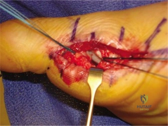
Candidate: The K-wire is usually removed at 6 weeks to protect the repair while allowing for early consolidation. It keeps the MTP joint in neutral or slight plantarflexion. The orthotic is then used to limit MTP dorsiflexion, usually with a stiff-soled shoe or carbon fiber insert, to prevent excessive strain on the healing plate during the return-to-sport phase.
Ignoring the rehabilitation phase. A candidate who suggests immediate mobilization without mentioning protected weight-bearing or orthotic support risks the examiner questioning the durability of the repair.
Structure the answer by healing biology: The K-wire is removed at 6 weeks to balance joint protection with the necessity of restoring ROM to prevent stiffness. The orthotic (rigid sole/carbon fiber) is critical for the "return to sport" phase to limit terminal dorsiflexion, which is the primary deforming force on the plantar plate repair. Mention the importance of intrinsic foot muscle strengthening (short foot exercises) to restore dynamic arch support.
