Bunionette Deformity (Tailor's Bunion): Etiology, Classification, Anatomy & Biomechanics
Key Takeaway
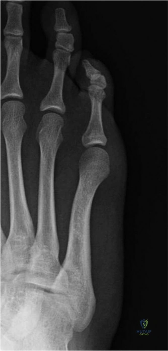
A bunionette, or tailor's bunion, is a painful prominence of the fifth metatarsal head. It's broadly classified into Type I (lateral exostosis), Type II (lateral bowing of the distal fifth metatarsal shaft), and Type III (increased 4-5 intermetatarsal angle). These classifications guide surgical planning by defining distinct anatomical abnormalities.
A 45-year-old female presents with a chronic, painful lateral prominence at the base of her 5th toe. She has failed 6 months of conservative management with wide-toe-box shoes and orthotics. An AP weight-bearing radiograph shows an increased 4th-5th intermetatarsal (IM) angle of 11 degrees with mild lateral bowing of the distal shaft.
Classify this deformity and describe the relationship between the anatomical classification and the selection of the surgical procedure.
Candidate: This is a Type III bunionette based on the Coughlin classification, given the increased 4th-5th IM angle. I would classify this as Type III because it involves a splayfoot component. My surgical plan would focus on a proximal osteotomy to correct the IM angle, rather than a simple exostectomy or distal osteotomy.
Candidates often jump straight to "distal chevron osteotomy." While popular, this ignores the biomechanical reality of a Type III deformity. Failing to address the proximal 4-5 IM angle in a patient with an 11-degree divergence leads to high recurrence rates. Also, failing to mention the risk to the lateral dorsal cutaneous nerve is a significant oversight.
This is a Coughlin Type III bunionette. Classification/Management Logic: 1. Type I (Exostosis): Treat with simple lateral condylectomy. 2. Type II (Lateral Bowing): Treat with distal/diaphyseal osteotomy. 3. Type III (Increased 4-5 IM Angle): Requires correction at the source—the proximal metatarsal—via crescentic or closing-wedge osteotomy. For this patient (11-degree IM angle), a proximal osteotomy is superior to a distal one to avoid lateral translation that fails to reduce the base divergence. I would also note the need to protect the lateral dorsal cutaneous nerve (sural branch) dorsolaterally and ensure robust fixation (plate/screws) given the higher biomechanical stress at the base.
During your surgical approach for a distal osteotomy, you are concerned about injury to the lateral dorsal cutaneous nerve. What are the specific anatomical risks, and how do you mitigate this during your exposure?
Candidate: The lateral dorsal cutaneous nerve, a branch of the sural nerve, runs along the dorsolateral aspect of the 5th metatarsal. I would be careful during the incision to identify it and retract it either dorsally or plantarly to avoid iatrogenic neuroma.
Treating this as a simple "don't cut the nerve" question. The candidate fails to emphasize that the nerve is "highly susceptible to compression or traction injury" even if not transected. A top-tier answer must mention the clinical consequence of an iatrogenic neuroma and the importance of blunt dissection.
The lateral dorsal cutaneous nerve is at high risk due to its superficial position over the lateral metatarsal head and shaft. Mitigation Strategy: 1. Incision Planning: Use a longitudinal or dorsolateral incision; avoid overly aggressive retraction that causes traction neurapraxia. 2. Identification: Perform meticulous sharp dissection in the subcutaneous layer to visualize the nerve early. 3. Protection: Once identified, use blunt dissection to mobilize the nerve and retract it out of the working field (typically plantarward is safer for the dorsolateral approach). 4. Salvage: If transected, perform primary repair or bury the proximal stump in deep tissue to prevent a symptomatic painful neuroma.
