Total Knee Arthroplasty: Biomechanics, Component Selection, and Surgical Principles
Key Takeaway
Total knee arthroplasty (TKA) remains one of the most successful orthopedic interventions. This comprehensive guide synthesizes decades of biomechanical research and clinical outcomes to detail component selection, kinematic principles, and surgical techniques. From cruciate-retaining versus posterior-stabilized designs to the nuances of gap balancing and polyethylene wear, mastering these evidence-based principles is essential for orthopedic surgeons striving to optimize implant survivorship and restore native knee kinematics in patients with end-stage osteoarthritis.
Introduction to Total Knee Arthroplasty
Total Knee Arthroplasty (TKA) represents a pinnacle of modern orthopedic surgery, offering profound pain relief and functional restoration for patients suffering from end-stage osteoarthritis, rheumatoid arthritis, and post-traumatic arthropathy. The evolution of TKA is deeply rooted in a rigorous understanding of knee biomechanics, tribology, and kinematic function. From the early polycentric designs of Gunston to the highly sophisticated, kinematically optimized implants of the modern era, the success of TKA relies entirely on the surgeon’s ability to balance soft tissues, restore mechanical alignment, and select the appropriate component design for the individual patient.
This masterclass synthesizes decades of foundational research—spanning in vivo kinematic studies, polyethylene wear analyses, and long-term survivorship data—to provide a comprehensive, textbook-level guide to the biomechanics, component selection, and surgical execution of total knee arthroplasty.
Biomechanics and Kinematics of the Reconstructed Knee
Understanding the kinematics of the native knee is paramount to replicating its function through arthroplasty. The knee is not a simple hinge; it is a complex polycentric joint that exhibits a combination of rolling, gliding, and axial rotation.
Femoral Rollback and Cruciate Function
In the native knee, the posterior cruciate ligament (PCL) acts as the primary restraint to posterior tibial translation. During deep flexion, the PCL drives femoral rollback—the posterior translation of the femoral condyles on the tibial plateau. This rollback is critical because it clears the posterior femoral cortex from impinging on the posterior margin of the tibia, thereby maximizing the arc of flexion and improving the extensor mechanism's mechanical advantage.
In TKA, restoring or substituting this kinematic profile is a primary design objective. Fluoroscopic in vivo kinematic studies have demonstrated that traditional TKA designs often fail to perfectly replicate native rollback. Instead of rolling back, some cruciate-retaining (CR) knees exhibit paradoxical anterior translation of the femur on the tibia during deep flexion, which can limit maximum flexion and increase polyethylene wear.
Clinical Pearl: Paradoxical anterior sliding in a CR TKA is often the result of an overly tight PCL or an unbalanced flexion gap. Proper PCL balancing—sometimes requiring a partial recession—is mandatory to achieve physiologic rollback and prevent accelerated posterior polyethylene wear.
Patellofemoral Kinematics
The patellofemoral joint is subjected to immense compressive forces, often exceeding multiple times body weight during stair ascent and descent. The Q-angle, patellar height, and the rotational alignment of the femoral and tibial components dictate patellar tracking. Internal rotation of the femoral component lateralizes the trochlear groove relative to the tibial tubercle, increasing the Q-angle and predisposing the patient to lateral patellar subluxation, tilt, and accelerated wear.
Component Design and Selection
The selection of TKA components must be tailored to the patient's ligamentous integrity, bone stock, and functional demands. The orthopedic surgeon must navigate several critical design dichotomies.
Cruciate-Retaining (CR) vs. Posterior-Stabilized (PS) Designs
The debate between retaining or sacrificing the PCL is one of the most enduring in joint reconstruction.
- Cruciate-Retaining (CR) TKA: Relies on an intact, properly tensioned PCL to provide posterior stability and drive femoral rollback. Proponents argue that CR designs preserve native proprioception, require less bone resection (as no intercondylar box is needed), and avoid the complications associated with a cam-post mechanism. However, CR knees are highly sensitive to joint line alterations. Elevating the joint line alters the PCL's origin-to-insertion distance, leading to kinematic dysfunction.
- Posterior-Stabilized (PS) TKA: Sacrifices the PCL and utilizes a tibial polyethylene post that engages a femoral cam to mechanically enforce femoral rollback during flexion. PS designs are indicated in cases of PCL insufficiency, severe deformity (e.g., severe varus/valgus or flexion contractures), or prior patellectomy. While PS knees reliably achieve rollback and often yield excellent flexion, they introduce specific risks, including cam-post wear, anterior tibial post impingement in hyperextension, and "patellar clunk syndrome."
Surgical Warning: Patellar clunk syndrome occurs in PS knees when a fibrotic nodule forms at the superior pole of the patella and catches in the intercondylar box during extension. Modern femoral components with a deepened, smoothed trochlear transition have significantly reduced the incidence of this complication.
Mobile-Bearing vs. Fixed-Bearing Articulations
Mobile-bearing knees (e.g., the New Jersey Low-Contact-Stress [LCS] system) were designed to decouple the conflicting requirements of high articular conformity (to reduce contact stress and wear) and rotational freedom (to accommodate native kinematics). By allowing the polyethylene insert to rotate on a highly polished tibial baseplate, mobile bearings theoretically reduce cross-shear forces on the polyethylene.
Despite the biomechanical rationale, long-term randomized controlled trials have shown no significant difference in survivorship or clinical outcome scores between mobile-bearing and fixed-bearing designs. Furthermore, mobile-bearing designs carry a unique risk of bearing spin-out or dislocation if the flexion and extension gaps are not perfectly balanced.
Polyethylene Tribology and Osteolysis
Ultra-high-molecular-weight polyethylene (UHMWPE) wear remains a primary limiting factor in the longevity of TKA. Wear debris generates a macrophage-mediated inflammatory response, leading to periprosthetic osteolysis and aseptic loosening.
Historically, polyethylene sterilized by gamma irradiation in air was subject to oxidative degradation, leading to catastrophic delamination and subsurface cracking. Modern highly cross-linked polyethylene (HXLPE), melted or annealed to eliminate free radicals, and sterilized in inert environments, has drastically reduced wear rates. Furthermore, optimizing articular conformity and ensuring a minimum polyethylene thickness of 8 mm (to prevent high subsurface shear stresses) are critical tenets of component selection.
Unicompartmental Knee Arthroplasty (UKA)
For patients with isolated medial or lateral compartment osteoarthritis, Unicompartmental Knee Arthroplasty (UKA) offers a less invasive alternative to TKA. UKA preserves both the anterior and posterior cruciate ligaments, resulting in kinematics that closely mirror the native knee.
Indications for UKA:
* Isolated unicompartmental osteoarthritis (most commonly medial).
* Intact ACL and PCL.
* Correctable varus or valgus deformity (intra-articular deformity, not extra-articular).
* Flexion contracture < 15 degrees.
* Flexion arc > 90 degrees.
UKA can be performed using fixed-bearing or mobile-bearing (meniscal bearing) designs. The Oxford mobile-bearing UKA has demonstrated excellent long-term survivorship when strict indications are followed. However, UKA is technically demanding; overcorrection of the mechanical axis into valgus (in a medial UKA) will rapidly accelerate disease progression in the preserved lateral compartment.
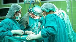
Surgical Technique: Step-by-Step Approach
Meticulous surgical technique is the bedrock of TKA survivorship. The following outlines the standard approach for a primary TKA using measured resection and gap balancing principles.
1. Preoperative Planning and Positioning
- Imaging: Weight-bearing anteroposterior (AP), lateral, and skyline patellar radiographs, alongside full-length standing alignment films, are mandatory. The mechanical axis, anatomic axis, and the angle between them (typically 5-7 degrees) must be calculated.
- Positioning: The patient is positioned supine. A tourniquet is applied to the proximal thigh. A lateral post or leg holder is utilized to allow stable positioning of the knee at 90 degrees of flexion and full extension.
2. Surgical Approach
- Incision: A straight midline longitudinal incision is made from the superior pole of the patella to the medial border of the tibial tubercle.
- Arthrotomy: A standard medial parapatellar arthrotomy is performed. The incision extends through the medial retinaculum, leaving a 2-3 mm cuff of tissue on the vastus medialis obliquus (VMO) for robust closure.
- Exposure: The patella is everted or subluxated laterally. The ACL is excised. If a PS design is chosen, the PCL is also excised. Medial and lateral menisci are resected, and peripheral osteophytes are meticulously removed to prevent soft-tissue tethering.
3. Bone Preparation and Alignment
The goal is to create rectangular, symmetric flexion and extension gaps while restoring the mechanical axis to neutral (0 degrees).
- Femoral Preparation: An intramedullary alignment rod is introduced into the distal femur. The distal femoral resection is typically set at 5 to 7 degrees of valgus relative to the anatomic axis.
- Femoral Rotation: Establishing correct femoral rotation is critical for patellar tracking and flexion gap symmetry. Rotation is determined using three anatomical landmarks:
- The Transepicondylar Axis (TEA): The clinical TEA connects the lateral epicondylar prominence to the medial epicondylar sulcus.
- Whiteside’s Line: The AP axis of the trochlea. The femoral component should be placed perpendicular to this line.
- Posterior Condylar Axis: Typically, the femur is externally rotated 3 degrees relative to the posterior condylar axis to account for the native medial tibial slope.
- Tibial Preparation: An extramedullary alignment guide is typically used. The tibial resection is made perpendicular to the mechanical axis of the tibia in the coronal plane. In the sagittal plane, a posterior slope of 3 to 7 degrees is incorporated, depending on the implant design and whether the PCL is retained.
Clinical Pearl: Never internally rotate the tibial component to improve coverage. The tibial component should be aligned with the medial third of the tibial tubercle. Internal rotation of the tibia or femur will lead to lateral patellar tracking and catastrophic patellofemoral complications.
4. Gap Balancing
Once the initial resections are made, spacer blocks or laminar spreaders are used to assess the extension and flexion gaps.
* Extension Gap: Defined by the distal femoral resection and the proximal tibial resection. If the gap is tight medially, a medial release (deep MCL) is performed.
* Flexion Gap: Defined by the posterior femoral resection and the proximal tibial resection.
* Both gaps must be rectangular and equal in magnitude. If the extension gap is tight but the flexion gap is balanced, additional distal femur must be resected.
5. Patellar Resurfacing
The decision to resurface the patella remains controversial. Routine resurfacing eliminates the risk of anterior knee pain from native patellofemoral arthritis, while selective resurfacing preserves bone stock. If resurfacing is performed, the patella is resected symmetrically to restore its native composite thickness. The component is medialized to optimize tracking. The "no thumb" test is performed: the patella should track centrally in the trochlear groove during a full range of motion without being held by the surgeon's thumb.
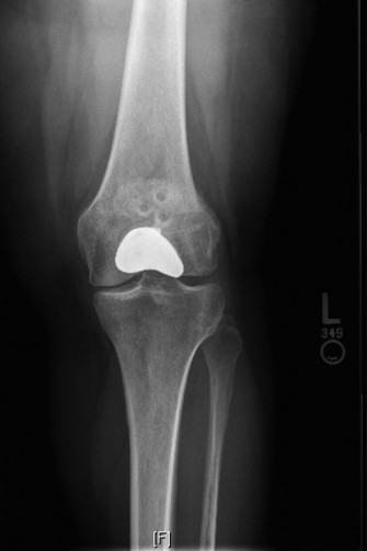
6. Component Implantation
The bone surfaces are thoroughly irrigated and dried using pulsatile lavage. High-viscosity polymethylmethacrylate (PMMA) bone cement is applied to the bone and the components. The implants are impacted into position, and the knee is brought into full extension to pressurize the cement during polymerization. Excess cement is meticulously removed, particularly from the posterior compartments.
Postoperative Protocol and Complication Management
Analgesia and VTE Prophylaxis
Modern TKA recovery relies heavily on multimodal analgesia. Preemptive analgesia (gabapentin, acetaminophen, NSAIDs), intraoperative periarticular injections (bupivacaine, epinephrine, ketorolac), and regional anesthesia (adductor canal blocks) have largely replaced epidural anesthesia, facilitating rapid mobilization.
Venous thromboembolism (VTE) prophylaxis is mandatory. Depending on patient risk stratification, agents ranging from enteric-coated aspirin to low-molecular-weight heparin (LMWH) or direct oral anticoagulants (DOACs) are utilized for 2 to 4 weeks postoperatively.
Rehabilitation
Early mobilization is the cornerstone of preventing arthrofibrosis and deep vein thrombosis. Patients are mobilized on the day of surgery. Physical therapy focuses on achieving full active extension (to prevent flexion contractures) and maximizing the flexion arc.
Conclusion
Total knee arthroplasty is a highly reproducible, life-altering procedure when executed with precision. Mastery of TKA requires an intimate understanding of knee kinematics, a rigorous approach to component selection, and an uncompromising dedication to surgical technique. By adhering to the evidence-based principles of mechanical alignment, gap balancing, and modern tribology, the orthopedic surgeon can consistently deliver durable, high-functioning joint reconstructions.
📚 Medical References
- total knee arthroplasty in patients who have a long-standing fusion of the hip, J Bone Joint Surg 71A:1355, 1989.
- Haber D, Goodman SB: Total hip arthroplasty in juvenile chronic arthritis, J Arthroplasty 13:259, 1998.
- Haidukewych GJ, Berry DJ: Hip arthroplasty for salvage of failed treatment of intertrochanteric
You Might Also Like