Optimizing TKR in Valgus: Expert Strategies for Complex Cases
Key Takeaway
In this comprehensive guide, we discuss everything you need to know about Optimizing TKR in Valgus: Expert Strategies for Complex Cases. TKR in valgus involves total knee arthroplasty for a valgus (knock-knee) deformity, commonly associated with rheumatoid arthritis or osteoarthritis. Key considerations include thorough preoperative assessment of ligament competency and deformity. Intraoperatively, surgeons address challenges like deficient lateral femoral condyles, femoral/tibial rotation, and soft tissue imbalance, often requiring specific adjustments or a lateral retinacular release for optimal patella tracking.
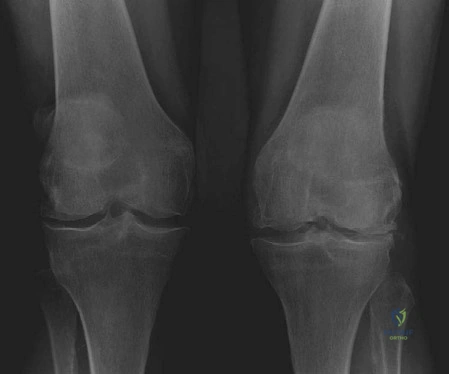
This is a 72-year-old lady with chronic bilateral knee pain. She is referred for consideration of total knee arthroplasty (TKA). Describe the radiographic findings in Figure 3.1 and outline the associated clinical patterns.
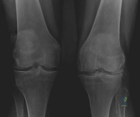
Candidate: These are weightbearing AP views showing significant joint space narrowing and subchondral sclerosis predominantly in the lateral compartments, with relative preservation medially. There is a clear valgus deformity. This pattern is often seen in rheumatoid arthritis, post-traumatic settings (e.g., previous lateral meniscectomy), or primary OA in women.
Candidates often fail to describe the bilateral nature or the specific location of the joint space narrowing. Simply saying "arthritis" is insufficient. A poor answer neglects the etiology—failing to differentiate between primary OA and inflammatory conditions like RA, which significantly impacts systemic surgical planning.
State: "Bilateral weightbearing AP radiographs demonstrating asymmetric joint space loss with lateral compartment predominance and secondary valgus alignment. Crucially, I would evaluate for inflammatory markers, as this presentation—particularly if progressive—is a hallmark of inflammatory arthropathy or severe primary degenerative valgus knee. The differential includes post-traumatic deformity, previous high tibial osteotomy overcorrection, or neurogenic arthropathy."
During the TKA procedure, what are the primary structural challenges posed by the valgus knee, and how do you address the rotational alignment of the femoral component?
Candidate: The lateral femoral condyle is hypoplastic/deficient. Consequently, using posterior condylar referencing can lead to internal rotation of the femoral component. I would use the Whiteside line (AP axis) to determine rotation and manage the soft tissues by releasing the tight lateral structures.
Forgetting the specific effect of posterior condylar referencing in a deficient lateral condyle is a fatal flaw. Candidates often fail to mention that the tibia is effectively externally rotated and the vastus lateralis may require release to fix patellar tracking.
Systematically structure the response: 1) Anatomy: Lateral condyle deficiency and contracted lateral/posterior structures. 2) Rotational Alignment: Avoid standard 3-degree posterior referencing as it results in component internal rotation; use the Whiteside line or transepicondylar axis. 3) Patellar Tracking: Mention the role of lateral retinacular release. 4) Gaps: Acknowledge that lateral/posterior releases significantly increase the extension gap, potentially requiring thicker implants or constrained liners.
You have performed a significant soft tissue correction for a severe valgus deformity. What specific neurological complication must you monitor for postoperatively, and why?
Candidate: I must monitor for common peroneal nerve palsy. This is caused by the sudden lengthening of the soft tissues on the lateral side of the knee as the valgus deformity is corrected.
Candidates often identify the nerve but fail to discuss the management of the "at-risk" patient. A high-scoring candidate should mention intraoperative knee flexion or the role of checking the nerve if the deformity correction is >20 degrees.
Identify the Common Peroneal Nerve (CPN). Explain the mechanism: traction injury due to rapid correction of chronic valgus. Mitigation strategy: If a severe deformity is corrected, keep the knee in some flexion postoperatively, avoid over-lengthening, and perform a formal pre- and post-op neurovascular examination. If persistent stretch is anticipated, consider prophylactic nerve decompression.
