Complex Patellar Fracture: A Case Study in Surgical Management & Rehabilitation
Key Takeaway
High-energy patellar fractures require meticulous management, as demonstrated in this case study of a 45-year-old carpenter. Key considerations include assessing injury mechanism and clinical presentation to guide surgical repair. Post-operative rehabilitation is crucial to restore knee extensor mechanism function, ensuring optimal recovery for demanding occupations and preventing long-term disability.
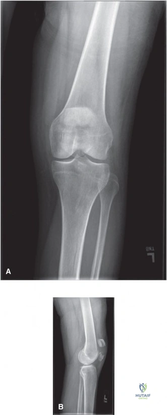
Mr. Davies presents with a high-energy injury to his knee. You are presented with his lateral knee radiograph. Describe the findings and explain the implications for the extensor mechanism.
Candidate: The radiograph shows a comminuted, displaced fracture of the patella. There is clear superior pole retraction and loss of articular congruity. Given the patient cannot perform a straight leg raise, this confirms a complete disruption of the extensor mechanism.
Failing to mention the specific classification (AO/OTA 34-C3), ignoring the soft tissue status (even if inferred as closed), or failing to quantify the displacement/articular step-off which dictates the surgical urgency and complexity.
Systematically describe the image: "This is a lateral view showing a comminuted, intra-articular patellar fracture (AO/OTA 34-C3). There is significant cephalad retraction of the superior fragment due to quadriceps tension, and a posterior articular step-off >2mm. Clinically, this correlates with a functional failure of the extensor mechanism. My management strategy would involve urgent open reduction and internal fixation to restore the length of the patella, anatomical articular congruity, and retinacular integrity."
You have decided to proceed to theatre. Why is simple Tension Band Wiring (TBW) potentially insufficient for this specific fracture pattern, and how would you augment your fixation?
Candidate: Simple TBW relies on the tension band principle to convert distractive forces to compression. However, in highly comminuted fractures, it provides poor rotational and shear stability. I would augment this with a low-profile plate and interfragmentary screws.
Suggesting TBW is "wrong"—it is a recognized technique, but the candidate fails to understand *why* it fails in comminution. Failing to mention retinacular repair as a critical component of the construct stability is also a major oversight.
The candidate should state: "In comminuted fractures, the bone fragments are too small to support pure TBW, leading to fracture collapse and potential hardware failure. I would use a 'hybrid' construct: 1. Meticulous articular reduction using K-wires and cerclage as a containment frame. 2. Application of a pre-contoured small fragment (2.7/3.5mm) plate to provide multi-planar stability against shear and rotation. 3. Ensuring the retinacula are repaired with non-absorbable sutures to restore the full extensor retinacular apparatus, which is essential for tracking and preventing long-term subluxation."
