Comprehensive Introduction and Patho-Epidemiology
Fracture-subluxations and fracture-dislocations of the proximal interphalangeal (PIP) joint represent some of the most formidable and technically demanding intra-articular injuries managed by hand and orthopedic surgeons. The PIP joint is the primary arc of motion for digital flexion, contributing approximately 85% of the total flexion arc of the finger. Consequently, any disruption to its congruent articular surface or supporting ligamentous structures can lead to devastating functional impairment, characterized by severe stiffness, chronic pain, and early post-traumatic osteoarthritis. These injuries predominantly affect young, active individuals, manual laborers, and athletes, often resulting from a high-energy axial load combined with forced hyperextension.
The pathomechanics of this injury typically involve a shear or impaction fracture of the volar base of the middle phalanx. When an axial load is applied to a hyperextended digit, the volar lip of the middle phalanx is driven into the condyles of the proximal phalanx. If the applied force exceeds the structural integrity of the subchondral bone, the volar base fractures. The critical threshold for instability is widely recognized as 30% to 40% involvement of the volar articular surface. When the fracture fragment exceeds this size, the primary volar stabilizing structures—the volar plate and the accessory collateral ligaments—remain attached to the fractured fragment rather than the intact shaft of the middle phalanx. Deprived of this volar tether, the remaining intact middle phalanx base is subjected to the unopposed dorsal and proximal pull of the extensor mechanism, leading to dorsal subluxation or frank dislocation.
Historically, the management of these complex injuries was fraught with complications. Static dorsal block splinting, while effective for minor, stable fractures, inevitably leads to profound joint stiffness and flexion contractures when utilized for prolonged periods in unstable injuries. Conversely, open reduction and internal fixation (ORIF) with screws or plates is often technically impossible due to the severe comminution of the volar fragment (the "pilon" variant) and carries a high risk of devascularizing the small articular fragments, leading to avascular necrosis and further stiffness.
To navigate the treacherous dichotomy between instability and stiffness, dynamic external splint reduction—most notably pioneered by Agee with his force-couple technique, and later modified by Suzuki with the pins and rubber band (PRB) traction system—has become a cornerstone of operative management. These techniques rely on a shared, elegant biomechanical principle: coupling longitudinal distraction (ligamentotaxis) with volarly directed forces across the joint. By neutralizing the deforming forces of the extensor mechanism, the dynamic external splint maintains concentric joint reduction during the critical phases of bone and soft tissue healing while permitting immediate active range-of-motion (ROM) exercises. This early mobilization is paramount; it enhances cartilage nutrition through synovial fluid diffusion, prevents restrictive tendon adhesions, and dynamically molds the healing fracture callus to the contour of the proximal phalanx condyles.
Detailed Surgical Anatomy and Biomechanics
A profound understanding of the osseous architecture, ligamentous restraints, and dynamic forces acting upon the PIP joint is an absolute prerequisite for the successful application of a dynamic external splint. The PIP joint is a classic ginglymus (hinge) joint. The head of the proximal phalanx is bicondylar, featuring an intercondylar groove that articulates with the biconcave base of the middle phalanx. This geometric congruency provides inherent osseous stability, but it is the surrounding soft tissue envelope that dictates the joint's kinematic behavior.
The primary static stabilizers of the PIP joint are the proper collateral ligaments (PCL), the accessory collateral ligaments (ACL), and the volar plate. The PCL originates from the dorsal aspect of the proximal phalanx head and inserts onto the volar-lateral aspect of the middle phalanx base, remaining taut in flexion to resist lateral deviation. The ACL originates just volar to the PCL and inserts directly into the lateral margins of the volar plate. The volar plate itself is a thick, fibrocartilaginous structure distally where it inserts into the volar base of the middle phalanx, but becomes membranous proximally where it attaches to the proximal phalanx, allowing it to fold upon itself during joint flexion. Together, the volar plate and ACL form a robust "check-rein" mechanism that prevents hyperextension. In a dorsal fracture-dislocation, this entire volar complex is avulsed along with the volar fracture fragment.
The dynamic forces acting across the PIP joint are generated by the extrinsic and intrinsic musculotendinous units. The flexor digitorum superficialis (FDS) bifurcates and inserts onto the volar aspect of the middle phalanx, exerting a volar and proximal force. Dorsally, the central slip of the extensor tendon inserts onto the dorsal base of the middle phalanx, while the lateral bands bypass the joint to form the terminal tendon. In the setting of a volar base fracture, the loss of the volar buttress allows the central slip and lateral bands to exert an unopposed dorsal and proximal pull. This dynamic imbalance is the primary driver of dorsal subluxation.
The biomechanical genius of the Agee force-couple splint lies in its ability to counteract these specific deforming forces through a precise mechanical linkage. The system relies on the concept of the instant center of rotation (ICR). In the PIP joint, the ICR is located exactly at the center of the concentric circles formed by the proximal phalanx condyles when viewed on a true lateral radiograph. By placing Kirschner wires (K-wires) perfectly through the ICR of the PIP and distal interphalangeal (DIP) joints, the surgeon creates a dynamic hinge that perfectly mimics the joint's native kinematics.
The force-couple achieves reduction through two distinct mechanical vectors:
1. Longitudinal Distraction: Tension generated by the rubber band connecting the proximal and distal K-wires unloads the articular surface. This utilizes the principle of ligamentotaxis—tensioning the intact soft tissue envelope to partially reduce the comminuted volar fracture fragments and restore the length of the digit.
2. Volar Translation: The mechanical linkage is intentionally designed so that the tension of the rubber band creates a volar vector on the middle phalanx. This active volar force directly resists the dorsal pull of the extensor mechanism, preventing the middle phalanx from riding up over the proximal phalanx condyles.
The ultimate goal of this biomechanical construct is to achieve a "parallel gliding motion." As the patient actively flexes and extends the digit, the biconcave base of the middle phalanx must glide concentrically along the condyles of the proximal phalanx. If the pins are placed eccentrically (missing the true ICR), the joint will exhibit a "rocking motion." This non-concentric hinge is catastrophic; it subjects the articular cartilage to exceptionally high focal surface pressures, leading to rapid cartilage necrosis, secondary traumatic arthritis, and inevitable recurrent joint subluxation.
Exhaustive Indications and Contraindications
The decision to utilize a dynamic external splint must be based on a rigorous evaluation of the injury pattern, the integrity of the remaining soft tissues, and the physiological profile of the patient. Proper patient selection is the most critical determinant of a successful outcome.
Indications
Dynamic external splinting is primarily indicated for acute dorsal fracture-subluxations and fracture-dislocations of the PIP joint where the volar articular defect exceeds 30% to 40% of the joint surface. At this threshold, the joint is universally unstable and will inevitably subluxate dorsally if treated with simple extension block splinting. Furthermore, this technique is the treatment of choice for highly comminuted, "pilon-type" fractures of the middle phalanx base. In these scenarios, the volar fragments are too small, numerous, and osteopenic to hold internal fixation (screws or plates). Attempting ORIF in such cases often results in iatrogenic fragmentation, devascularization of the articular pieces, and catastrophic failure. The dynamic splint bypasses the need for direct fixation, relying instead on ligamentotaxis to mold the comminuted fragments while maintaining joint reduction.
Additionally, dynamic external fixation is highly valuable for injuries that demonstrate persistent instability after an attempted closed reduction and dorsal block splinting. It is also utilized in the management of chronic dorsal fracture-subluxations (typically presenting more than 3 to 4 weeks post-injury). In these delayed presentations, the splint is applied in conjunction with an open surgical approach to release contracted collateral ligaments, clear intra-articular fibrosis, and mobilize the malunited volar fragments.
Contraindications
The contraindications for dynamic external splinting are absolute and must be strictly respected to avoid severe iatrogenic complications. The most critical contraindication is a volar fracture-dislocation. Because the force-couple mechanism is specifically engineered to generate a volar translation vector to counteract dorsal subluxation, applying this device to a volar dislocation will actively exacerbate the deformity, driving the middle phalanx further palmarward.
Another absolute contraindication is severe, irreparable collateral ligament deficiency. The dynamic splint relies on the intact portions of the collateral ligaments to provide lateral stability and to act as a tether against which the volar vector can pull. If the collateral ligaments are completely destroyed, the volar vector of the force-couple will simply pull the middle phalanx straight through the joint, converting a dorsal dislocation into an iatrogenic palmar dislocation.
Patient compliance is also a paramount consideration. The success of this device relies heavily on the patient's ability to perform rigorous, active ROM exercises multiple times an hour and to maintain meticulous pin site hygiene. Patients with severe cognitive impairment, active substance abuse issues, or those who are uncooperative with rehabilitation protocols are poor candidates. Finally, severe osteoporosis or metabolic bone disease is a relative contraindication, as the K-wires may cut through the soft cancellous bone of the phalangeal heads under the continuous tension of the rubber bands, leading to loss of reduction and frame failure.
Summary of Indications and Contraindications
| Category | Specific Conditions | Rationale / Biomechanical Implication |
|---|---|---|
| Indications | Acute dorsal fracture-subluxations (>30% articular loss) | Restores concentric reduction; counteracts unopposed extensor pull. |
| Indications | Comminuted "pilon-type" volar base fractures | Avoids devascularization from ORIF; utilizes ligamentotaxis to mold fragments. |
| Indications | Failure of closed reduction / dorsal block splinting | Provides rigid dynamic stability when static methods fail to maintain reduction. |
| Indications | Chronic dorsal fracture-subluxations (with open release) | Maintains reduction after extensive soft tissue releases and joint mobilization. |
| Contraindications | Volar fracture-dislocations | Absolute: The device's volar vector will actively worsen a volar dislocation. |
| Contraindications | Complete, irreparable collateral ligament destruction | Absolute: Loss of lateral tether will result in iatrogenic palmar dislocation. |
| Contraindications | Non-compliant patients (cognitive, psychiatric, substance) | Absolute: Device relies entirely on patient-driven active ROM and pin care. |
| Contraindications | Severe osteoporosis / metabolic bone disease | Relative: High risk of K-wire cut-out through the phalangeal axis of rotation. |
Pre-Operative Planning, Templating, and Patient Positioning
Meticulous preoperative planning is the foundation of a flawless surgical execution. The evaluation begins with high-quality, perfectly positioned radiographs. True anteroposterior (AP) and true lateral radiographs of the affected individual digit (not the entire hand) are mandatory. The true lateral view is particularly critical; the condyles of the proximal phalanx must overlap perfectly to form a single concentric circle. It is on this view that the surgeon assesses the size of the volar fragment, the degree of comminution, and the presence of the "V" sign—a dorsal opening of the PIP joint space indicating dorsal subluxation of the middle phalanx. In cases of complex, highly comminuted pilon fractures, a dedicated computed tomography (CT) scan with 3D reconstructions may be warranted to fully appreciate the articular topography and plan potential open fragment mobilization.
Templating involves identifying the precise axis of rotation on the lateral radiograph. The surgeon must mentally map the insertion points for the axis pins. The proximal pin must pass exactly through the center of the proximal phalanx condyles, while the distal pin must pass through the center of the middle phalanx condyles (at the DIP joint).
The choice of anesthesia is a critical strategic decision that directly impacts the surgical technique. For acute injuries where a closed percutaneous reduction is anticipated, the procedure is strongly preferred to be performed under a precise digital block anesthesia (e.g., using 1% lidocaine or 0.5% bupivacaine without epinephrine). This is not merely for patient comfort; it is a vital surgical tool. A digital block provides excellent local anesthesia to the digit while preserving the function of the extrinsic flexor and extensor muscles located in the forearm. This allows the patient to act as an active participant intraoperatively, demonstrating the joint’s active range of motion under fluoroscopy to confirm concentric reduction and the absence of joint rocking.
Conversely, for chronic injuries requiring an open approach, extensive soft tissue releases, and potential osteotomies, a digital block is insufficient. These procedures require an axillary block or regional anesthesia to ensure complete patient comfort and to allow the use of a pneumatic arm tourniquet, providing the absolute bloodless field necessary for meticulous intra-articular work.
Patient positioning is standardized. The patient is placed supine with the affected arm extended on a radiolucent hand table. A mini C-arm fluoroscopy unit is positioned parallel to the floor, allowing the surgeon to obtain true lateral views by simply rotating the patient's forearm, rather than awkwardly maneuvering the heavy C-arm. The surgical tray must be equipped with smooth 0.045-inch (1.14 mm) K-wires, heavy-duty wire cutters, specialized wire benders, a selection of sterile orthodontic rubber bands, and needle drivers for precise frame assembly.
Step-by-Step Surgical Approach and Fixation Technique
The application of a dynamic external splint is an exercise in geometric precision. A deviation of even a few millimeters in pin placement will alter the joint kinematics, leading to failure. The technique varies depending on whether the injury is acute (amenable to closed reduction) or chronic (requiring open release).
Closed Percutaneous Application (Acute Injuries)
When the injury is acute (typically less than 10-14 days old) and the fracture fragments are mobile, the force-couple splint is applied percutaneously.
Step 1: Axis Identification and Pin Placement
Under fluoroscopic guidance, the surgeon must obtain a perfect true lateral view of the PIP joint. The condyles of the proximal phalanx must perfectly overlap, appearing as a single, flawless concentric circle. Using a mini-driver, a smooth 0.045-inch K-wire is inserted strictly perpendicular to the longitudinal axis of the phalanx, passing exactly through the center of rotation of the proximal phalanx head. This is the most critical step of the operation. A second smooth K-wire is then inserted in an identical fashion through the center of rotation of the middle phalanx head at the DIP joint.
Step 2: Mechanical Linkage Assembly
Once the axis pins are confirmed to be perfectly central on both AP and lateral fluoroscopic views, the mechanical linkage is assembled. The K-wires are bent at precise 90-degree angles at an equal distance from the skin on both sides of the digit. The wires are fashioned into interlocking hooks or loops, depending on the specific frame design (Agee vs. Suzuki).
Crucial Step: The surgeon must ensure there is adequate clearance (at least 5-7 mm) between the wire linkage and the skin throughout the entire arc of motion. Postoperative edema is inevitable, and insufficient clearance will lead to devastating pressure necrosis of the digital skin.
Step 3: Applying the Force-Couple
A sterile orthodontic rubber band is placed across the linkage hooks. The tension of the rubber band is the engine of the device. The surgeon must apply tension adequate to maintain reduction and counteract the dorsal subluxation.
Avoid excessive tension: Over-distraction is a common and severe technical error. It will lead to a widened joint space, preventing union of the fracture fragments, and causing severe, intractable joint stiffness. The tension should be calibrated to be just enough to maintain a concentric reduction of the middle phalanx on the proximal phalanx.
Step 4: Intraoperative Dynamic Assessment
With the frame assembled and tensioned, the surgeon asks the patient (who is under digital block) to actively flex and extend the finger. The quality of joint reduction is determined dynamically using continuous fluoroscopy. The surgeon must examine the flexion and extension lateral radiographs closely to ensure that the intact dorsal base of the middle phalanx is concentrically reduced. The surgeon is looking for a smooth, parallel gliding motion of the articular surfaces.
⚠️ Surgical Warning: The "Rocking" Joint
If fluoroscopy reveals a rocking motion, hinging, or opening of the joint space during active flexion or extension, the pins are definitively not in the true axis of rotation, or the rubber band tension is vastly incorrect. This cannot be accepted. The surgeon must immediately remove the pins, re-evaluate the axis, and re-insert them. Leaving a joint to "rock" guarantees early mechanical failure, rapid cartilage destruction, and severe traumatic arthritis.
Open Reduction (Chronic or Complex Injuries)
In chronic injuries (presenting >3-4 weeks post-injury), the fracture callus has begun to consolidate, the collateral ligaments are severely contracted, and the joint is filled with dense fibrotic tissue. Closed reduction is impossible, and an open approach is mandatory.
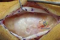
Step 1: Surgical Approach
With the patient under regional anesthesia and tourniquet control, a midlateral incision is utilized on the most affected side of the digit. This approach avoids the volar flexor creases and provides excellent access to the collateral ligaments. The neurovascular bundle is carefully identified and retracted volarly. The transverse retinacular ligament is identified and divided to expose the underlying collateral ligament complex.
Step 2: Soft Tissue Releases
To mobilize the dorsally subluxated middle phalanx, the dorsal portion of the collateral ligament and the adjacent dorsal joint capsule must be sharply divided. In chronic cases, this unilateral release is rarely sufficient. The surgeon must frequently perform a sequential release of the contralateral collateral ligament. This is best accomplished through a separate, smaller midlateral incision on the opposite side of the digit to avoid excessive, devascularizing soft tissue stripping over the dorsum of the joint.
Step 3: Joint Preparation and Fragment Mobilization
Once the joint is accessed, a blunt probe and small curettes are used to clear the palmar side of the joint, excising fibrotic tissue and organizing hematoma. A small, sharp osteotome is carefully introduced to mobilize the malunited or avulsed fracture fragment from the palmar base of the middle phalanx. It is absolutely critical to preserve any soft tissue attachments (specifically the volar plate) to this fragment to maintain its tenuous blood supply.
🛑 Pitfall: Collateral Ligament Incompetence
During the open release, the surgeon must continuously evaluate the remaining integrity of the collateral ligaments. If the portions of the collateral ligaments necessary for adequate lateral stability and volar tethering cannot be maintained, the dynamic splint cannot be used. Without the collateral ligament restraint, the volar vector of the force-couple will simply pull the middle phalanx palmarward, converting the dorsal dislocation into an iatrogenic palmar dislocation. In such cases, the surgeon must immediately pivot to alternative salvage treatments, such as a volar plate arthroplasty, a hemi-hamate autograft reconstruction, or a primary joint arthrodesis.
Step 4: Frame Application and Closure
With adequate mobilization achieved, the intact dorsal base of the middle phalanx is manually reduced concentrically onto the proximal phalanx condyles. The force-couple splint is then applied exactly as described in the closed technique. The smooth transverse K-wires may exit directly through the surgical incision if necessary, though separate percutaneous stab incisions are preferred if the anatomy allows. The soft tissues and volar plate are repaired if possible. The skin is closed meticulously with non-absorbable sutures, and final dynamic fluoroscopic views are obtained to confirm flawless concentric reduction.
Complications, Incidence Rates, and Salvage Management
While dynamic external splinting is a highly effective and elegant solution, it is not without significant risks. The device requires meticulous surgical precision and rigorous postoperative management. Complications can compromise the functional outcome and occasionally require complex salvage procedures.
| Complication | Estimated Incidence | Pathophysiology & Prevention | Management & Salvage Strategy |
|---|---|---|---|
| Pin Tract Infection | 15% - 25% | Patho: Bacterial colonization of the K-wire skin interface. Prevention: Daily pin site care with chlorhexidine or antibiotic ointment; avoiding skin tension around pins. | Management: Superficial infections resolve with oral antibiotics (e.g., cephalexin). Deep infections with osteomyelitis require immediate frame removal, IV antibiotics, and joint immobilization. |
| Over-Distraction | 5% - 10% | Patho: Excessive rubber band tension widening the joint space, preventing fracture union. Prevention: Fluoroscopic calibration of tension; using the thinnest rubber band that maintains reduction. | Management: Immediate reduction of rubber band tension in the clinic. If non-union occurs, may require late open bone grafting or arthrodesis. |
| Loss of Reduction / Recurrent Subluxation | 10% - 15% | Patho: Missed axis of rotation (causing joint rocking), incompetent collateral ligaments, or pin cut-out in osteoporotic bone. Prevention: Perfect lateral fluoroscopy; avoiding use in severe osteoporosis. | Management: If recognized early, frame adjustment or re-pinning. If chronic, salvage requires hemi-hamate arthroplasty, volar plate arthroplasty, or PIP joint fusion. |
| Pin Impingement / Skin Necrosis | 5% - 8% | Patho: Inadequate clearance between the K-wire frame and the skin during flexion, exacerbated by postoperative edema. Prevention: Leaving 5-7mm of clearance during frame assembly. | Management: The surgeon must manually bend the wires outward in the clinic using heavy needle drivers. If full-thickness necrosis occurs, local wound care or small local flaps may be necessary. |
| Traumatic Osteoarthritis | 20% - 40% (Long-term) | Patho: Initial cartilage impact damage at the time of injury, exacerbated by any residual joint incongruity or rocking motion. Prevention: Achieving absolute concentric reduction and early active ROM for cartilage nutrition. | Management: Conservative management with NSAIDs and corticosteroid injections. End-stage salvage requires PIP joint arthroplasty (silicone or surface replacement) or arthrodesis. |
Phased Post-Operative Rehabilitation Protocols
The entire philosophy of the dynamic external splint rests on the facilitation of early active motion. The frame itself is the splint; therefore, traditional cast immobilization is strictly contraindicated. A coordinated effort between the surgeon, the certified hand therapist, and a compliant patient is essential.
Immediate Postoperative Care (Days 1-2)
In the immediate postoperative period, the primary goal is the management of surgical edema and pain. A soft, bulky, non-compressive dressing is applied to the finger and hand. This dressing is designed to absorb any immediate postoperative bleeding and protect the pin sites. The dressing should be worn for a maximum of 1 or 2 days. Strict elevation of the hand above the level of the heart is enforced to minimize swelling, which could otherwise lead to skin impingement against the external frame.
Early Rehabilitation (Days 3 to Week 5)
By day 3, all restrictive bulky dressings are completely removed. To permit unobstructed active range-of-motion exercises, no secondary splints or dressings are used. The patient is instructed in meticulous pin site hygiene, applying an antibiotic ointment or chlorhexidine solution daily to the pin-skin interfaces to prevent superficial infection.
The patient is instructed to perform active flexion and extension exercises of the PIP and DIP joints for 10 minutes every hour while awake. Passive range of motion by the therapist is generally avoided in the early phases to prevent over-stressing the healing fracture fragments. During weekly clinic visits, the surgeon must carefully inspect the frame, adjusting the smooth K-wire limbs as needed to keep them perfectly centered on the finger, strictly avoiding any pressure on the swelling skin. Interval radiographs (AP and lateral) are obtained weekly for the first 3 weeks to ensure that concentric reduction is maintained under the dynamic load and to monitor the progression of bone and soft tissue healing.
Device Removal (Weeks 5 to 8)
The dynamic frame is maintained for an absolute minimum of 5 weeks. Injuries with advancing degrees of comminution, severe initial instability, or those requiring open releases often require the frame to be left in place for 6 to 8 weeks to ensure adequate consolidation of the volar buttress.
Once bone and soft tissue healing is judged to be adequate clinically (absence of pain at the fracture site) and radiographically (visible callus formation and maintenance of joint space), the effect of the force couple is tested. The rubber band is detached in the clinic, removing the dynamic tension.
Critical Step: The surgeon must obtain active flexion and extension lateral radiographs without the rubber band tension. This confirms the intrinsic osseous and ligamentous stability of the joint. If the joint remains concentrically reduced and demonstrates a parallel glide without the rubber band, the K-wires are pulled in the clinic. If the joint subluxates without tension, the rubber band is reapplied for an additional 2 weeks.
Late Rehabilitation (Weeks 8 and Beyond)
Following frame removal, the patient continues aggressive active ROM exercises. If residual stiffness is present (particularly extension contractures, which are common), static progressive splinting or dynamic extension splinting may be initiated by the hand therapist. Strengthening exercises (e.g., putty squeezing) are gradually introduced after week 8, once the fracture is solidly united.
Summary of Landmark Literature and Clinical Guidelines
The evolution of dynamic external splinting for PIP joint fracture-subluxations is well-documented in the orthopedic hand literature, reflecting a paradigm shift from static immobilization to dynamic restoration of kinematics.
The foundational principle was established by Agee in 1978, who first described the force-couple splint. Agee's seminal work demonstrated that coupling longitudinal traction with a volarly directed force could effectively counteract the extensor mechanism, maintaining reduction while allowing motion. His original paper remains required reading for orthopedic residents, establishing the geometric importance of the instant center of rotation.
In 1994, Suzuki et al. published their landmark paper on the pins and rubber band (PRB) traction system. The Suzuki frame simplified the application of dynamic traction, utilizing a more modular construct that was easier to assemble percutaneously. Their long-term follow-up studies demonstrated that patients could achieve an average PIP joint arc of motion of 70 to 85 degrees, a vast improvement over the historical outcomes of static splinting or complex ORIF in highly comminuted injuries.
More recent comparative literature, such as studies by Slater et al. and various meta-analyses, have consistently reinforced the superiority of dynamic external
