Comprehensive Introduction and Patho-Epidemiology
The overarching domain of pediatric hip and pelvic reconstruction encompasses some of the most biomechanically demanding and technically unforgiving pathologies in orthopedic surgery. Among these, congenital and developmental coxa vara, alongside severe pelvic dysplasias such as those seen in bladder exstrophy, represent critical challenges that require a profound understanding of skeletal maturation, physeal dynamics, and complex multiplanar deformity correction.
The term congenital coxa vara encompasses a highly specific spectrum of proximal femoral deformities observed in infancy and early childhood. Clinically and prognostically, it is imperative to subdivide this entity into two distinct typologies. The first type is truly present at birth, is exceedingly rare, and is typically syndromic. This variant is frequently associated with severe congenital anomalies such as proximal femoral focal deficiency (PFFD), campomelic dysplasia, or systemic skeletal dysplasias like cleidocranial dysostosis. The embryological insult in these cases occurs between the 4th and 6th weeks of gestation, resulting in profound structural deficiencies that extend beyond isolated angular deformities.
The second type, more accurately termed developmental coxa vara (or infantile coxa vara), is significantly more common, though still a rare entity with an estimated incidence of 1 in 25,000 live births. It is usually insidious in its presentation, remaining completely undetected until the child begins to ambulate independently. Unlike the syndromic variant, developmental coxa vara is generally an isolated anomaly. The only potential concurrent abnormality is a congenitally short femur, which may manifest as a progressive limb-length discrepancy. The etiology of developmental coxa vara remains a subject of debate, but it is widely considered to be a localized primary dysplasia of the medial aspect of the proximal femoral physis, leading to a failure of normal endochondral ossification.
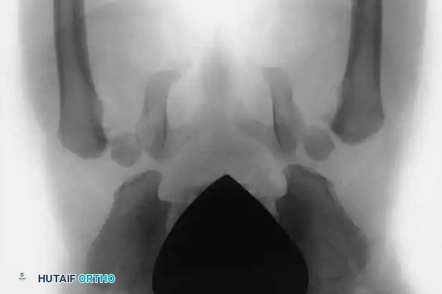
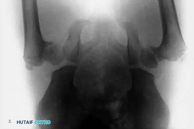
Parallel to proximal femoral deformities, pediatric orthopedic surgeons frequently manage complex pelvic anomalies, particularly those involving the innominate bones in the setting of bladder exstrophy. Bladder exstrophy is a severe congenital malformation, occurring in approximately 1 in 30,000 to 50,000 live births. It is characterized by a catastrophic defect in the anterior abdominal wall and bladder, accompanied by profound diastasis of the symphysis pubis. The skeletal hallmark of this condition is the severe external rotation and lateral displacement of the innominate bones, fundamentally altering the biomechanics of the pelvic ring and the acetabular orientation. Addressing these dual pathologies—proximal femoral varus and pelvic diastasis—requires a masterful command of corrective osteotomies to restore physiological load transmission and prevent lifelong disability.
Histological Pathophysiology of Coxa Vara
The hallmark anatomical lesion in developmental coxa vara is a distinct, localized defect in the medial aspect of the femoral neck. Microscopic analysis of this medial defect reveals a disorganized matrix of dysplastic cartilage. The normal columnar arrangement of chondrocytes, which is essential for longitudinal growth, is highly irregular and fragmented. Endochondral ossification within this specific zone is profoundly atypical, closely resembling a dysplastic or abnormal physis that is incapable of sustaining physiological loads.
The adjacent metaphyseal bone is markedly osteoporotic, characterized by atrophic, thinned trabeculae that occasionally harbor large, ectopic islands of undifferentiated cartilage cells. This structural void creates a locus of mechanical weakness precisely at the region of the proximal femur that experiences the highest compressive loads during the single-leg stance phase of gait. As the child grows, this localized dysplasia acts as a hinge point, allowing the femoral head to progressively slip into varus and retroversion relative to the femoral shaft.
Detailed Surgical Anatomy and Biomechanics
The successful surgical management of coxa vara and exstrophy-related pelvic dysplasia hinges upon an intimate understanding of the native and pathological biomechanics governing the hip joint and the pelvic ring. The proximal femur and the pelvis operate as a coupled biomechanical system; alterations in one invariably affect the load-bearing characteristics of the other.
Proximal Femoral Biomechanics and the Heuter-Volkmann Principle
Coxa vara, which is frequently bilateral, is pathognomonically characterized by a progressive decrease in the angle between the femoral neck and the femoral shaft (the neck-shaft angle), accompanied by progressive limb shortening. In a normal pediatric hip, the neck-shaft angle is approximately 135 to 145 degrees, and the proximal femoral physis is oriented horizontally, subjecting it primarily to compressive forces. These compressive forces stimulate normal physeal growth according to the Heuter-Volkmann principle.
In the dysplastic hip of a child with coxa vara, a vicious biomechanical cycle is established. When the child begins walking, the physiological load across the hip joint increases exponentially. Because the dysplastic medial femoral neck is structurally compromised, the normal compressive forces are converted into pathological shear forces across the increasingly verticalized physis. This pathological shear stress actively inhibits normal ossification and accelerates the progressive varus deformity. The abductor moment arm is severely compromised as the greater trochanter migrates proximally.
As the patient ages and gains weight, the mechanical disadvantage worsens. The deformity progresses relentlessly until the greater trochanter eventually overrides the femoral head, leading to severe abductor insufficiency, clinically manifesting as a profound Trendelenburg gait. If left untreated, the unrelenting shear forces can culminate in a frank pseudarthrosis of the femoral neck. In neglected adult cases, the greater trochanter may migrate several inches superior to the femoral head, resulting in wide separation of the head and neck fragments and catastrophic joint failure.
Biomechanical Implications of the Exstrophy Pelvis
In the context of bladder exstrophy, the pelvic ring is entirely disrupted anteriorly. The skeletal deformity is characterized by a massive diastasis of the symphysis pubis, which can measure anywhere from 4 to 12 centimeters depending on the age of the patient. This diastasis is not merely a linear separation; it is the result of a complex, three-dimensional outward rotation of the innominate bones.
The sacroiliac joints act as the posterior hinge for this deformity. The iliac wings are externally rotated, and the anterior superior iliac spines are displaced laterally. This anatomical distortion results in a retroverted, laterally displaced, and shallow acetabulum. If left uncorrected, the child will develop a pathognomonic wide-based, waddling, and externally rotated gait. Furthermore, this pelvic dysplasia is frequently associated with other orthopedic anomalies, including developmental dysplasia of the hip (DDH) and iatrogenic hip subluxation. From a general surgical perspective, attempting soft-tissue closure of the bladder and abdominal wall without addressing the skeletal diastasis places immense, unsustainable tension on the fascial repair. This invariably leads to catastrophic complications, including wound dehiscence, fistula formation, and complete bladder prolapse.
Exhaustive Indications and Contraindications
The decision to proceed with surgical intervention in these complex pediatric deformities requires a rigorous evaluation of clinical progression, radiographic parameters, and the overall physiological status of the child. Early diagnosis is critical, particularly in coxa vara, as the likelihood of achieving a functionally normal hip diminishes precipitously after 8 years of age due to the loss of remodeling potential.
Indications for Subtrochanteric Valgus Osteotomy
The definitive treatment for developmental coxa vara is a subtrochanteric valgus osteotomy. The primary biomechanical goal is to reorient the femoral neck and head into an extreme valgus position relative to the femoral shaft, thereby converting pathological shear forces back into physiological compressive forces across the physis. According to the classic criteria established by Beals, surgical intervention is strictly indicated under specific, measurable conditions. The most critical radiographic parameter is the Hilgenreiner epiphyseal angle (HEA). The HEA is the angle subtended by a horizontal line drawn through the triradiate cartilages (Hilgenreiner's line) and a line drawn parallel to the proximal femoral physis. A normal HEA is less than 25 degrees; an angle greater than 60 degrees is an absolute indication for surgical intervention due to a documented 100% rate of progression.
Indications for Pelvic Osteotomies in Bladder Exstrophy
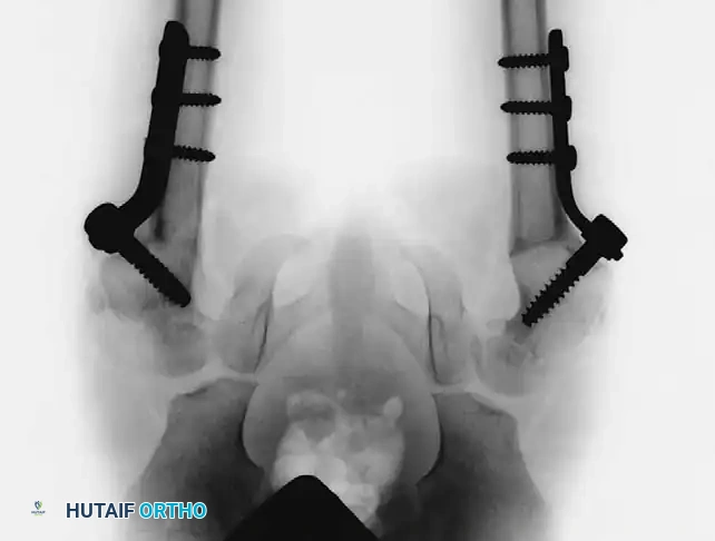
For bladder exstrophy, orthopedic intervention is required to mobilize the innominate bones and close the pelvic ring, facilitating tension-free urological and abdominal wall reconstruction. Anterior iliac osteotomies are generally indicated for primary closures in children older than 72 hours of age, or for any secondary (revision) closure regardless of age, as the maternal relaxin has cleared the infant's system and the pelvis has become rigid.
| Parameter | Indications for Surgical Intervention | Absolute & Relative Contraindications |
|---|---|---|
| Developmental Coxa Vara | - HEA consistently > 60 degrees. - Neck-shaft angle < 110 degrees. - Documented radiographic progression. - Clinical pain or severe Trendelenburg gait. - Unilateral deformity causing severe pelvic obliquity. |
- HEA < 45 degrees (observe closely). - Active hip joint infection. - Severe, uncorrectable PFFD (relative). - Medical comorbidities precluding major surgery. |
| Bladder Exstrophy (Pelvic Osteotomy) | - Primary closure in infants > 72 hours old. - Symphyseal diastasis > 4 cm preventing tension-free closure. - Any secondary/revision bladder closure. - Severe external rotation gait causing functional impairment. |
- Infants < 72 hours old with flexible pelvis (can often be closed without osteotomy). - Active pelvic or systemic infection. - Severe neurological compromise precluding rehab. |
Pre-Operative Planning, Templating, and Patient Positioning
Meticulous preoperative planning is the cornerstone of successful deformity correction. The surgeon must transition from conceptual understanding to precise geometric execution.
Radiographic Evaluation and Templating for Coxa Vara
Radiographic evaluation must include high-quality, weight-bearing anteroposterior (AP) and frog-leg lateral views of the pelvis.
The AP radiograph is utilized to measure the preoperative neck-shaft angle and the HEA. Templating is required to determine the exact angle of the lateral closing wedge needed to achieve a postoperative neck-shaft angle of 140 to 150 degrees, and, most crucially, a postoperative HEA of less than 38 degrees. The surgeon must trace the proximal femur, draw the intended osteotomy line at the level of the lesser trochanter, and simulate the wedge resection. The angle of the wedge to be removed is exactly equal to the desired correction angle. For example, if the preoperative neck-shaft angle is 90 degrees and the goal is 140 degrees, a 50-degree lateral wedge must be resected.
Furthermore, the surgeon must select the appropriate internal fixation device during the templating phase. Pediatric locking plates, specialized pediatric blade plates (e.g., 100-degree or 110-degree plates), or dynamic hip screws may be utilized depending on the child's age and bone stock. While early intervention is ideal to harness the remodeling potential of the proximal femur, surgery is often strategically delayed until the child is 4 to 5 years old. This delay allows for sufficient ossification and increased bone stock in the proximal femur, which drastically improves the purchase and stability of internal fixation devices.
Planning for Anterior Iliac Osteotomies
For bladder exstrophy, a 3D reconstructed CT scan of the pelvis is invaluable for assessing the true spatial orientation of the innominate bones and the exact magnitude of the diastasis. The surgical plan requires bilateral anterior iliac osteotomies, typically performed superior to the acetabulum, extending from the anterior inferior iliac spine (AIIS) to the greater sciatic notch.
Patient Positioning and Setup
For both procedures, the patient is placed supine on a radiolucent operating table. For coxa vara, a bump is placed under the ipsilateral hip. For bilateral pelvic osteotomies, the patient is positioned completely flat. Strict biplanar fluoroscopy (image intensification) must be available and verified prior to draping. The entire lower extremity and hemipelvis (or bilateral lower extremities and full pelvis/abdomen for exstrophy) are prepped and draped free to allow for intraoperative manipulation and assessment of range of motion.
Step-by-Step Surgical Approach and Fixation Technique
The execution of these osteotomies demands meticulous soft tissue handling, precise bone cuts, and rigid internal fixation to withstand the massive deforming forces postoperatively.
Surgical Technique: Valgus Osteotomy for Developmental Coxa Vara
Step 1: Soft Tissue Release. Begin by performing a percutaneous or mini-open adductor tenotomy through a small medial incision in the groin crease. This is a critical, non-negotiable step to relieve medial tension, prevent lateral subluxation or frank dislocation of the femoral head during the extreme valgus correction, and reduce compressive forces across the hip joint.
Step 2: Surgical Approach. Expose the greater trochanter and the proximal shaft of the femur through an 8- to 10-cm direct lateral, longitudinal incision. Incise the fascia lata in line with the skin incision. Elevate the vastus lateralis off the lateral intermuscular septum and reflect it anteriorly to expose the subtrochanteric region of the femur. Place Hohmann retractors anteriorly and posteriorly to protect the soft tissues.
Step 3: Guidewire and Implant Insertion. If utilizing a pediatric screw and side-plate device (or a specialized pediatric blade plate), insert the primary guidewire into the midline of the femoral neck under strict biplanar fluoroscopic guidance.
* Trajectory: Insert the wire as close as possible to the trochanteric apophysis without violating it, to avoid iatrogenic growth arrest. The angle of insertion relative to the femoral shaft must match the pre-calculated angle of the implant to achieve the desired valgus.
* Depth: Ideally, center the screw in the femoral neck distal to the abnormal physis. If the neck is too short, dysplastic, or structurally inadequate, the screw must be advanced across the physis and centered securely within the femoral head to achieve adequate purchase.
Step 4: The Osteotomy. Perform a transverse osteotomy slightly distal to the entry point of the screw, approximately at the level of the lesser trochanter, using an oscillating saw. Ensure the saw blade is coplanar with the fluoroscopic beam to avoid oblique cuts.
Step 5: Deformity Correction. Resect the pre-calculated lateral wedge of bone from the distal fragment. Reduce the osteotomy by bringing the femoral shaft into abduction to close the wedge. This maneuver corrects the neck-shaft angle to the targeted 140 to 150 degrees and horizontalizes the physis.
Step 6: Fixation. Secure the side plate to the femoral shaft using cortical screws in standard compression fashion. Use an articulated tension device if necessary to compress the osteotomy site. Ensure rigid internal fixation.
Step 7: Closure. Thoroughly irrigate the surgical bed. Close the vastus lateralis over the plate. Close the fascia lata, subcutaneous tissues, and skin in anatomical layers. Insert a subfascial irrigation-suction drain if significant dead space or bleeding is present.
Surgical Technique: Anterior Iliac Osteotomies for Bladder Exstrophy
Historically, O’Phelan championed bilateral posterior iliac osteotomies combined with symphyseal approximation. While effective, this required intraoperative repositioning (flipping) of the patient, massively increasing anesthesia time, airway risk, and infection risk. Currently, the gold standard, as recommended by Sponseller et al., is the use of bilateral anterior iliac osteotomies.
Step 1: Approach. Make a bikini-line incision over the anterior iliac crests bilaterally. Expose the inner and outer tables of the ilium down to the greater sciatic notch.
Step 2: The Osteotomy. Using a Gigli saw or an osteotome, create a vertical cut extending from the iliac crest, passing just anterior to the sacroiliac joint, and exiting into the apex of the greater sciatic notch.
Step 3: Mobilization. Carefully mobilize the anterior segment of the innominate bone. This allows the pubic rami to swing medially like a gate on a hinge.
Step 4: Symphyseal Approximation. Following the urological repair of the bladder, the orthopedic team returns to place heavy, nonabsorbable sutures (e.g., #2 or #5 Ticron) or specialized orthopedic pins through the pubic bones. The symphysis is forcibly approximated and tied securely, closing the pelvic ring over the delicate urological repair and eliminating fascial tension.
Complications, Incidence Rates, and Salvage Management
Despite meticulous surgical technique, complication rates in these complex reconstructive procedures remain notable. Surgeons must be prepared to identify and manage these complications aggressively.
Complications of Valgus Osteotomy for Coxa Vara
Regardless of the osteotomy technique or fixation method, developmental coxa vara carries a notorious risk of recurrence. Children must be monitored clinically and radiographically at regular intervals until skeletal maturity. Furthermore, a significant cohort of these children present with concurrent femoral hypoplasia. Progressive limb-length discrepancy must be monitored, as it may ultimately necessitate contralateral epiphysiodesis or ipsilateral limb lengthening procedures using intramedullary magnetic nails or external fixators.
Complications of Pelvic Osteotomies in Exstrophy
In a massive retrospective review of 624 bladder exstrophy repairs, Okubadejo, Sponseller, and Gearhart reported an overall orthopedic complication rate of 4% (26 patients). The most notable orthopedic complication was transient palsy of the femoral nerve, likely due to traction or compression during the medial swing of the innominate bones.
| Complication | Incidence Rate | Etiology / Risk Factors | Salvage Management |
|---|---|---|---|
| Recurrence of Coxa Vara | Up to 50% (if HEA > 38°) | Failure to correct HEA to < 38 degrees; insufficient valgus. | Revision subtrochanteric valgus osteotomy; optimization of fixation. |
| Premature Physeal Closure | 5% - 10% | Iatrogenic violation of the capital femoral physis with hardware. | Monitor for LLD; contralateral epiphysiodesis if discrepancy > 2 cm. |
| Femoral Nerve Palsy | ~1% - 2% (Exstrophy) | Traction injury during anterior iliac osteotomy and symphyseal closure. | Usually transient. Observation, physical therapy, AFO for foot drop. |
| Wound Dehiscence / Bladder Prolapse | 4% (Exstrophy) | Inadequate pelvic osteotomy; excessive tension on the anterior abdominal wall. | Emergent return to OR; revision of pelvic fixation; external fixation application. |
| Hardware Failure / Nonunion | < 5% | Inadequate bone stock; early weight-bearing; technical error. | Revision open reduction internal fixation (ORIF) with bone grafting. |
Phased Post-Operative Rehabilitation Protocols
The postoperative management is as critical as the surgical execution. The goal is to protect the osteotomy sites while promoting bone healing and eventually restoring full functional mobility.
Phase I: Strict Immobilization (Weeks 0 to 8)
Immediately following wound closure, both coxa vara and exstrophy patients require rigid immobilization.
* Coxa Vara: Apply a one-and-one-half spica cast in the operating room. The operative leg is casted to the toes, and the non-operative leg is casted to just above the knee. The hips are placed in slight abduction (15-20 degrees) and neutral rotation to neutralize rotational forces and protect the proximal femoral fixation during the early healing phase.
* Bladder Exstrophy: Patients are typically placed in modified spica casts or specialized external fixators with cross-bars to maintain the approximation of the symphysis pubis. Strict supine positioning is often required to protect the urological repair.
Phase II: Early Mobilization and Radiographic Verification (Weeks 8 to 12)
At 8 weeks, the patient returns to the operating room or clinic for cast removal and radiographic evaluation. AP and lateral radiographs are scrutinized for the presence of bridging callus across the osteotomy sites.
* If union is confirmed, the cast is permanently removed.
* Patients are transitioned to a wheelchair or allowed toe-touch weight-bearing with assistive devices. Aggressive, passive range-of-motion (ROM) exercises are initiated to combat capsular stiffness and muscle atrophy.
Phase III: Strengthening and Functional Restoration (Months 3 to 6)
Once full weight-bearing is authorized, the focus shifts to aggressive physical therapy.
* Abductor Strengthening: For coxa vara patients, restoring the strength of the gluteus medius and minimus is paramount to eliminating the Trendelenburg gait.
* Gait Training: Exstrophy patients require extensive gait retraining to overcome the habitual external rotation and wide-based stance dictated by their preoperative anatomy.
Long-term follow-up is mandatory. Children must be evaluated annually until skeletal maturity to monitor for recurrence of varus, progressive limb-length discrepancies, or late-onset hardware complications.
Summary of Landmark Literature and Clinical Guidelines
The evolution of surgical management for these complex conditions is deeply rooted in landmark clinical studies that have defined current best practices.
Coxa Vara and the Hilgenreiner Epiphyseal Angle:
A landmark study by Carroll, Coleman, and Stevens evaluated 26 patients undergoing valgus osteotomies for coxa vara and found a 50% overall recurrence rate. Crucially, they determined that etiology, age at surgery, osteotomy type, and implant choice had no bearing on recurrence. The single defining prognostic factor was the postoperative Hilgenreiner epiphyseal angle (HEA).
* 95% of patients whose HEA was corrected to less than 38 degrees experienced no recurrence of varus.
* The traditional head-shaft angle proved highly unreliable for determining the adequacy of correction; two-thirds of patients corrected to >135 degrees still recurred, while one-third corrected to <135 degrees maintained satisfactory results. This paradigm shift cemented the HEA as the absolute gold standard for intraoperative templating and postoperative assessment.
Bladder Exstrophy and Multidisciplinary Determinants of Success:
The management of bladder exstrophy requires seamless coordination between pediatric urology and orthopedic surgery. Achieving a successful primary closure is highly complex. Kasat and Borwankar identified critical factors necessary for optimal outcomes in exstrophy reconstruction, emphasizing that anterior approximation of the pubic bones is mandatory to allow deep placement of the bladder and urethra within the true pelvis.
Furthermore, Sponseller's extensive series on anterior iliac osteotomies revolutionized the approach to the exstrophy pelvis. By demonstrating that anterior osteotomies provide superior mobility of the pubic rami, allow for greater correction of the diastasis, and completely avoid the need to turn the patient under anesthesia, the anterior approach became the definitive standard of care. Long-term data from these cohorts indicates that children who undergo precise, tension-free closures supported by robust orthopedic stabilization maintain their skeletal correction better over time, drastically reducing the incidence of catastrophic urological failures.
Through meticulous preoperative planning, precise execution of complex osteotomies, adherence to biomechanical principles, and rigorous multidisciplinary postoperative care, orthopedic surgeons can successfully alter the natural history of both congenital coxa vara and the exstrophy pelvis, providing these children with stable, functional, and pain-free ambulation for the duration of their lives.
