ATLS Protocol: Pelvic Fracture Workup & Management Explained
Key Takeaway
Here are the crucial details you must know about ATLS Protocol: Pelvic Fracture Workup & Management Explained. The ATLS protocol for initial trauma workup includes AP chest, AP pelvis, and lateral C-spine radiographs. For suspected unstable pelvic ring injuries, immediate management involves placing a pelvic binder centered over the greater trochanters. Chest injuries are the most common associated injury with pelvic fractures (up to 63%), emphasizing the importance of a thorough workup and management of multiply injured patients.
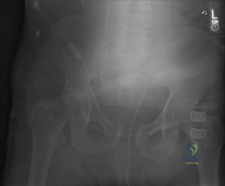
A 32-year-old male is brought to the trauma bay following a high-speed motorcycle accident. He is hemodynamically unstable. An AP pelvis radiograph is obtained during the primary survey. Describe your findings and the immediate management steps.
Candidate: The radiograph shows a widened pubic symphysis consistent with an 'open-book' injury. Given the patient is unstable, I would ensure he is managed under ATLS guidelines. I'd apply a pelvic binder at the level of the greater trochanters to reduce pelvic volume, resuscitate with blood products using a massive transfusion protocol, and perform a FAST exam to rule out intra-abdominal bleeding. If he remains unstable, I would consider pelvic packing or angiography.
Candidates often fail to describe the injury formally (e.g., APC II vs III), place the binder at the iliac crests (which is ineffective or may worsen distraction), or focus purely on the orthopedic injury while ignoring the multidisciplinary requirement for hemorrhage control.
A structured response: 1. Description: Radiograph shows significant pubic symphysis diastasis (>2.5cm) with possible sacroiliac joint involvement, suggesting an APC II/III injury. 2. Immediate ATLS action: Hemodynamic resuscitation (Massive Transfusion Protocol), pelvic binder application (at greater trochanters), and assessment for "life over limb." 3. Hemorrhage control: Explicitly mention the algorithm: If responding to binder/fluids -> CT scan. If not responding -> Preperitoneal Pelvic Packing (PPP) or Interventional Radiology for embolization. 4. Urgency: Acknowledge that this is a potential source of catastrophic venous and arterial hemorrhage.
You have stabilized the patient hemodynamically. You are now planning definitive fixation of the posterior pelvic ring. What are the specific risks associated with the placement of an S1 iliosacral screw?
Candidate: The main risks are injury to the L5 nerve root, which lies anterior to the sacral ala. Also, if the screw is too long or placed too anteriorly, it can injure the iliac vessels. I would use fluoroscopic views—inlet, outlet, and lateral sacral—to ensure the trajectory is safe within the S1 corridor.
Forgetting the specific anatomical landmarks or failing to mention the L5 nerve root, which is the most critical structure at risk due to its proximity to the sacral ala. Also, failing to mention the lateral sacral view to ensure the screw stays within the 'safe zone' of the S1 body.
Structure by anatomy and technique: 1. Neurological: L5 nerve root (anterior-superior to the sacral ala) is the most significant risk. 2. Vascular: Injury to the internal iliac artery/vein if the screw breaches the anterior cortex. 3. Technical/Radiographic: Emphasize the 'safe corridor' for S1. Mention the need for Inlet view (to avoid anterior/posterior breach), Outlet view (to avoid superior breach into the canal), and Lateral sacral view (to ensure the screw is within the alar-sacral body complex). 4. Nuance: Mention that S2 screws are an option, but note that the risk profile changes compared to S1.
