High-Energy Hawkins Type III Talus Neck Fracture: A Detailed Case Study
Key Takeaway
A Hawkins Type III talus neck fracture involves severe displacement and dislocation from high-energy trauma. Diagnosis requires a meticulous clinical exam assessing neurovascular status and deformity. Urgent X-rays reveal specific fracture patterns; CT imaging is crucial for comprehensive assessment of displacement, guiding initial management.
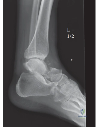
You are on-call in the Emergency Department. A 32-year-old male presents following a 15-foot fall. He has a deformed ankle with skin tenting and diminished distal pulses. Plain radiographs are shown below. Describe the injury and your immediate management priorities.
Candidate: This is a complex talar neck fracture with dislocation of the talar body from the ankle mortise and the subtalar joint. Based on the imaging and the Hawkins classification, this is a Type III fracture. My immediate priorities are to perform an urgent closed reduction to relieve tension on the skin and restore neurovascular status, followed by stabilization, neurovascular reassessment, and arranging for definitive CT imaging and urgent ORIF.
Failing to emphasize the "urgent" nature of the reduction. A common mistake is to suggest "admitting for elective surgery tomorrow" or focusing solely on the fracture classification without addressing the limb-threatening soft tissue and vascular compromise (skin tenting/diminished pulses).
A structured, safety-first response: 1) Clinical assessment: Confirm neurovascular status and prioritize decompression of tented skin. 2) Emergency intervention: Perform immediate closed reduction under sedation to protect skin and restore perfusion. 3) Definitive imaging: Order a CT scan for 3D fracture mapping once stable. 4) Classification: Identify it as a Hawkins Type III and clearly state the high prognostic risk of AVN (70-100%). 5) Surgical Planning: Propose dual approaches (anteromedial/anterolateral) and warn that a medial malleolar osteotomy may be required for anatomic reduction.
You have reduced the fracture and taken the patient to the operating theatre. During your planning, you are asked about the vascular supply of the talus and why these injuries have such a high incidence of Avascular Necrosis (AVN). How do you explain this to a junior trainee?
Candidate: The talus has a tenuous blood supply, derived mainly from the artery of the tarsal canal (branch of the posterior tibial), the artery of the tarsal sinus, and the deltoid artery. In high-energy fractures, these vessels are frequently disrupted. The Hawkins Type III injury involves dislocation of the talar body, which effectively devascularizes it, leading to the high AVN risk. I would also mention the 'Hawkins Sign'—a subchondral lucency on the talar dome visible on X-ray at 6-8 weeks—which indicates bone resorption and implies that the talus is revascularized.
Listing the arteries without explaining the clinical correlation. A poor candidate forgets to mention the Deltoid branch (which supplies the medial body) or ignores the prognostic value of the Hawkins sign entirely.
Demonstrate anatomical depth: Define the vascular supply (Posterior Tibial → Tarsal Canal; Anterior Tibial/Peroneal → Tarsal Sinus; Deltoid branch → Medial aspect). Explain that the retrograde flow makes it vulnerable. Correctly identify the "Hawkins sign" as a positive clinical indicator, and mention that its absence is a reliable indicator of AVN, necessitating long-term follow-up.
