Part of the Master Guide

AAOS Basic Science MCQs (Set 4): Bone Physiology, Biomechanics & Ortho Pharmacology | ABOS Board Prep

Mastering Orthopedic Basic Science: FRCS Exam MCQ Engine & Revision

23 Apr 2026 66 min read 130 Views
Basic science oral core topics Introduction

Key Takeaway

Preparing for FRCS Orthopedic Basic Science involves mastering core concepts through targeted practice. Our interactive MCQ engine provides exam-style questions covering fracture healing, biomechanics, anatomy, and physiology. Detailed explanations accompany each question, identifying critical factors like interfragmentary strain in fracture repair, crucial for deep understanding and effective revision towards exam success.
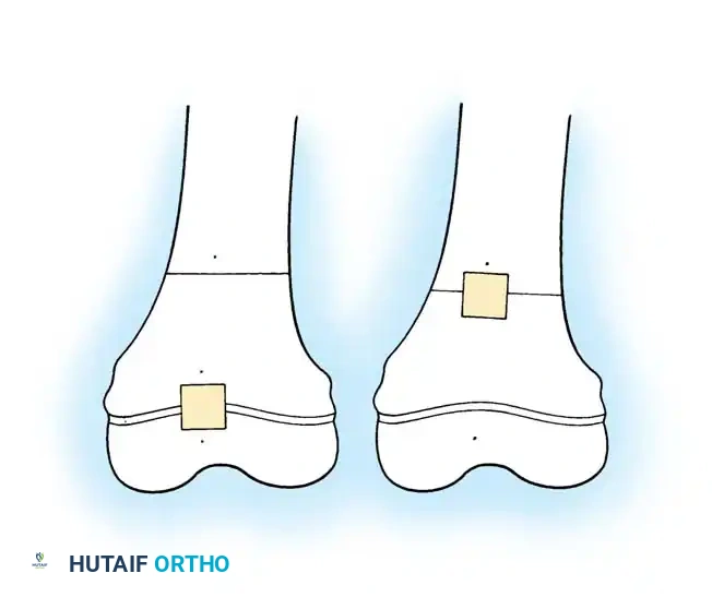
Comprehensive Exam


00:00

Start Quiz

Question 1

A 45-year-old male sustains a comminuted diaphyseal tibia fracture. Which of the following factors is MOST critical in determining the rate of secondary fracture healing, assuming adequate reduction and stabilization?


Explanation

The degree of interfragmentary strain is the most critical factor influencing secondary fracture healing, also known as callus healing. Too much strain (macromotion) disrupts the forming callus and inhibits healing, leading to non-union. Too little strain (excessive rigidity, as in primary healing) may prevent sufficient callus formation required for secondary healing. The optimal strain environment allows for the progression from hematoma to granulation tissue, soft callus, hard callus, and finally remodeling. While an intact periosteal sleeve is important for osteogenic cells, and immobilization rigidity contributes to managing strain, the degree of strain itself is the direct biomechanical determinant. Vitamin D and BMD are systemic factors but less direct than local biomechanical factors.

Question 2

Regarding the biomechanics of bone, cortical bone is optimally designed to resist which type of force?


Explanation

Cortical bone, with its dense, anisotropic structure, is strongest in compression. It has high stiffness and can withstand significant compressive loads before failure. It is weaker in tension, and even weaker in shear and torsion. Bending creates both tension and compression, so while it resists bending, its primary strength lies in resisting direct compression.

Question 3

Which of the following statements MOST accurately describes the role of osteocytes in bone remodeling?


Explanation

Osteocytes, entrapped within the bone matrix, are the primary mechanosensors of bone. They sense mechanical strain and fluid flow through their lacunar-canalicular network. This mechanotransduction initiates signaling cascades (e.g., sclerostin, RANKL) that regulate the activity of osteoblasts (bone formation) and osteoclasts (bone resorption), thereby orchestrating bone remodeling. Osteoblasts synthesize new matrix, osteoclasts resorb bone, and progenitor cells are distinct (e.g., mesenchymal stem cells). While they indirectly contribute to calcium homeostasis through remodeling, they don't directly release calcium as their primary role.

Question 4

In the context of articular cartilage, which proteoglycan is predominantly responsible for its remarkable ability to withstand compressive loads?


Explanation

Aggrecan is the major proteoglycan in articular cartilage and is critical for its compressive stiffness. Aggrecan molecules aggregate with hyaluronic acid, forming large complexes that trap water within the collagen network. This creates a high osmotic swelling pressure, which resists compression by forcing water out, providing load-bearing capacity and resilience. Decorin, Biglycan, Versican, and Fibromodulin are smaller proteoglycans with different roles, such as collagen fibril organization or cell signaling, but not the primary role in compressive strength.

Question 5

A patient undergoes total hip arthroplasty. The choice of bearing surface is critical. Which material pairing typically exhibits the lowest wear rate in modern hip arthroplasty?


Explanation

Ceramic-on-ceramic (CoC) bearings generally exhibit the lowest wear rates among the options provided, particularly with modern advanced ceramics. This is due to their excellent hardness, scratch resistance, and hydrophilicity. While highly cross-linked polyethylene (HXLPE) significantly improved wear compared to conventional polyethylene, CoC often still has superior wear characteristics. Metal-on-metal has fallen out of favor due to concerns regarding metal ion release and pseudotumor formation. Conventional polyethylene has high wear, and conventional ceramic-on-polyethylene is better than conventional metal-on-polyethylene, but CoC and HXLPE-on-metal are generally superior.

Question 6

Which of the following principles BEST explains why a long intramedullary nail is effective in stabilizing a diaphyseal fracture?


Explanation

An intramedullary nail acts as a load-sharing device. It shares the load with the bone, rather than rigidly fixing it and completely shielding the bone from stress (as a plate might, leading to stress shielding). This load sharing allows for controlled micromotion at the fracture site, which is conducive to secondary (callus) bone healing. Nails are very effective at resisting bending and torsional forces along the length of the diaphysis. While they promote biological fixation and periosteal callus (secondary healing), 'load-sharing' is the fundamental biomechanical principle distinguishing their function from rigid plating.

Question 7

The primary role of Type X collagen in the growth plate is associated with which zone?


Explanation

Type X collagen is specifically expressed by chondrocytes in the hypertrophic zone of the growth plate. It plays a crucial role in cartilage maturation, mineralization, and vascular invasion, signaling the transition from cartilage to bone. It is considered a marker of terminal chondrocyte differentiation and hypertrophy. The other zones have different primary collagen types and functions (e.g., Type II in resting and proliferative zones).

Question 8

Regarding peripheral nerve injury, Wallerian degeneration typically begins how long after axon transection?


Explanation

Wallerian degeneration, the process of axonal degeneration distal to a site of injury, typically begins within 24-48 hours after axon transection. While some changes might be observed earlier, the complete breakdown of the axon and myelin sheath becomes evident within this timeframe. This process clears the debris to allow for potential regeneration, especially in the peripheral nervous system.

Question 9

What is the primary function of the annulus fibrosus in an intervertebral disc?


Explanation

The annulus fibrosus consists of concentric lamellae of collagen fibers (predominantly Type I) arranged obliquely. Its primary function is to contain the nucleus pulposus and provide significant tensile strength, especially against torsional and bending forces. While the nucleus pulposus bears axial compressive loads through hydrostatic pressure, the annulus resists the radial expansion of the nucleus under compression and helps stabilize the vertebral segment by resisting tensile forces in various directions. It is the outer containment system.

Question 10

Which cytokine is a potent stimulator of osteoclastogenesis and bone resorption, and is targeted by denosumab?


Explanation

RANKL (Receptor Activator of Nuclear Factor kappa-B Ligand) is a key cytokine that binds to RANK receptors on pre-osteoclasts and mature osteoclasts, stimulating their differentiation, activation, and survival, thereby promoting bone resorption. Denosumab is a monoclonal antibody that targets and inhibits RANKL, making it a powerful antiresorptive agent. TGF-β and BMP-2 are more involved in bone formation, IL-6 has diverse inflammatory roles, and PTHrP is involved in endochondral ossification and hypercalcemia of malignancy.

Question 11

A surgeon plans to use a cannulated screw for an intra-articular fracture. What is the primary biomechanical advantage of a cannulated screw compared to a solid screw of the same external diameter?


Explanation

The primary biomechanical advantage of a cannulated screw mentioned here is the ability to guide the screw with a K-wire. This allows for precise placement, especially in intra-articular fractures where anatomical reduction and specific screw trajectories are critical. Cannulation reduces torsional and bending strength compared to a solid screw of the same external diameter because it removes material from the core. Pullout strength is related to thread design and bone quality, not cannulation directly.

Question 12

Regarding polymethylmethacrylate (PMMA) bone cement, which of the following statements is FALSE?


Explanation

The addition of barium sulfate (or zirconium dioxide) to PMMA bone cement is for radiopacity, allowing the cement to be visible on X-ray. It does not significantly increase the strength of the cement; in fact, it can slightly decrease mechanical properties. PMMA functions via mechanical interlock (macro- and micro-interlock) with trabecular bone. Its polymerization is an exothermic reaction. Antibiotics are commonly mixed in for prophylaxis or treatment of infection. The monomer (methyl methacrylate) is a known irritant and allergen.

Question 13

The 'creep' phenomenon in viscoelastic materials like articular cartilage refers to:


Explanation

Creep is the time-dependent increase in deformation (strain) of a material under a constant applied load (stress). In articular cartilage, this is due to the slow exudation of fluid from the matrix under sustained compression. Viscoelastic materials also exhibit stress relaxation (decrease in stress under constant strain) and hysteresis. The rapid deformation is instantaneous elastic response, recovery is elasticity/viscoelasticity, and constant deformation regardless of load is incorrect. Decrease in stress over time under constant deformation is stress relaxation.

Question 14

Which type of collagen is the most abundant in mature, healthy bone and provides its primary tensile strength?


Explanation

Type I collagen is the most abundant protein in the human body and constitutes approximately 90% of the organic matrix of bone. Its fibrous structure provides the bone's primary tensile strength and flexibility, complementing the compressive strength provided by the mineralized hydroxyapatite crystals. Type II collagen is found predominantly in cartilage, Type III in highly extensible tissues like skin and blood vessels (and early wound healing), and Types IX and X are associated with cartilage and the growth plate, respectively.

Question 15

The 'zone of Ranvier' in the growth plate is primarily responsible for:


Explanation

The zone of Ranvier is a periosteal ring located circumferentially around the periphery of the growth plate. It contains chondrocytes and osteoblasts and is primarily responsible for appositional bone growth, contributing to the increase in the width of the bone metaphysis. Longitudinal growth occurs through the proliferation and hypertrophy of chondrocytes within the growth plate itself. Vascular invasion and mineralization occur in the zone of calcification/ossification. The zone of Ranvier is distinct from articular cartilage repair.

Question 16

Which statement regarding peripheral nerve regeneration is MOST accurate?


Explanation

After peripheral nerve injury and Wallerian degeneration, Schwann cells dedifferentiate, proliferate, and form 'Büngner bands' or basal lamina tubes. These tubes provide a scaffold and release neurotrophic factors (e.g., NGF, BDNF) that guide the regenerating axonal sprouts from the proximal stump toward the target. Schwann cells promote regeneration. Regeneration is much slower and less successful in the CNS. Myelin debris contains inhibitory factors. Axonal growth rate is typically 1-3 mm/day, not 10 mm/day.

Question 17

In the context of bone grafts, which property describes the ability of the graft to provide a scaffold for new bone formation?


Explanation

Osteoconduction refers to the ability of a bone graft material to serve as a scaffold for the ingrowth of new bone-forming cells and capillaries from the host bone. This allows for creeping substitution, where new bone is laid down on the graft surface. Osteoinduction is the ability to stimulate undifferentiated mesenchymal cells to differentiate into osteoblasts and form bone. Osteogenesis is the formation of new bone by cells within the graft (e.g., autograft). Osteointegration describes direct structural and functional connection between living bone and the surface of a load-bearing implant.

Question 18

A patient presents with pain and swelling over the Achilles tendon. Histological examination reveals angiofibroblastic hyperplasia and disorganized collagen fibers, but no inflammatory cells. This pathology is most consistent with:


Explanation

This description is classic for tendinosis (or tendinopathy), which is a degenerative process characterized by disorganized, degenerated collagen, increased ground substance, vascular proliferation (angiofibroblastic hyperplasia), and a notable absence of significant inflammatory cells. Acute tendinitis implies active inflammation. Tenosynovitis is inflammation of the tendon sheath. Partial rupture is a macroscopic tear. Gout involves urate crystal deposition.

Question 19

The mechanical behavior of articular cartilage is highly dependent on the interactions between collagen, proteoglycans, and water. What is the primary role of water in enabling cartilage's load-bearing function?


Explanation

Water, held within the aggrecan-hyaluronic acid complexes, creates a significant osmotic (swelling) pressure within the cartilage matrix. When compressive loads are applied, water is exuded, but the trapped proteoglycans resist this outflow, creating a fluid-pressurization effect that supports the load. This fluid-solid interaction is fundamental to cartilage's ability to resist compression and allows for its viscoelastic properties. While water aids lubrication and nutrient transport, its primary load-bearing role is via osmotic pressure.

Question 20

Regarding implant materials, what is the primary advantage of titanium alloys (e.g., Ti-6Al-4V) over stainless steel (e.g., 316L) for orthopedic implants?


Explanation

Titanium alloys are generally preferred for long-term orthopedic implants due to their superior corrosion resistance and excellent biocompatibility compared to stainless steel. They form a stable, passive oxide layer (TiO2) that prevents ion release. Titanium also has a lower modulus of elasticity than stainless steel (closer to bone), which can reduce stress shielding, making 'A' a plausible distractor but 'B' is a more encompassing primary advantage. Stainless steel often has higher ultimate tensile strength but is less bioinert. Titanium wear resistance is not universally superior; for example, ceramic is superior.

Question 21

Which of the following cellular events is characteristic of the 'soft callus' stage of fracture healing?


Explanation

The soft callus stage is characterized by the differentiation of mesenchymal stem cells from the periosteum and bone marrow into chondroblasts, which then produce cartilage. This cartilage forms a soft, flexible bridge across the fracture gap. Direct bone apposition is primary healing. Vascularization and mineralization of cartilage occur in the hard callus stage, and remodeling is a later stage. Osteoclasts are active throughout but are not characteristic of the soft callus itself.

Question 22

The concept of 'stress shielding' in orthopedics primarily refers to:


Explanation

Stress shielding occurs when a rigid implant (e.g., a stiff plate or femoral stem) carries a disproportionately high amount of the load, thereby shielding the adjacent bone from its normal physiological stresses. According to Wolff's Law, bone adapts to the loads it experiences. When shielded from stress, the bone undergoes resorption and decreases in density, which can lead to implant loosening or periprosthetic fracture. It is a detrimental phenomenon, not a protective one leading to increased density.

Question 23

Which physiological process is critically dependent on cyclic loading and fluid flow for maintaining bone health and stimulating remodeling?


Explanation

Osteocytes, through their extensive lacunar-canalicular network, are exquisitely sensitive to mechanical stimuli, particularly fluid flow within the canaliculi induced by cyclic loading. This mechanotransduction is the primary mechanism by which bone senses its mechanical environment and initiates remodeling cascades (involving both osteoblasts and osteoclasts) to adapt its structure to applied loads, maintaining bone health. The other options are related to bone but not directly dependent on cyclic loading and fluid flow for their function in this context.

Question 24

A fracture is stabilized using a compression plate. What is the primary mechanism by which this implant promotes healing?


Explanation

Compression plating aims to achieve absolute stability at the fracture site. By compressing the fracture fragments, it reduces interfragmentary motion to a negligible level (less than 2%). This environment promotes direct bone healing (primary bone healing), where osteons directly bridge the fracture gap without significant callus formation. This contrasts with intramedullary nails, which are load-sharing and promote secondary healing.

Question 25

Which of the following statements about the mechanical properties of cancellous (trabecular) bone is MOST accurate?


Explanation

Cancellous bone, like cortical bone, is anisotropic. Its strength and stiffness vary significantly depending on the direction of loading, due to the orientation of its trabecular network along lines of stress. It is stronger in compression than tension. Its mechanical properties are highly dependent on bone density (volume fraction) and architecture. It is more compliant (lower modulus) and ductile than cortical bone, but this option isn't the most accurate overall descriptor of its specific mechanical nature regarding anisotropy and density dependence. It is not primarily designed to resist high torsional forces, which are better handled by cortical bone.

Question 26

What is the primary function of the A1 pulley in the hand?


Explanation

The A1 pulley is the most proximal of the annular pulleys in the finger. Its primary function is to hold the flexor tendons close to the bone (metacarpal or phalanx) and prevent bowstringing, thereby maintaining the mechanical efficiency of the flexor system during finger flexion. Dysfunction or inflammation of the A1 pulley is characteristic of trigger finger. The other options describe roles of different structures.

Question 27

Which growth factor plays a crucial role in initiating the cascade of endochondral ossification, particularly in chondrogenesis and osteogenesis?


Explanation

Bone Morphogenetic Proteins (BMPs) are a family of growth factors that are potent osteoinductive agents. They play a critical role in bone and cartilage formation, differentiation of mesenchymal stem cells into chondroblasts and osteoblasts, and are essential for both embryonic development and fracture repair. VEGF is critical for angiogenesis, IGF for cell proliferation, EGF and FGF have broader mitogenic roles but are not as specific to initiating osteochondral differentiation as BMPs.

Question 28

A 60-year-old patient with rheumatoid arthritis develops cervical myelopathy. From a biomechanical perspective, what is the MOST concerning feature of the atlantoaxial joint in this condition?


Explanation

In rheumatoid arthritis, chronic inflammation can lead to the destruction and laxity of ligaments, particularly the transverse atlantal ligament (TAL). The TAL is crucial for stabilizing the atlantoaxial joint by holding the odontoid process (dens) against the anterior arch of C1. Its destruction allows anterior translation of C1 on C2, leading to atlantoaxial subluxation, which can compress the spinal cord (myelopathy) or brainstem. While odontoid erosion occurs, TAL destruction is the direct cause of instability.

Question 29

Regarding the vascular supply of the femoral head, which artery is MOST susceptible to disruption in a femoral neck fracture, leading to osteonecrosis?


Explanation

The medial circumflex femoral artery (MCFA) is the predominant blood supply to the femoral head in adults, particularly via its retinacular branches that ascend along the femoral neck. Fractures of the femoral neck, especially displaced ones, frequently disrupt these retinacular vessels, leading to ischemia and subsequent osteonecrosis of the femoral head. The artery of the ligamentum teres contributes to blood supply in childhood and some adults but is often insufficient on its own. The LCFA supplies the anterior thigh, and profundra femoris is a main thigh artery. Superior gluteal supplies gluteal muscles.

Question 30

Which factor is LEAST likely to promote angiogenesis at a fracture site?


Explanation

Sclerostin is a protein produced by osteocytes that inhibits bone formation by antagonizing Wnt signaling. It is not directly involved in promoting angiogenesis; in fact, its primary role is related to bone remodeling balance. Hypoxia is a potent stimulus for VEGF production, which is a key promoter of angiogenesis. VEGF and FGF are major pro-angiogenic growth factors. IL-1 is a pro-inflammatory cytokine that can indirectly contribute to angiogenesis in the context of tissue repair, although its primary role is not angiogenesis.

Question 31

The primary biomechanical role of the menisci in the knee joint is to:


Explanation

The menisci are C-shaped fibrocartilaginous structures that sit on the tibial plateau. Their primary biomechanical roles include increasing the contact area between the femoral condyles and tibial plateau, which significantly reduces contact stress on the articular cartilage. They also contribute to joint stability, shock absorption, and some lubrication, but increasing congruity and reducing stress are their most critical functions. Ligaments limit hyperextension and provide varus/valgus stability.

Question 32

In muscle physiology, what is the primary role of tropomyosin?


Explanation

Tropomyosin is a regulatory protein that, in a relaxed muscle, wraps around the actin filaments and covers the myosin-binding sites, preventing the myosin heads from binding to actin. When calcium ions (released from the sarcoplasmic reticulum) bind to troponin, troponin undergoes a conformational change that pulls tropomyosin away from the myosin-binding sites, allowing cross-bridge formation and muscle contraction. Troponin binds calcium, myosin forms thick filaments and generates force, and acetylcholine release is at the NMJ.

Question 33

The 'fatigue life' of an orthopedic implant refers to its ability to:


Explanation

Fatigue life is a critical material property for orthopedic implants. It refers to the number of stress cycles an implant can withstand before fracturing. Implants in the body are subjected to millions of loading cycles (e.g., walking, running) at stresses below their ultimate tensile strength. Fatigue failure is a common mode of implant failure, making high fatigue life a crucial design consideration. Plastic deformation without fracture is ductility; elastic deformation is elasticity.

Question 34

Which histological feature is characteristic of a benign osteochondroma?


Explanation

An osteochondroma is characterized by an osseous projection from the bone surface, capped by hyaline cartilage. Crucially, the cartilage cap of the osteochondroma is continuous with the periosteum, and the underlying trabecular bone of the lesion is continuous with the medullary cavity of the parent bone. The cartilage cap has an organized growth plate-like structure (enchondral ossification), which is benign. Atypical chondrocytes and invasion are signs of malignancy (e.g., chondrosarcoma). Giant cells suggest giant cell tumor. Disorganized woven bone without a cap might be fibrous dysplasia or other benign bone lesions, but not osteochondroma.

Question 35

The 'creep' phenomenon in a cancellous bone graft, if significant, could lead to:


Explanation

Creep in a bone graft refers to its time-dependent deformation under sustained load. If a cancellous bone graft, which has viscoelastic properties, is subjected to significant sustained compressive forces, it can slowly deform and collapse over time. This 'creep' can lead to a loss of initial reduction, settling of the construct, and potential instability, especially in reconstructive surgery where structural integrity is paramount. It does not directly affect revascularization, stability, osteoinduction, or infection risk in this manner.

Question 36

What is the primary mechanism by which non-steroidal anti-inflammatory drugs (NSAIDs) can potentially impair fracture healing?


Explanation

NSAIDs exert their anti-inflammatory and analgesic effects by inhibiting cyclooxygenase (COX) enzymes, particularly COX-2. COX-2 is crucial for the synthesis of prostaglandins, which play a vital role in the early inflammatory phase of fracture healing, including callus formation, angiogenesis, and chondrogenesis. By inhibiting prostaglandin synthesis, NSAIDs can impair or delay fracture healing, especially in the early stages, though the clinical significance of this effect can vary.

Question 37

Which of the following ligaments is considered the primary static stabilizer against anterior translation of the tibia relative to the femur?


Explanation

The Anterior Cruciate Ligament (ACL) is the primary static stabilizer that prevents anterior translation of the tibia on the femur, particularly in knee extension and near extension. It also resists internal rotation and hyperextension. The PCL prevents posterior translation, and the MCL and LCL primarily resist valgus and varus forces, respectively.

Question 38

Regarding imaging principles, which modality is BEST suited for visualizing cortical bone breaches and complex fracture patterns, especially in intra-articular fractures?


Explanation

Computed Tomography (CT) scans provide excellent cross-sectional imaging with high spatial resolution and contrast for bone. It is superior to plain radiographs for demonstrating complex fracture patterns, cortical bone breaches, impaction, and intra-articular involvement, aiding in surgical planning. MRI is superior for soft tissue, cartilage, and marrow edema. Ultrasound has limited utility for bone visualization due to acoustic shadowing. Bone scintigraphy shows metabolic activity.

Question 39

The 'creep' property of a material refers to its time-dependent deformation under constant stress. In the context of orthopedic implants, a material with high creep resistance is generally preferred for components subjected to:


Explanation

High creep resistance is desirable for implants subjected to sustained, constant loads, such as the acetabular component of a total hip arthroplasty, where the body weight can exert a continuous load. Creep deformation over time under these constant loads can lead to dimensional changes, implant loosening, or failure. Fatigue resistance is more relevant for cyclic loads, and yield strength/ultimate tensile strength for acute high-impact loads.

Question 40

What is the primary mechanism of action for bisphosphonates in treating osteoporosis?


Explanation

Bisphosphonates are potent antiresorptive agents. They are taken up by osteoclasts during bone resorption and then disrupt the osteoclast's mevalonate pathway, leading to impaired osteoclast function, reduced survival, and ultimately apoptosis (programmed cell death). This effectively decreases the rate of bone resorption, allowing osteoblast-mediated formation to catch up, leading to increased bone mineral density. They do not directly stimulate osteoblasts, inactivate PTH, or increase gut calcium absorption.

Question 41

The 'tidemark' in articular cartilage represents the interface between which two zones?


Explanation

The tidemark is a wavy, basophilic line that demarcates the boundary between the deep radial zone (uncalcified cartilage) and the calcified cartilage layer within articular cartilage. It is an important histological landmark and represents a region of active remodeling, playing a role in the integrity of the cartilage-bone interface. The calcified cartilage, in turn, interfaces with the subchondral bone, separated by a cement line.

Question 42

Regarding the structural hierarchy of a tendon, what is the smallest organizational unit that contains collagen fibrils?


Explanation

The hierarchical organization of a tendon is: Tropocollagen molecules aggregate to form collagen microfibrils, which aggregate to form collagen fibrils. Collagen fibrils then form fibril bundles. Multiple fibril bundles make up a collagen fiber. Collagen fibers are grouped into fascicles, which are surrounded by endotenon. The entire tendon is surrounded by epitenon. So, the fibril bundle is the smallest unit containing multiple collagen fibrils.

Question 43

What is the main advantage of dynamic hip screw (DHS) fixation over multiple cancellous screws for intertrochanteric hip fractures?


Explanation

A Dynamic Hip Screw (DHS) system is designed to allow controlled collapse and impaction at the fracture site. The sliding barrel and lag screw construct permits controlled shortening of the femoral neck, allowing the fracture fragments to dynamically impact. This impaction increases the stability of the fracture and promotes secondary bone healing. It does not provide absolute stability; rather, it provides relative stability. While it provides good rotational stability, 'controlled collapse and impaction' is its unique and primary advantage for this fracture type.

Question 44

Which of the following is an example of an allograft in orthopedic surgery?


Explanation

An allograft is tissue (e.g., bone) harvested from a genetically non-identical individual of the same species (i.e., from a cadaveric donor to a living human recipient). Demineralized bone matrix (DBM) from a cadaver is a common type of allograft. Autologous grafts use the patient's own tissue (autograft). Synthetic materials are non-biological. Xenografts are from a different species.

Question 45

In bone tumors, what is the significance of a 'Codman triangle' observed on plain radiographs?


Explanation

A Codman triangle (or Codman's triangle) is a triangular area of new bone formation at the angle where the elevated periosteum meets the cortical bone. It indicates that an aggressive lesion (often a malignant tumor like osteosarcoma, but also aggressive infection or trauma) is rapidly growing and lifting the periosteum faster than it can lay down new bone. It is a sign of rapid periosteal elevation, not necessarily pathognomonic but highly suggestive of an aggressive process, typically malignant.

Question 46

A patient undergoes a peripheral nerve repair. Which factor is LEAST likely to positively influence the outcome of nerve regeneration?


Explanation

Proximal nerve injuries (e.g., at the brachial plexus or root level) generally have a poorer prognosis for regeneration compared to distal injuries. This is because the distance for axonal regrowth to the target muscle or sensory receptor is much longer, increasing the likelihood of misdirection, neuroma formation, and muscle atrophy before reinnervation. Younger age, sharp transection (clean injury), short gaps, and tension-free primary repair all positively influence outcomes.

Question 47

The use of locked plating systems in fracture fixation primarily aims to:


Explanation

Locked plating systems (e.g., Locking Compression Plates - LCPs) utilize screws that lock into the plate, creating a fixed-angle construct. This angular stability makes the plate-screw construct function as an internal fixator, independent of plate-to-bone compression. This is particularly advantageous in osteoporotic bone, comminuted fractures, or metaphyseal/epiphyseal fractures where traditional compression plating might not achieve stable purchase. It aims for relative stability and minimizes periosteal stripping, but its primary distinction is angular stability.

Question 48

Which type of joint receptor is primarily responsible for sensing extreme joint positions and potential tissue damage?


Explanation

Free nerve endings are ubiquitous in joint capsules, ligaments, synovium, and fat pads. They are polymodal, sensing noxious stimuli, inflammation, and extremes of mechanical deformation, thus acting as nociceptors and providing a sense of 'danger' or tissue damage. Ruffini endings sense static joint position and joint movement. Pacinian corpuscles detect dynamic changes in joint movement and pressure. Golgi-Mazzoni corpuscles are similar to Pacinian corpuscles. Merkel endings are primarily cutaneous mechanoreceptors.

Question 49

In spinal biomechanics, what is the significance of the 'neutral zone'?


Explanation

The neutral zone is a concept describing the small, central range of intervertebral motion where there is minimal resistance from passive spinal structures (ligaments, capsules). It represents the region of greatest laxity or 'play' in the spinal segment. An increase in the neutral zone is an indicator of spinal instability, as the segment becomes less constrained by passive structures and relies more on active muscle control. It is where minimal passive resistance is offered, not maximal.

Question 50

What is the primary mechanism by which parathyroid hormone (PTH) initially increases serum calcium levels?


Explanation

Parathyroid hormone (PTH) primarily increases serum calcium levels by stimulating osteoclast activity, which leads to the breakdown of bone matrix and release of calcium into the bloodstream. While PTH also acts on the kidneys to increase calcium reabsorption and stimulate calcitriol synthesis (which then enhances gut absorption), its immediate and direct effect on bone involves stimulating osteoclasts (indirectly, via osteoblasts, but resulting in resorption) to raise serum calcium. Osteoblasts are responsible for bone formation.

Question 51

Which type of bone graft has both osteoconductive and osteoinductive properties, along with osteogenic potential?


Explanation

Autogenous cancellous bone graft is considered the 'gold standard' because it possesses all three essential properties for bone healing: osteogenesis (living osteoblasts and mesenchymal stem cells within the graft), osteoinduction (bone morphogenetic proteins and other growth factors), and osteoconduction (a scaffold for new bone growth). Allografts are primarily osteoconductive and osteoinductive (if DBM), but lack living cells (osteogenic). Synthetics are typically only osteoconductive.

Dr. Mohammed Hutaif
Medically Verified Content by
Prof. Dr. Mohammed Hutaif
Consultant Orthopedic & Spine Surgeon
Chapter Index