Bioabsorbable Materials in Orthopaedic Surgery: Biomechanics, Biomaterials, and Clinical Application
Key Takeaway
Bioabsorbable materials, including polyglycolic acid (PGA) and polylactic acid (PLA), offer significant advantages in orthopaedic surgery by enabling gradual load transfer and eliminating hardware removal. However, their use requires a deep understanding of polymer biomechanics, degradation profiles, and potential complications such as aseptic osteolysis. This guide details the biochemical properties, surgical indications, and intraoperative handling techniques essential for optimizing clinical outcomes with bioabsorbable fixation devices.
Comprehensive Introduction and Patho-Epidemiology
The evolution of orthopaedic fixation has increasingly embraced bioabsorbable materials, driven by a paradigm shift aimed at eliminating secondary hardware removal surgeries, mitigating stress shielding, and facilitating unhindered postoperative advanced imaging. Historically, the socio-economic burden of routine hardware removal has been substantial, carrying inherent risks of iatrogenic nerve injury, infection, and refracture through residual stress risers. Polyglycolic acid (PGA) was the first totally synthetic bioabsorbable suture developed, which subsequently paved the way for Vicryl—a copolymer consisting of 92% PGA and 8% polylactic acid (PLA)—and polydioxanone (PDS). PDS holds the historical distinction of being the first bioabsorbable material successfully manufactured into rigid orthopaedic screws, fundamentally altering the trajectory of biomaterials in musculoskeletal surgery.
Currently, the primary alpha polyesters utilized for bioabsorbable implants in contemporary orthopaedic practice include PGA, PDS, polylevolactic acid (PLLA), and racemic poly(D, L)-lactic acid (PDLLA). While these materials offer the distinct theoretical advantage of gradual load transfer to healing tissue—thereby promoting secondary bone healing via physiological micromotion—their successful application demands a rigorous, uncompromising understanding of their degradation kinetics, biomechanical limitations, and potential for inciting adverse biological responses. The patho-epidemiology of bioabsorbable implant failure is inextricably linked to a mismatch between the polymer's degradation profile and the biological healing timeline of the host tissue.
The degradation of bioabsorbable alpha polyesters occurs primarily through bulk hydrolysis, a complex biochemical process wherein interstitial water molecules cleave the ester bonds of the polymer backbone. This leads to a progressive, predictable reduction in molecular weight, followed by a precipitous loss of mechanical strength, and ultimately, total mass loss. PGA is highly crystalline and hydrophilic, leading to rapid hydrolytic degradation. It is hydrolyzed primarily into pyruvic acid, which subsequently enters the tricarboxylic acid (TCA) cycle and is excreted physiologically as carbon dioxide via respiration and water. Conversely, the addition of a methyl group makes PLA significantly more hydrophobic than PGA, drastically slowing its degradation rate. PDLLA is similarly hydrolyzed via the TCA cycle, while PLLA exhibits the slowest degradation profile of the alpha polyesters, maintaining its mechanical integrity for extended periods—a characteristic highly advantageous for ligamentous reconstructions requiring prolonged mechanical support. PDS is uniquely hydrolyzed such that its degradation byproducts are primarily excreted in the urine rather than through the respiratory system.
Clinical Implications of Polymer Selection
The choice of bioabsorbable polymer must be meticulously matched to the expected healing time of the target tissue. Utilizing a rapidly degrading PGA implant for a slow-healing cortical fracture will invariably result in catastrophic fixation failure before clinical union is achieved. Conversely, utilizing a highly crystalline, slow-degrading PLLA implant in a rapidly healing metaphyseal fracture may expose the patient to unnecessary long-term risks of late foreign body reactions and osteolysis. The orthopaedic surgeon must act as an applied biochemist, selecting an implant whose strength-degradation curve perfectly intersects with the strength-acquisition curve of the healing bone or soft tissue.
Detailed Surgical Anatomy and Biomechanics
The biomechanical behavior of bioabsorbable polymers is fundamentally divergent from that of traditional metallic implants such as titanium alloys or surgical-grade stainless steel. Their material properties are heavily influenced by chemical composition, manufacturing processes, physical dimensions, environmental factors (such as pH and temperature), and time in vivo. A critical limitation of biodegradable implants is their high glass transition temperature ($T_g$)—the specific thermodynamic temperature at which the amorphous regions of the polymer transition from a hard, glassy state to a softer, rubbery, pliable state. Because the $T_g$ of these orthopaedic polymers is well above human body temperature and standard operating room ambient temperatures, biodegradable implants cannot be contoured or bent intraoperatively. Attempting to forcefully bend a bioabsorbable plate or pin at room temperature will invariably result in catastrophic brittle failure and micro-fracturing of the polymer matrix.
To overcome the inherent mechanical weakness of raw, unadulterated polymers, manufacturers employ sophisticated "self-reinforcement" techniques. By drawing the polymer through a specialized die under precise thermal and pressure conditions, the internal polymer chains and micro-fibers are oriented parallel to the longitudinal axis of the implant. This self-reinforcement process (resulting in materials like SR-PGA and SR-PLLA) dramatically increases the tensile, shear, and flexural strength of the device, elevating it from a simple temporary tether to a construct suitable for specific load-bearing applications in orthopaedic surgery. However, despite this reinforcement, absorbable polymers remain highly viscoelastic, making them exceptionally subject to creep (progressive deformation under a constant load) and stress relaxation (a decrease in stress under a constant strain).
Claes and colleagues elegantly demonstrated that self-reinforced PLA (SR-PLA) and PDLLA-PLLA screws lost up to 20% of their initial compressive force within just 20 minutes of insertion. In a more natural, warm saline environment mimicking the human body, this loss of compressive force was even more rapid and pronounced. Consequently, surgeons must be acutely aware that the initial interfragmentary compression achieved across a fracture or osteotomy site will diminish rapidly in the immediate postoperative period. This viscoelastic relaxation dictates that bioabsorbable screws cannot be relied upon to maintain dynamic compression in the same manner as a fully threaded metallic cortical screw. Furthermore, because these implants are absorbable, they lose mechanical strength relatively rapidly compared to their mass loss. For instance, SR-PGA rods retain only 50% of their initial strength at 2 weeks, and a mere 13% at 4 weeks, whereas PLLA exhibits the slowest degradation and loss of strength, making it the preferred material for interference screws in anterior cruciate ligament (ACL) reconstruction.
Loading Modes and Stress-Strain Characteristics
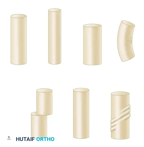
Understanding how bioabsorbable implants respond to various physiological loading modes is essential for preventing premature mechanical failure. In vivo, implants may be subjected to complex, multi-planar forces including tension, compression, bending, shear, torsion, or a combination thereof.
When evaluating the material properties of these polymers, a single-cycle stress-strain curve is traditionally utilized. Testing is usually performed in a fixed sample with stress applied under tension. The curve delineates the elastic region (where deformation is completely reversible), the yield stress (the point of permanent plastic deformation), the ultimate stress (maximum load capacity), and the breaking stress (the point of catastrophic structural failure). The modulus of elasticity (stiffness) of bioabsorbables is significantly closer to that of cortical and cancellous bone than metallic implants. This closer modulus matching theoretically reduces stress shielding—a phenomenon where rigid metallic implants bypass physiological loads, leading to localized osteopenia—and promotes secondary bone healing through the stimulation of callus formation via controlled micromotion.
Exhaustive Indications and Contraindications
The most common and universally accepted orthopaedic application of bioabsorbable implants is for the secure attachment of soft tissue to bone. This is predominantly seen in sports medicine and arthroscopic procedures, including shoulder surgeries (rotator cuff repair, superior labrum anterior and posterior [SLAP] repairs, and Bankart lesions) and knee surgeries (ACL and PCL reconstruction utilizing bioabsorbable interference screws). In these applications, the implant serves to hold the soft tissue graft or native tendon in intimate contact with a prepared bony bed just long enough for biological integration (Sharpey's fiber formation) to occur. However, the use of bioabsorbables in rigid fracture fixation is expanding, particularly in scenarios where minimal physiological load is applied until radiographic healing is evident.
Established indications for absorbable fixation devices in the realm of trauma and reconstructive surgery include specific applications in the foot and ankle, such as metatarsal osteotomies (e.g., Chevron or Scarf osteotomies for hallux valgus correction), metacarpal and metatarsal fusions, and select malleolar fractures. In pediatric orthopaedics, bioabsorbables are highly advantageous for the fixation of physeal (epiphyseal) fractures, radial head fractures, and olecranon fractures, where avoiding a secondary operation for hardware removal is highly desirable to prevent psychological trauma to the child and reduce healthcare costs. Furthermore, in the realm of advanced biologics, bioabsorbable matrices are utilized as drug delivery systems, cell transplantation scaffolds (e.g., Dermagraft), nerve reconstruction conduits (e.g., Neurotube), and adhesion prevention barriers.
Despite these expanding indications, bioabsorbable bone implants currently must be strictly limited to applications where there is minimal shear or bending load applied until healing is evident. They are absolutely contraindicated in diaphyseal fractures of major load-bearing long bones (e.g., femur, tibia, humerus) due to their inability to withstand the immense bending moments and torsional forces experienced during early rehabilitation. Furthermore, they are contraindicated in osteoporotic bone where screw purchase is inherently poor, and in comminuted fractures where the implant must bridge a gap and bear the entirety of the mechanical load.
Clinical Indications and Contraindications Matrix
| Anatomical Region / Subspecialty | Accepted Indications for Bioabsorbables | Preferred Polymer Profile | Strict Contraindications |
|---|---|---|---|
| Sports Medicine (Knee) | ACL/PCL interference screws, Meniscal repair darts, OCD fragment fixation | PLLA or PDLLA (Slow degradation, high initial strength) | Multi-ligamentous knee injuries requiring rigid early mobilization |
| Sports Medicine (Shoulder) | Rotator cuff anchors, Bankart/SLAP repair anchors | PEEK/PLLA composites, PLLA | Massive, retracted, irreparable cuff tears under high tension |
| Foot and Ankle | Hallux valgus osteotomies, Syndesmotic fixation, Medial malleolar fractures | SR-PGA, PDLLA (Moderate degradation) | Charcot arthropathy, severe comminuted pilon fractures |
| Hand and Upper Extremity | Metacarpal/phalangeal fractures, UCL ruptures (thumb), Radial head fractures | SR-PGA, PDS (Rapid to moderate degradation) | Diaphyseal radius/ulna fractures (both-bone forearm fractures) |
| Pediatric Orthopaedics | Salter-Harris physeal fractures, Osteochondritis dissecans | SR-PGA, PDLLA (Avoids secondary removal surgeries) | Pathological fractures secondary to aggressive bone cysts/tumors |
| Trauma (General) | Periarticular fractures protected by casts, Non-load-bearing osteotomies | Polymer matched to expected union time | Diaphyseal long bone fractures (Femur, Tibia, Humerus) |
Pre-Operative Planning, Templating, and Patient Positioning
Pre-operative planning for the utilization of bioabsorbable implants requires a meticulous, multi-modal imaging approach and a profound understanding of the specific implant's biomechanical limitations. High-resolution magnetic resonance imaging (MRI) is essential when planning for osteochondritis dissecans (OCD) fragment fixation or meniscal repairs to assess the viability of the osteochondral fragment and the quality of the surrounding host bed. For periarticular fractures, fine-cut computed tomography (CT) with 3D reconstructions is mandated to fully appreciate the fracture geometry, assess the degree of comminution, and plan the exact trajectory of the bioabsorbable screws. Because bioabsorbable screws lack the robust pull-out strength of metallic screws in osteopenic bone, the surgeon must pre-operatively template the screw trajectories to maximize purchase in the densest available subchondral or cortical bone.
Templating must also account for the physical dimensions of the bioabsorbable hardware. Unlike metallic implants, which can often be inserted with a degree of angular tolerance, bioabsorbable screws require absolute precision. The surgeon must ensure that the specific inventory of exact-match drill bits, specialized taps, and custom drivers are available and sterile prior to the induction of anesthesia. Furthermore, the pre-operative plan must include a contingency for metallic fixation; if the fracture pattern is found to be more complex intraoperatively, or if the bioabsorbable implant fails during insertion, the surgical team must seamlessly transition to a traditional metallic construct without compromising the patient's outcome.
Patient positioning is dictated by the anatomical region being addressed but must be optimized to allow for perfect orthogonal fluoroscopic imaging. For shoulder arthroscopy utilizing bioabsorbable anchors, the patient may be positioned in either the beach chair or lateral decubitus position, ensuring that the traction setup allows for unhindered access to the glenoid rim. For ankle and foot osteotomies, the patient is positioned supine with a bump under the ipsilateral hip to internally rotate the leg, bringing the medial malleolus and first ray into a neutral, easily accessible plane. A radiolucent table is mandatory, and the C-arm must be positioned to allow for rapid acquisition of true anteroposterior, lateral, and mortise views without requiring manipulation of the fractured extremity.
Equipment and Instrumentation Considerations
The instrumentation required for bioabsorbable fixation is highly specialized and unforgiving of technical errors. The surgical setup must include a hot-wire cutting device or specialized, sharp trimming forceps. Bioabsorbable pins and screws cannot be cut with standard heavy wire cutters, as the crushing force will cause longitudinal splintering and micro-fracturing of the polymer matrix, drastically reducing its pull-out strength and creating sharp, prominent edges that irritate adjacent soft tissues. Additionally, the pre-operative checklist must verify the presence of the manufacturer-specific tap; attempting to use a standard metallic screw tap will result in a mismatched thread pitch, leading to immediate thread stripping and loss of fixation upon insertion of the bioabsorbable screw.
Step-by-Step Surgical Approach and Fixation Technique
The surgical application of bioabsorbable screws, pins, and anchors requires a meticulous, deliberate technique that differs significantly from standard metallic hardware insertion. The margin for error is exceedingly narrow; failure to adhere to these biomechanical principles almost universally results in intraoperative implant breakage, stripping of the screw head, or loss of fracture reduction. The surgeon must approach the insertion of a bioabsorbable device with patience and a deep respect for the material's viscoelastic properties.
The first and most critical step is achieving absolute anatomical reduction. The fracture or osteotomy must be provisionally held in perfect alignment using metallic K-wires, reduction forceps, or a specialized compression device. Bioabsorbable implants cannot be used to "lag" or pull a poorly reduced fracture together with the same aggressive force as a metallic screw. If a gap remains between the fracture fragments, attempting to close it by over-tightening a bioabsorbable screw will exceed the polymer's ultimate tensile strength, causing the screw head to shear off from the shaft. The reduction must be complete and statically compressed before the bioabsorbable implant is even introduced to the surgical field.
Once reduction is secured, the pilot hole is drilled using the exact drill bit specified by the manufacturer. Because bioabsorbables do not possess the sharp cutting flutes of self-tapping metallic screws, the hole must be perfectly sized to accommodate the minor diameter of the screw. Following drilling, tapping the drill tract is a mandatory, non-negotiable step. The tract must be tapped through the entire depth of the planned screw insertion, including the far cortex if bicortical fixation is planned. Bioabsorbable screws cannot cut their own thread path in dense cortical bone; attempting to force an untapped or under-tapped insertion will result in catastrophic torsional failure of the screw shaft.
Insertion, Countersinking, and Trimming
Insertion of the bioabsorbable screw must be performed using the specialized, manufacturer-provided driver. The surgeon must apply steady, axial pressure and turn the driver slowly and deliberately. High-torque, rapid insertion must be strictly avoided. Rapid rotation generates significant friction against the surrounding bone, which can cause localized frictional melting of the polymer. This thermal degradation not only damages the screw's structural integrity but also induces thermal necrosis in the adjacent osteocytes, compromising biological integration and predisposing the site to aseptic loosening.
Finally, the head of the implant must be meticulously countersunk beneath the articular cartilage or flush with the cortical bone surface. Prominent bioabsorbable hardware is a primary catalyst for severe localized synovitis and mechanical irritation of overlying tendons. If utilizing bioabsorbable pins, they must be cut flush with the bone surface using a specialized hot wire or dedicated cutting forceps to avoid leaving sharp, prominent edges or inducing longitudinal splintering of the pin shaft. Copious irrigation of the surgical site is mandatory prior to closure to remove any microscopic polymer debris generated during insertion, thereby minimizing the risk of postoperative foreign body reactions.
Complications, Incidence Rates, and Salvage Management
While bioabsorbable implants successfully eliminate the need for routine hardware removal, their degradation process is not entirely benign. The body's biological response to the hydrolytic byproducts can lead to several well-documented, sometimes severe, complications. The degradation of these polymers relies heavily on local macrophage activity and tissue perfusion. When the rate of polymer degradation exceeds the local tissue's capacity to buffer and clear the acidic byproducts, significant clinical sequelae arise.
PGA, due to its rapid degradation profile, has been heavily implicated in aseptic inflammation and sterile sinus track formation. As the PGA polymer rapidly hydrolyzes, the local clearance mechanisms can become overwhelmed by the sudden influx of pyruvic acid, leading to a precipitous localized drop in pH. This highly acidic environment triggers a robust, nonspecific foreign body reaction, characterized by a massive influx of polymorphonuclear leukocytes and giant cells, which can ultimately result in aggressive osteolysis around the implant site. In intra-articular applications, such as the fixation of osteochondral lesions, severe aseptic synovitis has been reported with both PGA and PLLA implants. This is believed to be a direct result of the sheer volume of biodegradation debris shed into the confined joint space. If an implant breaks off into a joint space, the resulting loose body can cause rapid, severe arthropathy, often necessitating emergent arthroscopic retrieval and extensive synovectomy.
Despite these significant biological concerns, large-scale epidemiological studies demonstrate highly favorable overall complication profiles when bioabsorbables are used in appropriate, load-protected indications. In a comprehensive review of more than 2,500 fractures fixed with bioabsorbable implants, the rate of bacterial wound infections was 3.6%, nonspecific foreign body reactions occurred in 2.3% of cases, and catastrophic failures of fixation were noted in only 3.7%. Furthermore, in a massive comparative study of 3,111 ankle fractures, deep infection was slightly less frequent with bioabsorbable fixation (3.2%) than with traditional metallic fixation (4.1%). However, caution is heavily warranted in specific anatomical regions with poor soft tissue envelopes; a report on absorbable plate-and-screw fixation for metacarpal fractures noted severe foreign body reactions in 33% of patients, all of whom required subsequent surgical débridement.
Complications and Salvage Management Matrix
| Complication | Associated Polymer / Scenario | Estimated Incidence | Salvage / Management Strategy |
|---|---|---|---|
| Sterile Sinus Tract Formation | PGA (Rapid degradation, pH drop) | 2% - 5% | Local wound care, observation. Surgical débridement if persistent or if secondary bacterial infection is suspected. |
| Aseptic Synovitis | Intra-articular PGA/PLLA debris | 1% - 4% | Aggressive arthroscopic lavage, extensive synovectomy, removal of unresorbed polymer fragments. |
| Intraoperative Screw Breakage | Failure to tap, excessive torque | Highly variable (Technique dependent) | Over-drill the retained fragment, replace with a larger diameter metallic rescue screw, or alter fixation trajectory. |
| Loss of Fixation / Creep | SR-PLA under dynamic load | 3% - 5% | Revision open reduction and internal fixation (ORIF) utilizing standard metallic plates and screws. |
| Osteolysis / Cystic Changes | Slow-degrading PLLA in metaphyseal bone | 5% - 10% (Often asymptomatic) | Observation if asymptomatic. Curettage and bone grafting if mechanically compromising the joint or causing pain. |
Phased Post-Operative Rehabilitation Protocols
Postoperative rehabilitation protocols following bioabsorbable fixation must be heavily modified to account for the rapid loss of mechanical strength inherent to these viscoelastic polymers. Unlike metallic fixation, which provides rigid stability that often allows for immediate, aggressive range of motion and early weight-bearing, bioabsorbable constructs demand a more conservative, phased approach. The rehabilitation timeline must respect the intersection of the polymer's degradation curve and the host tissue's biological healing curve.
Joints and fractures fixed with bioabsorbable implants typically require a significantly longer period of rigid external immobilization (e.g., fiberglass casting, rigid fracture boots, or specialized thermoplastic splinting) compared to those fixed with metallic implants. This external support is absolutely critical to protect the surgical construct during the highly vulnerable 2-to-6-week postoperative window. During this phase, the polymer's mechanical strength drops precipitously due to hydrolysis, yet the biological callus or Sharpey's fibers have not yet matured sufficiently to withstand physiological loads independently. Premature discontinuation of external immobilization during this nadir of mechanical stability will almost certainly result in creep, loss of reduction, and clinical failure.
A major clinical advantage of these implants is their complete radiolucency, which facilitates unobstructed postoperative radiographic, CT, and MRI evaluation of fracture healing, joint congruity, and soft tissue integrity. Surgeons can accurately assess the formation of bridging callus without the obscuring artifact generated by metallic plates and screws. However, surgeons must be acutely aware of the long-term radiographic realities of bioabsorbable materials. While the implants do eventually dissolve and lose their mass, they are frequently not replaced by structurally sound woven or lamellar bone. Postoperative CT scans and MRIs of bones previously fixed with bioabsorbable implants often reveal empty, sclerotic tracts or cystic voids at the original screw site years after the fractures have clinically healed. These voids represent areas of fibrous encapsulation rather than true osteoconduction, a factor that must be considered if future revision surgery or arthroplasty is anticipated in that specific anatomical region.
Progression of Weight Bearing and Motion
The progression of weight-bearing and active range of motion must be meticulously titrated. For lower extremity periarticular fractures (e.g., medial malleolus), patients are typically maintained strictly non-weight-bearing in a rigid cast for a minimum of 4 to 6 weeks. Transition to a controlled ankle motion (CAM) boot with progressive partial weight-bearing is only initiated once hard radiographic evidence of bridging callus is confirmed. In sports medicine applications, such as rotator cuff repairs utilizing bioabsorbable anchors, passive range of motion may be initiated early to prevent adhesive capsulitis, but active, resisted motion is delayed until 8 to 12 weeks to ensure the soft tissue-to-bone interface has achieved sufficient biological strength to compensate for the degrading anchor.
Summary of Landmark Literature and Clinical Guidelines
The integration of bioabsorbable materials into standard orthopaedic practice is supported by a robust and expanding body of landmark literature and clinical trials. Early skepticism regarding the mechanical viability of these polymers has been largely mitigated by advancements in self-reinforcement technologies and a more profound understanding of indication-specific applications. The prospective, randomized comparison by Bostman et al., evaluating PLA screws versus stainless steel screws for displaced medial malleolar fractures, serves as a foundational text in this domain. This study definitively demonstrated no statistically significant differences in operative or postoperative complications, union rates, or functional outcomes between the two cohorts, thereby validating the use of bioabsorbables in protected, low-load fracture patterns.
Furthermore, the massive retrospective analyses of over 3,000 ankle fractures highlighted earlier underscore the safety profile of bioabsorbables regarding deep infection rates, which are comparable, if not slightly superior, to traditional metallic hardware. However, clinical guidelines derived from these studies emphasize strict adherence to patient selection criteria. The literature universally condemns the use of bioabsorbables in diaphyseal long bone fractures, highly comminuted patterns lacking cortical contact, and in patients with severe osteoporosis where the initial purchase of the non-self-tapping threads is compromised.
The future horizons of bioabsorbable materials in orthopaedics extend far beyond simple, inert mechanical fixation. Because these alpha polyesters can be precisely engineered with specific, tunable degradation profiles, they are ideal candidates for use as sophisticated delivery vehicles. Currently, bioabsorbable implants are being heavily researched and clinically utilized as carriers for osteoinductive biochemicals, most notably Bone Morphogenetic Protein 2 (BMP-2). By embedding these potent proteins within the polymer matrix, the implant can provide a sustained, localized, and controlled release of growth factors directly to the fracture site as the polymer hydrolyzes. Additionally, their emerging use in localized antibiotic delivery for osteomyelitis, stem cell transplantation matrices for avascular necrosis, and nerve reconstruction conduits represents the absolute cutting edge of orthopaedic biomaterial science. This convergence of biomechanics and advanced biologics potentially makes targeted biochemical delivery the ultimate, most impactful application for bioabsorbable technology in the 21st century.
