Comprehensive Introduction and Patho-Epidemiology
The evolution of ankle arthroscopy represents one of the most significant paradigm shifts in modern orthopedic foot and ankle surgery. Historically, the tibiotalar joint was considered a "tight" articulation, relatively inaccessible to early endoscopic techniques without significant iatrogenic morbidity. However, with the advent of specialized 2.7-mm and 4.0-mm short-barrel arthroscopes, refined non-invasive distraction techniques, and a profoundly enhanced understanding of periarticular neurovascular anatomy, ankle arthroscopy has transitioned from a purely diagnostic novelty to a primary therapeutic modality. Today, it is the gold standard for managing a vast spectrum of intra-articular pathologies, offering the distinct advantages of minimal soft tissue disruption, accelerated rehabilitation, and direct visualization of the articular surfaces that often surpasses traditional open arthrotomy.
Understanding the patho-epidemiology of the tibiotalar joint is essential for any orthopedic surgeon undertaking this procedure. The ankle is a highly congruent, constrained hinge joint subjected to tremendous biomechanical forces—often bearing up to five times the body's weight during normal ambulation and significantly more during athletic activities. This unique biomechanical environment predisposes the joint to specific patterns of injury. Acute inversion injuries, the most ubiquitous of athletic traumas, frequently result in lateral ligamentous complex attenuation, leading to chronic ankle instability (CAI). Over time, the altered kinematics of an unstable ankle precipitate repetitive microtrauma to the articular cartilage and the surrounding soft tissue envelope, culminating in reactive synovitis, the formation of osteophytes, and ultimately, anterior or anterolateral impingement syndromes.
Osteochondral lesions (OCLs) of the talus represent another major pathologic entity frequently addressed via arthroscopy. These lesions, classically categorized by Berndt and Harty, are predominantly traumatic in origin, particularly those located on the anterolateral talar dome, which typically result from shear forces during inversion and dorsiflexion. Conversely, posteromedial lesions are often deeper, morphologically cup-shaped, and may have a more insidious or ischemic etiology, though trauma remains a primary catalyst. The ability to directly visualize, probe, and treat these lesions via debridement, curettage, and marrow stimulation techniques (such as microfracture) has revolutionized the treatment algorithm, sparing the patient the morbidity of a medial malleolar osteotomy in many cases.
Furthermore, the scope of ankle arthroscopy has expanded to include the management of degenerative and inflammatory arthritides. In the setting of the "frozen ankle" or advanced osteoarthritis, arthroscopy facilitates aggressive synovectomy, removal of osteochondral loose bodies, and excision of restrictive capsular fibrosis, providing significant symptomatic relief and delaying the need for definitive arthrodesis or arthroplasty. Even when end-stage degeneration mandates joint sacrifice, arthroscopically assisted ankle arthrodesis has emerged as a highly effective technique, yielding fusion rates comparable to open procedures but with a markedly reduced incidence of wound complications and a faster time to clinical union.
Detailed Surgical Anatomy and Biomechanics
Mastery of ankle arthroscopy is inextricably linked to a profound, three-dimensional understanding of the surrounding surgical anatomy. The tibiotalar joint is encased within a dense, complex network of tendons, vessels, and nerves. Unlike the knee or shoulder, where portals are established through relatively thick muscular or tendinous planes, ankle portals traverse a thin subcutaneous layer directly adjacent to critical neurovascular structures. The margin for error is measured in millimeters, and a superficial knowledge of anatomy is a direct precursor to catastrophic iatrogenic injury.
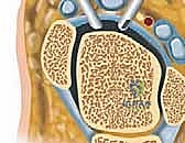
Anterior Ankle Anatomy and Portal Considerations
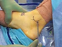
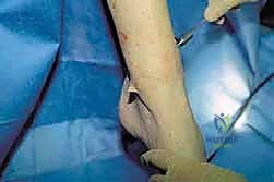
The anterior compartment of the ankle is the standard starting point for most diagnostic and therapeutic arthroscopic interventions. The standard approach utilizes the anteromedial and anterolateral portals, which, when properly placed, provide comprehensive access to the anterior joint space, the medial and lateral gutters, and the anterior half of the talar dome. The anteromedial portal is typically established first. It is located precisely at the level of the joint line, immediately medial to the tibialis anterior tendon. The primary structures at risk here are the great saphenous vein and the saphenous nerve. The saphenous nerve, a terminal sensory branch of the femoral nerve, courses anterior to the medial malleolus alongside the vein. To mitigate risk, the skin incision must be strictly superficial, followed by blunt dissection using a mosquito hemostat to spread the subcutaneous tissues down to the capsule.
The anterolateral portal is placed at the joint line, just lateral to the peroneus tertius tendon (or the extensor digitorum longus tendons if the peroneus tertius is absent). The critical structure at risk in this region is the superficial peroneal nerve (SPN), specifically its intermediate dorsal cutaneous branch. The SPN bifurcates proximal to the ankle joint, and its branches course directly over the anterolateral joint capsule. Injury to this nerve is the most common complication of ankle arthroscopy, leading to debilitating neuromas and complex regional pain syndrome (CRPS). Transillumination of the skin from the intra-articular arthroscope in the anteromedial portal can often highlight the superficial venous network, and by proxy, the adjacent nerve branches, allowing for safe localization of the anterolateral portal.
Anterior portal anatomy highlighting the relationship of superficial nerves to the joint line.
It is paramount to understand the "danger zone" associated with the antero-central portal. Located between the extensor hallucis longus (EHL) and extensor digitorum longus (EDL) tendons, this portal directly overlies the deep peroneal nerve and the anterior tibial artery. Due to the unacceptably high risk of catastrophic neurovascular injury, the antero-central portal is universally condemned in modern routine ankle arthroscopy and should only be considered in extraordinary circumstances by highly experienced surgeons utilizing meticulous open dissection techniques.
Detailed topographical mapping of anterior neurovascular structures.
Posterior Ankle Anatomy and Portal Considerations
When addressing pathology in the posterior half of the tibiotalar joint, the subtalar joint, or extra-articular posterior hindfoot structures (such as os trigonum excision or flexor hallucis longus tenolysis), the anterior approach is insufficient. The advent of the two-portal posterior endoscopic approach, pioneered by C. Niek van Dijk, revolutionized access to this region. This technique utilizes posterolateral and posteromedial coaxial portals, which lie parallel to the bimalleolar axis, allowing instruments to converge at the posterior aspect of the talus.
The posterolateral portal is the primary viewing portal. It is established just lateral to the Achilles tendon, approximately 1.5 to 2.0 cm proximal to the distal tip of the lateral malleolus. The primary structure at risk is the sural nerve, which courses distally along the posterolateral aspect of the calf, communicating with the small saphenous vein. Cadaveric studies demonstrate that the sural nerve lies, on average, a mere 6.6 mm from the standard posterolateral portal. Consequently, the incision must be meticulously placed, and the initial entry should be directed anteriorly toward the fibula to avoid plunging laterally into the nerve's trajectory.
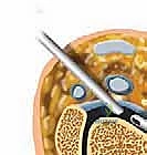
FIG 4 • Coaxial posteromedial portal anatomy.
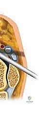
FIG 4 • Coaxial posteromedial portal anatomy.
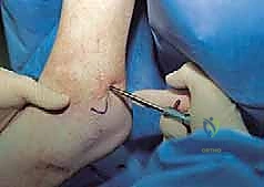
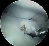
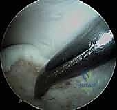
The posteromedial portal is established under direct arthroscopic visualization from the posterolateral portal. The skin incision is made just medial to the Achilles tendon, at the same horizontal level as the lateral portal. The trocar is directed toward the arthroscope shaft, aiming for the posterior process of the talus. The critical anatomic hazard here is the posterior tibial neurovascular bundle (comprising the posterior tibial artery, vein, and nerve), which lies medial to the flexor hallucis longus (FHL) tendon. The FHL tendon serves as a crucial anatomic landmark and a protective barrier; all arthroscopic work must remain lateral to the FHL tendon to absolutely ensure the safety of the medial neurovascular bundle.
FIG 2 • Coaxial portal cross-sectional anatomy. Notice the tight arrangement of the posterior tibial artery and nerve.
Posterior portal trajectory aiming toward the posterior talar process.
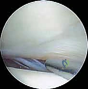
Endoscopic view of the FHL tendon, the crucial landmark protecting the medial neurovascular bundle.
Exhaustive Indications and Contraindications
Patient selection is the cornerstone of successful ankle arthroscopy. The procedure is a highly effective surgical tool, but it is not a panacea for all sources of ankle pain. A meticulous clinical examination, combined with advanced cross-sectional imaging, is required to correlate the patient's subjective symptoms with objective intra-articular pathology. The surgeon must distinguish between extra-articular sources of pain (e.g., peroneal tendinopathy, superficial nerve entrapment) and true intra-articular derangement.
The primary indications for ankle arthroscopy are broad but well-defined. Anterior ankle impingement, characterized by anterior joint pain exacerbated by dorsiflexion, is a classic indication. This is frequently caused by hypertrophic synovial tissue (soft tissue impingement) or the formation of kissing osteophytes on the anterior distal tibia and the talar neck (bony impingement). Arthroscopic debridement and osteophyte resection yield excellent clinical outcomes. Osteochondral lesions (OCLs) of the talus or tibial plafond are also prime indications. Arthroscopy allows for precise staging of the cartilage damage, removal of unstable fragments, curettage of the subchondral cyst, and marrow stimulation to promote fibrocartilage infill.
Other robust indications include the extraction of intra-articular loose bodies, which can cause mechanical locking and accelerated chondral wear. In the setting of chronic lateral ankle instability, arthroscopy is frequently employed adjunctively prior to open or percutaneous ligamentous reconstruction to address concomitant intra-articular pathology, which is present in up to 90% of these patients. Furthermore, arthroscopically assisted ankle arthrodesis is indicated for end-stage osteoarthritis, particularly in patients with minimal deformity, offering the benefits of high fusion rates with significantly less soft tissue stripping than open techniques.
Contraindications, while relatively few, must be strictly respected to avoid disastrous outcomes. Absolute contraindications include localized soft tissue infection or active cellulitis overlying the portal sites, which risks introducing pathogens into the sterile joint space. Severe peripheral vascular disease is another absolute contraindication, as the tourniquet application and fluid extravasation can critically compromise an already tenuous limb perfusion. Relative contraindications include severe, end-stage osteoarthritis with complete obliteration of the joint space, making safe portal entry and instrument maneuverability nearly impossible without causing extensive iatrogenic damage.
| Category | Specific Conditions | Rationale / Clinical Note |
|---|---|---|
| Primary Indications | Anterior/Posterior Impingement | Excision of osteophytes and hypertrophic synovium restores impingement-free ROM. |
| Osteochondral Lesions (OCLs) | Allows for staging, debridement, and marrow stimulation (microfracture). | |
| Loose Bodies / Chondromatosis | Minimally invasive extraction prevents mechanical locking and further cartilage wear. | |
| Arthrodesis Preparation | Denudation of articular cartilage with minimal soft tissue morbidity for end-stage OA. | |
| Unexplained Chronic Joint Pain | Diagnostic arthroscopy following exhaustive non-operative management and inconclusive MRI. | |
| Absolute Contraindications | Active Localized Infection | High risk of iatrogenic septic arthritis. |
| Severe Peripheral Vascular Disease | Tourniquet use and fluid extravasation may precipitate critical limb ischemia. | |
| Uncorrectable Coagulopathy | High risk of uncontrollable intra-articular hemarthrosis and compartment syndrome. | |
| Relative Contraindications | Severe End-Stage OA (Non-Fusion) | Obliterated joint space prevents safe instrument insertion; high risk of iatrogenic scuffing. |
| Severe Edema / Soft Tissue Compromise | Increases distance from skin to joint, complicating portal placement and fluid management. | |
| Charcot Arthropathy (Active Phase) | Altered anatomy and hyperemic bone make arthroscopy unpredictable and risky. |
Pre-Operative Planning, Templating, and Patient Positioning
Thorough preoperative planning is the invisible framework upon which surgical success is built. This process begins in the clinic with a meticulous review of imaging modalities. Weight-bearing orthogonal radiographs are standard for assessing joint space narrowing, osteophyte formation, and overall coronal and sagittal alignment. Magnetic Resonance Imaging (MRI) is indispensable for evaluating the integrity of the ligamentous complexes, identifying occult osteochondral lesions, and assessing the extent of reactive synovitis or bone marrow edema. For complex bony impingement or preoperative templating of large OCLs, a Computed Tomography (CT) scan provides unparalleled three-dimensional bony detail, dictating whether an anterior or posterior arthroscopic approach is required.
Anesthetic management plays a critical role in the intraoperative environment and the patient's postoperative recovery trajectory. We strongly advocate for the use of regional anesthesia, specifically a popliteal sciatic nerve block combined with a saphenous nerve block, administered preoperatively under ultrasound guidance. This approach provides profound intraoperative analgesia, allowing for lighter systemic sedation or general anesthesia. More importantly, the sympathectomy effect of the regional block induces vasodilation, which, when combined with controlled hypotensive anesthesia, significantly reduces intraoperative bleeding, thereby optimizing arthroscopic visualization and reducing the reliance on high tourniquet pressures.
General Setup and Tourniquet Placement
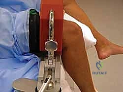
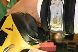
Once the patient is anesthetized, a comprehensive Examination Under Anesthesia (EUA) is performed to dynamically assess ligamentous laxity (Anterior Drawer and Talar Tilt tests) without the confounding factor of muscle guarding. The patient is then positioned on the operating table. The application of a pneumatic tourniquet is standard practice, though its location depends heavily on the planned positioning and distraction method. If a standard supine position is utilized without a thigh holder, a well-padded tourniquet is applied to the proximal thigh. However, if an external distractor requiring a thigh holder is used, the tourniquet must be placed carefully to avoid compounding pressure.
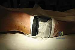
TECH FIG 7 • Distractor set-up: tourniquet placement.
Alternative view of thigh tourniquet placement, ensuring adequate padding.
Anterior and Posterior Positioning Strategies
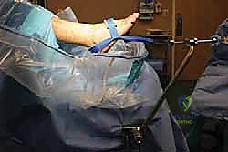
For isolated anterior pathology, the patient is positioned supine. A simple, yet highly effective method involves placing a firm, rolled towel or sandbag beneath the ipsilateral calf, allowing the heel to float freely off the edge of the bed. This permits the surgeon to utilize the weight of the foot, combined with manual plantarflexion, to passively distract the anterior joint space. Alternatively, the distal end of the operating table can be lowered, and the thigh placed in a standard arthroscopy knee holder. This setup restricts hip and thigh motion but allows the surgeon to freely manipulate the lower leg and ankle, optimizing dynamic assessment during the procedure.
Supine positioning with the leg secured in a standard arthroscopic thigh holder.
When posterior portals are anticipated, the patient is transitioned to the prone position. The operative leg is slightly elevated on a bump, and the foot is allowed to hang freely over the edge of the table. This positioning provides unimpeded access to the posterior heel and Achilles region. The contralateral leg must be meticulously padded to prevent pressure sores or neurapraxia. In the prone position, gravity assists in draining extravasated fluid away from the posterior portal sites, maintaining a clear visual field.
Ankle Distractor Placement and Optimization
The tibiotalar joint is inherently tight, and adequate distraction is the key to preventing iatrogenic chondral injury (scuffing) during instrument insertion. While manual distraction by an assistant is possible, it is physically demanding and often inconsistent. Therefore, mechanical distraction is heavily favored. Invasive skeletal distractors (utilizing pins in the tibia and calcaneus) provide powerful distraction but carry risks of pin tract infection, stress fractures, and neurovascular injury. Consequently, non-invasive external strap distractors have become the standard of care.
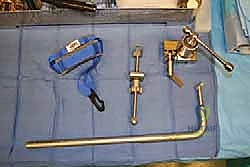
TECH FIG 6 • Distractor set-up: instruments required for non-invasive distraction.
TECH FIG 7 • Distractor set-up: tourniquet placement below the fibular head when using a thigh holder.
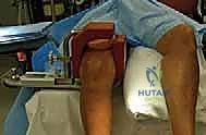
To apply the non-invasive distractor, the patient's hip is flexed to approximately 45-60 degrees, and the thigh is secured in a padded thigh holder. Crucially, the surgeon must ensure the thigh holder supports the posterior thigh musculature and does not rest directly in the popliteal fossa. Compression of the popliteal vein will cause venous engorgement distal to the holder, leading to intractable intra-articular bleeding that obliterates the arthroscopic view. The non-invasive strap is then applied in a figure-of-eight fashion around the hindfoot and midfoot, attaching to a tensioning device secured to the operating table.
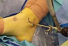
Application of the non-invasive distraction strap around the hindfoot.
Securing the tensioning device to the operating table.
Applying controlled, incremental traction via the distractor mechanism.
Final assessment of the distraction setup prior to prepping and draping.
Draped operative field with the non-invasive distractor engaged.
Step-by-Step Surgical Approach and Arthroscopic Technique
The execution of ankle arthroscopy demands a methodical, reproducible approach. Once the patient is prepped, draped,
Clinical & Radiographic Imaging Archive