Comprehensive Introduction and Patho-Epidemiology
Ankle arthroscopy has evolved from a purely diagnostic modality into a highly sophisticated, therapeutic cornerstone of modern operative orthopedics. Driven by exponential advancements in fiber-optic technology, specialized small-joint instrumentation, and a profoundly deeper understanding of ankle biomechanics, arthroscopic intervention now offers a minimally invasive solution to a broad spectrum of intraarticular pathologies. By minimizing soft tissue dissection, preserving the critical capsular envelope, and accelerating postoperative rehabilitation, ankle arthroscopy provides significant, evidence-based advantages over traditional open arthrotomy. The transition from large, morbid incisions to precise portal-based surgery has fundamentally altered the algorithm for treating chronic ankle pathology.
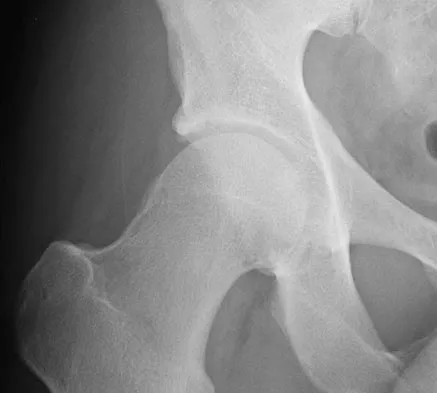
The patho-epidemiology of ankle joint disorders is heavily dominated by the sequelae of acute and repetitive trauma. The ankle is the most frequently injured joint in athletic populations, with lateral inversion sprains accounting for up to 85% of all ankle injuries. While the majority of these sprains resolve with conservative management, approximately 20% to 30% of patients will develop chronic ankle instability (CAI) or chronic recalcitrant pain. This chronic pain is frequently driven by intraarticular derangements that occur at the time of the initial injury, including osteochondral lesions of the talus (OCLs), chondral shear injuries, and capsular tears that subsequently heal with hypertrophic, impinging scar tissue.
Furthermore, repetitive microtrauma—particularly in athletes subjected to extreme ranges of dorsiflexion and plantarflexion, such as soccer players and ballet dancers—precipitates the development of anterior and posterior impingement syndromes. Anterior impingement is characterized by the formation of "kissing" osteophytes on the anterior distal tibia and the dorsal talar neck. These osteophytes are theorized to result from repetitive capsular traction or direct bony abutment during forced dorsiflexion. Posterior impingement often involves the compression of the posterior capsular structures, an os trigonum, or a prominent Stieda process between the posterior tibia and the calcaneus during forced plantarflexion.
Understanding the epidemiology and the underlying mechanical pathophysiology is essential for the orthopedic surgeon. The presence of chronic, unexplained ankle pain following an inversion injury should raise a high index of suspicion for intraarticular pathology. The modern orthopedic surgeon must synthesize the patient's kinematic history, precise clinical examination findings, and advanced imaging to determine the exact patho-anatomic lesion. This comprehensive understanding forms the foundation upon which successful arthroscopic intervention is built, ensuring that the surgical approach addresses the root mechanical failure rather than merely treating the symptoms.
Detailed Surgical Anatomy and Biomechanics
A profound, three-dimensional understanding of the osseous, ligamentous, and neurovascular anatomy surrounding the ankle joint is absolutely non-negotiable for the arthroscopic surgeon. The tibiotalar joint is a highly congruent, tightly constrained hinge joint. The osseous architecture consists of the tibial plafond and the medial and lateral malleoli, which together form a rectangular mortise that articulates with the trapezoidal talar dome. The talar dome is approximately 2 to 3 millimeters wider anteriorly than posteriorly. This anatomical nuance dictates that the mortise is maximally stable in dorsiflexion, where the wider anterior talus engages the mortise, and relatively less stable in plantarflexion, explaining the high propensity for inversion injuries when the foot is plantarflexed.
The ligamentous constraints of the ankle are divided into the lateral collateral complex, the medial deltoid complex, and the syndesmotic ligaments. The lateral complex comprises the anterior talofibular ligament (ATFL), the calcaneofibular ligament (CFL), and the posterior talofibular ligament (PTFL). The ATFL is intimately blended with the anterior capsule and is frequently visualized and evaluated during anterior arthroscopy. The distal fascicle of the anterior inferior tibiofibular ligament (AITFL), often referred to as Bassett’s ligament, can become pathologically thickened following repetitive trauma, leading to anterolateral soft tissue impingement. The robust deltoid ligament medially is rarely completely ruptured but can be a source of medial gutter impingement when hypertrophic.
The superficial neurovascular anatomy dictates the placement of arthroscopic portals and represents the greatest source of potential iatrogenic morbidity. The anterior ankle is traversed by several critical structures. Medially, the great saphenous vein and the saphenous nerve lie in close proximity to the anteromedial portal. Centrally, the anterior tibial tendon, extensor hallucis longus (EHL), deep peroneal nerve, and anterior tibial artery form a "danger zone" that must be strictly avoided; central portals are historically associated with high rates of neurovascular injury and are now largely obsolete. Laterally, the superficial peroneal nerve (SPN) and its arborization into the medial and intermediate dorsal cutaneous nerves cross the anterolateral joint line. The SPN is highly variable in its course and must be meticulously identified and protected during anterolateral portal establishment.
Biomechanically, the ankle joint is not a simple uniplanar hinge. The axis of rotation is oblique, passing from the anteromedial tip of the medial malleolus to the posterolateral tip of the lateral malleolus. This oblique axis allows for coupled motions: dorsiflexion is coupled with external rotation and slight eversion, while plantarflexion is coupled with internal rotation and inversion. During arthroscopy, the surgeon must manipulate the joint through these coupled kinematic pathways to fully visualize the articular surfaces and dynamically assess for impingement. The tight capsular constraints and the congruent osseous anatomy necessitate joint distraction—either gravity-assisted or mechanical—to safely introduce instruments and navigate the joint without causing iatrogenic scuffing of the delicate hyaline cartilage.
Exhaustive Indications and Contraindications
The success of ankle arthroscopy is intrinsically linked to meticulous patient selection and precise preoperative diagnosis. Purely diagnostic ankle arthroscopy, performed without a clear preoperative hypothesis, yields a notoriously low success rate and is generally discouraged in modern practice. Patients typically present with chronic, recalcitrant ankle pain that has failed to respond to an exhaustive course of conservative management, including physical therapy, bracing, and nonsteroidal anti-inflammatory drugs (NSAIDs) for a minimum of 3 to 6 months.
Primary indications for ankle arthroscopy revolve around impingement syndromes and chondral pathology. Soft tissue impingement, particularly anterolateral impingement involving hypertrophic synovium or a thickened Bassett’s ligament, is highly amenable to arthroscopic resection. Bony impingement, characterized by anterior tibial and talar osteophytes, can be definitively treated with arthroscopic burring, restoring terminal dorsiflexion. Osteochondral lesions of the talus (OCLs) represent a major indication; arthroscopy allows for direct visualization, probing of cartilage stability, meticulous débridement of the necrotic subchondral bone cyst, and execution of marrow stimulation techniques (e.g., microfracture or drilling) to promote fibrocartilage infill.
Secondary and expanding indications include the management of acute intraarticular fractures, where arthroscopy can assist in achieving anatomic reduction of the articular surface without the morbidity of a large arthrotomy. Arthroscopic-assisted lateral ligament stabilization (e.g., the arthroscopic Broström-Gould procedure) is gaining immense popularity for chronic ankle instability. Furthermore, arthroscopy is invaluable in the urgent management of septic arthritis, allowing for copious joint lavage, targeted synovectomy, and the breakdown of loculations. Arthrofibrosis following severe trauma or previous surgery can also be addressed via arthroscopic lysis of adhesions.
Contraindications must be strictly respected to avoid catastrophic complications. Absolute contraindications include localized active soft tissue infection or cellulitis over the portal sites, which risks introducing bacteria into the sterile joint space. Severe peripheral vascular disease is an absolute contraindication, as the use of a tourniquet and joint distension can compromise an already tenuous vascular supply, leading to ischemic necrosis or amputation. End-stage osteoarthritis with complete obliteration of the joint space represents a relative contraindication; while arthroscopic debridement (a "joint clean-out") may offer temporary palliative relief, it does not alter the natural history of the disease and often yields disappointing long-term results.
| Category | Specific Condition | Clinical Rationale and Considerations |
|---|---|---|
| Primary Indications | Anterolateral Soft Tissue Impingement | Excision of hypertrophic synovium or thickened Bassett's ligament provides excellent, predictable pain relief. |
| Primary Indications | Anterior Bony Impingement | Resection of tibial/talar osteophytes restores dorsiflexion and prevents further chondral abutment. |
| Primary Indications | Osteochondral Lesions of the Talus (OCLs) | Allows for staging, debridement of unstable cartilage, and marrow stimulation (microfracture). |
| Secondary Indications | Chronic Ankle Instability | Arthroscopic Broström procedures allow concurrent intraarticular pathology management and capsular plication. |
| Secondary Indications | Septic Arthritis | Enables urgent, high-volume lavage, synovectomy, and targeted antibiotic delivery. |
| Absolute Contraindications | Active Localized Cellulitis | High risk of iatrogenic inoculation of the sterile intraarticular space. |
| Absolute Contraindications | Severe Peripheral Vascular Disease | Tourniquet use and fluid extravasation can critically compromise distal perfusion. |
| Relative Contraindications | End-Stage Osteoarthritis | Obliterated joint space prevents safe instrument insertion; palliative debridement yields poor long-term outcomes. |
| Relative Contraindications | Severe Edema / Compromised Soft Tissue Envelope | Increases the risk of massive fluid extravasation, compartment syndrome, and wound healing failure. |
Pre-Operative Planning, Templating, and Patient Positioning
Thorough preoperative planning begins with a comprehensive clinical examination and advanced imaging. The surgeon must meticulously palpate the ankle to localize the point of maximal tenderness, differentiating between intraarticular pathology and extraarticular sources of pain (e.g., peroneal tendinopathy). Provocative maneuvers, such as forced dorsiflexion to elicit anterior impingement pain, are critical. If the diagnosis remains equivocal, a diagnostic intraarticular injection of a local anesthetic (e.g., lidocaine or bupivacaine) under ultrasound or fluoroscopic guidance is highly recommended. Significant, albeit temporary, pain relief strongly suggests an intraarticular pathological process amenable to arthroscopic intervention.
Imaging is paramount. Standard weight-bearing radiographs (anteroposterior, lateral, and mortise views) are evaluated for joint space narrowing, loose bodies, and the size and location of osteophytes. Magnetic Resonance Imaging (MRI) is the gold standard for evaluating soft tissue impingement, ligamentous integrity, and the exact dimensions of osteochondral lesions. For OCLs, the surgeon must template the lesion's size, depth, and the presence of subchondral cysts, as lesions larger than 1.5 cm² may require advanced cartilage restoration techniques (e.g., osteochondral autograft transfer) rather than simple microfracture. Computed Tomography (CT) is occasionally utilized for precise three-dimensional mapping of complex bony impingement or fracture patterns.
Optimal patient positioning is critical for adequate joint distraction, which is the absolute key to safe instrument insertion and comprehensive visualization. The supine gravity-assisted technique is widely favored. The patient is placed supine, and the proximal thigh of the operative extremity is secured in a well-padded leg holder. The end of the bed is dropped, flexing the hip to approximately 45 degrees and the knee to 90 degrees. The foot and lower leg hang free, utilizing the weight of the leg to provide a natural, dynamic distraction of the tibiotalar joint. This setup allows the surgeon to freely manipulate the ankle through a full range of motion, dynamically assessing impingement and facilitating access to the posterior recesses.
When gravity alone is insufficient to open a tightly constrained joint, noninvasive mechanical distraction is employed. A sterile, noninvasive distraction strap (e.g., a soft ankle hitch) is applied around the hindfoot and midfoot and connected to a tensioning device attached to the operating table. Controlled traction, typically between 15 to 30 pounds, is applied. However, the surgeon must be acutely aware of the pitfalls of mechanical distraction. Prolonged or excessive traction can lead to devastating neuropraxia of the superficial peroneal or sural nerves. Distraction time must be strictly monitored, and it is a fundamental rule that traction should be released periodically if the intraarticular portion of the procedure extends beyond 45 to 60 minutes.
Step-by-Step Surgical Approach and Fixation Technique
The execution of ankle arthroscopy demands a highly standardized, meticulous protocol to ensure both maximum therapeutic efficacy and patient safety. The procedure begins with precise preoperative marking prior to the inflation of the tourniquet. The surgeon must palpate and mark the medial and lateral malleoli, the anterior joint line, the tibialis anterior tendon, and the peroneus tertius. Crucially, the superficial peroneal nerve must be identified. By plantarflexing and inverting the foot while the patient actively extends the fourth toe, the SPN can often be visualized or palpated as a subcutaneous "bowstring." Its arborization pattern is marked with a surgical pen. The extremity is then elevated, exsanguinated using an Esmarch bandage, and the proximal thigh tourniquet is inflated to standard pressure (typically 250–300 mm Hg).
Joint insufflation is the next critical step. An 18-gauge spinal needle is introduced at the marked anteromedial portal site, directed slightly proximally and centrally toward the anatomic center of the tibiotalar joint. Approximately 15 to 20 mL of sterile normal saline is injected. Successful intraarticular placement is confirmed by the "dorsiflexion sign": as the anterior capsule becomes distended and tight, the foot will spontaneously dorsiflex. Furthermore, there should be minimal resistance to injection and immediate egress of saline when pressure on the plunger is released. Once insufflated, a precise, superficial skin incision is made using a #11 scalpel blade. This incision must be just large enough to accommodate the cannula. A blunt, straight hemostat is used to spread the subcutaneous tissues down to the capsule—the "nick and spread" technique—which gently sweeps the saphenous nerve and vein out of harm's way. The arthroscopic cannula with a blunt trocar is then introduced through the capsule until a distinct "pop" is felt, and the 30-degree arthroscope is inserted.
The anterolateral (AL) portal is always established under direct intraarticular visualization to prevent iatrogenic cartilage scuffing and to ensure optimal trajectory. With the arthroscope in the AM portal illuminating the lateral joint capsule, an 18-gauge spinal needle is inserted at the anticipated AL portal site. The surgeon visualizes the needle entering the joint, adjusting the trajectory to ensure it is parallel to the joint surface. Once confirmed, a superficial skin incision is made, blunt dissection is performed to protect the SPN, and the working cannula is introduced. A systematic, 21-point diagnostic sweep of the ankle joint is then performed, evaluating the anterior gutter, medial gutter, central joint (talar dome and tibial plafond), and lateral gutter to ensure no pathology is missed.
Therapeutic intervention proceeds based on the identified pathology. For soft tissue impingement, a 2.9-mm or 3.5-mm full-radius arthroscopic shaver is introduced to resect hypertrophic synovium and fibrotic bands. For bony impingement, an arthroscopic burr is utilized to resect anterior tibial or talar neck osteophytes, ensuring the resection is flush with the native cortical bone to restore impingement-free dorsiflexion. For OCLs, the unstable cartilage is debrided with a curette, and the necrotic subchondral bone is removed. Microfracture is then performed using arthroscopic awls, penetrating the subchondral bone plate at 3- to 4-millimeter intervals to a depth of approximately 4 millimeters, ensuring the release of marrow elements (fat droplets) into the defect. Following intervention, the joint is copiously irrigated, all fluid is evacuated, and the portals are meticulously closed with non-absorbable sutures (e.g., 4-0 nylon) using a horizontal mattress technique to definitively prevent postoperative synovial fistulas.
Complications, Incidence Rates, and Salvage Management
While ankle arthroscopy is generally considered a safe and minimally invasive procedure, it is not without risk. The tight spatial constraints of the tibiotalar joint, combined with the proximity of superficial neurovascular structures, create a unique environment where technical errors can lead to significant morbidity. The overall complication rate for ankle arthroscopy is historically reported in the orthopedic literature to be between 5% and 9%. The vast majority of these complications are minor and transient, but severe, limb-threatening complications, though rare, demand immediate recognition and expert salvage management.
Neurological injury represents the most frequent complication, accounting for nearly half of all adverse events. The superficial peroneal nerve (SPN) is the most commonly injured structure, typically damaged during the establishment of the anterolateral portal. Injuries range from transient neuropraxia due to excessive mechanical distraction or tourniquet pressure, to direct partial or complete transection by a scalpel or trocar. Saphenous nerve injury at the anteromedial portal and sural nerve injury at the posterolateral portal are also well-documented. Prevention relies heavily on the "nick and spread" technique and avoiding excessively deep skin incisions. Management of neuropraxia is expectant, often resolving within weeks to months. However, complete transection resulting in a painful neuroma may require subsequent surgical exploration, neuroma excision, and burying of the proximal nerve stump into deep muscle or bone.
Iatrogenic cartilage damage is a highly preventable but unfortunately common complication, particularly among less experienced arthroscopists. The convex talar dome is exquisitely vulnerable to scuffing or gouging during the blind insertion of sharp trocars or the aggressive manipulation of shavers and burrs in a tight joint space. To mitigate this risk, surgeons must mandate the use of blunt trocars for capsular penetration, ensure adequate joint distraction prior to instrument insertion, and always establish secondary working portals under direct intraarticular visualization. If significant iatrogenic cartilage damage occurs, it must be documented, and depending on the depth and size of the lesion, it may require immediate debridement or microfracture to stimulate fibrocartilage repair.
Infectious complications and wound healing issues, while less common, require aggressive management. Superficial portal site infections typically respond well to oral antibiotics and local wound care. Deep septic arthritis is exceedingly rare (incidence < 0.1%) but is a surgical emergency requiring immediate return to the operating room for arthroscopic lavage, synovectomy, and targeted intravenous antibiotic therapy. Synovial fistulas occur when the delicate subcutaneous tissues and skin fail to heal, allowing continuous egress of synovial fluid. This is directly correlated with poor portal closure techniques. Prevention requires meticulous, watertight closure of the skin, often utilizing mattress sutures, and the application of a compressive dressing. If a fistula develops, initial management consists of strict immobilization and compression; refractory cases necessitate operative debridement and layered closure.
| Complication Type | Estimated Incidence | Prevention Strategy | Salvage Management |
|---|---|---|---|
| Neurological Injury (SPN, Saphenous) | 3.0% - 5.0% | "Nick and spread" technique; limit mechanical distraction time to < 60 mins. | Expectant observation for neuropraxia; surgical excision and burying for painful neuromas. |
| Iatrogenic Cartilage Scuffing | 1.0% - 3.0% | Strict use of blunt trocars; establish secondary portals under direct visualization. | Document injury; debridement of loose flaps; microfracture if full-thickness defect occurs. |
| Synovial Fistula | 0.5% - 1.5% | Meticulous horizontal mattress suture closure; postoperative compressive dressing. | Immobilization and compression; operative over-sewing if refractory after 2 weeks. |
| Superficial Portal Infection | 0.5% - 1.0% | Strict aseptic technique; avoid operating through compromised skin. | Oral antibiotics (e.g., cephalexin); local wound care; suture removal if acting as a nidus. |
| Deep Septic Arthritis | < 0.1% | Preoperative prophylactic antibiotics; absolute respect for sterile boundaries. | Urgent surgical emergency: return to OR for arthroscopic lavage, synovectomy, and IV antibiotics. |
| Instrument Breakage | < 0.1% | Avoid levering shavers/burrs against dense cortical bone in tight joint spaces. | Immediate arthroscopic retrieval using graspers; may require arthrotomy if unretrievable. |
Phased Post-Operative Rehabilitation Protocols
The postoperative rehabilitation protocol following ankle arthroscopy is highly individualized and strictly dictated by the specific patho-anatomy addressed and the therapeutic interventions performed during the surgical procedure. A one-size-fits-all approach is inappropriate and can compromise surgical outcomes. Generally, rehabilitation is divided into distinct phases, progressing from tissue protection and edema control to the restoration of full range of motion, proprioception, and ultimately, sport-specific kinematic function. Communication between the orthopedic surgeon and the physical therapy team is critical to ensure the patient advances safely through these phases.
For patients who have undergone isolated soft tissue débridement, synovectomy, or resection of anterior bony impingement (osteophytes) without violation of the subchondral bone plate, the rehabilitation protocol is accelerated. Phase I (0 to 2 weeks) focuses on edema control and early mobilization. Patients are typically allowed immediate weight-bearing as tolerated in a supportive walking boot or stiff-soled surgical shoe. Early active range of motion (ROM) exercises—specifically focusing on dorsiflexion and plantarflexion—are initiated within 48 to 72 hours to prevent arthrofibrosis and capsular adhesions. Phase II (2 to 6 weeks) transitions the patient out of the boot and into standard footwear. Physical therapy aggressively targets the restoration of normal gait mechanics, peroneal and tibial strengthening, and intensive proprioceptive training using balance boards. Return to competitive sports or heavy manual labor is typically achieved between 4 to 8 weeks.
Conversely, if marrow stimulation techniques (e.g., microfracture, drilling, or abrasion arthroplasty) were performed for the treatment of osteochondral lesions of the talus (OCLs), the rehabilitation protocol is significantly more conservative. The biological imperative in Phase I is the protection of the fragile "superclot" that forms within the microfracture defect. Patients are generally kept strictly non-weight-bearing on crutches for 4 to 6 weeks to allow the mesenchymal stem cells to organize, differentiate, and mature into a stable fibrocartilage repair tissue. However, early, non-weight-bearing active ROM is highly encouraged, as the cyclical mechanical loading of the joint fluid provides essential nutrition to the developing chondrocytes.
Phase II for marrow stimulation patients (weeks 6 to 10) involves a graduated return to weight-bearing, typically starting at 25% of body weight and progressing weekly. Closed kinetic chain exercises are introduced to safely load the joint. Phase III (weeks 10 to 16+) focuses on advanced strengthening, eccentric loading of the Achilles and peroneal tendons, and dynamic stabilization. High-impact activities, plyometrics, and running are strictly prohibited until at least 12 to 16 weeks postoperatively, and only after clinical and radiographic evidence suggests adequate maturation of the fibrocartilage infill. Premature return to impact loading can result in the catastrophic failure of the microfracture clot, leading to the rapid recurrence of subchondral cyst formation and recalcitrant pain.
Summary of Landmark Literature and Clinical Guidelines
The evolution and standardization of ankle arthroscopy are deeply rooted in landmark orthopedic literature and rigorously developed clinical guidelines. A comprehensive understanding of these foundational texts is essential for the academic surgeon to practice evidence-based medicine. Early pioneers, such as Richard Ferkel, fundamentally defined the anatomical safe zones and portal placements that remain the gold standard today. Ferkel's seminal studies in the late 1980s and 1990s meticulously mapped the neurovascular anatomy of the anterior ankle, establishing the "nick and spread" technique as an absolute requirement to minimize the historically high rates of superficial peroneal and saphenous nerve injuries. His comprehensive reviews of complications in ankle arthroscopy serve as the baseline against which modern complication rates are measured.
In the realm of posterior ankle pathology, the paradigm was shifted by the groundbreaking work of C. Niek van Dijk. Prior to his contributions, accessing the posterior compartment was fraught with danger due to the proximity of the sural nerve, posterior tibial artery, and tibial nerve. Van Dijk introduced the two-portal endoscopic posterior approach, performed with the patient in the prone position. His landmark papers delineated the precise anatomical landmarks for the posteromedial and posterolateral portals, utilizing the Achilles tendon as the primary reference point. This technique revolutionized the treatment of posterior impingement syndrome, os trigonum excision, and posterior talar OCLs, offering a safe, reproducible, and highly effective alternative to extensive open posterior arthrotomies.
The management of osteochondral lesions of the talus has been heavily influenced by the work of Chuckpaiwong et al., who provided critical clinical guidelines regarding the size limitations of microfracture. Their retrospective analysis demonstrated that while microfracture yields excellent outcomes for OCLs smaller than 1.5 cm², the clinical failure rate increases exponentially for lesions exceeding this critical threshold. This 1.5 cm² cutoff has become a universally accepted clinical guideline, directing surgeons to utilize advanced cartilage restoration techniques—such as osteochondral autograft transfer system (OATS) or matrix-induced autologous chondrocyte implantation (MACI)—for larger, cystic lesions.
Modern consensus guidelines, published by organizations such as the International Society of Arthroscopy, Knee Surgery and Orthopaedic Sports Medicine (ISAKOS) and the American Orthopaedic Foot & Ankle Society (AOFAS), continuously synthesize this literature. Current guidelines strongly advocate for the judicious use of mechanical distraction, strictly limiting traction time to prevent neuropraxia, and emphasize the necessity of preoperative MRI for precise surgical templating. By adhering to these evidence-based guidelines and standing on the shoulders of these orthopedic pioneers, the modern surgeon can execute ankle arthroscopy with maximum precision, safety, and therapeutic efficacy.
