Comprehensive Introduction and Patho-Epidemiology
The evolution of reconstructive microsurgery has fundamentally altered the algorithmic approach to massive composite tissue defects in orthopaedic surgery. While conventional free tissue transfer effectively addresses soft tissue coverage and isolated diaphyseal bone loss, the reconstruction of articular surfaces, growing physes, and massive peripheral nerve gaps presents a uniquely complex biomechanical and physiological challenge. Conventional non-vascularized grafts rely entirely on the slow, unpredictable process of creeping substitution for bone, and plasmatic imbibition for nerves. In the context of massive defects or hostile recipient beds—such as those compromised by radiation, severe crush injury, or chronic infection—these non-vascularized grafts invariably fail, leading to nonunion, graft resorption, central ischemic necrosis, and catastrophic functional loss.
The clinical experiences of pioneer microsurgeons have definitively demonstrated that whole joints, epiphyseal plates, and large-caliber peripheral nerves, when transplanted on a meticulously preserved vascular pedicle, bypass the requisite phase of avascular necrosis. These composite tissues can survive, function, and grow without the rapid deterioration characteristic of avascular grafts. By immediately re-establishing arterial inflow and venous outflow, the cellular viability of chondrocytes, osteocytes, and Schwann cells is preserved. This immediate physiological perfusion maintains the structural integrity of the extracellular matrix and allows for continuous, uninterrupted biological activity, which is absolutely paramount when dealing with skeletally immature patients requiring longitudinal growth, or patients requiring massive nerve regeneration across scarred beds.
Epidemiologically, the demand for vascularized composite tissue transfers arises from some of the most devastating clinical scenarios encountered in orthopaedics. In the pediatric population, aggressive primary bone sarcomas (such as osteosarcoma and Ewing sarcoma) frequently necessitate wide intra-articular or trans-physeal resections. Reconstructing these massive defects in a growing child with a metallic endoprosthesis guarantees future limb-length discrepancies and multiple revision surgeries. Similarly, severe pediatric trauma resulting in physeal arrest, or profound congenital anomalies like radial longitudinal deficiency (radial club hand), demand a reconstructive solution that provides both immediate structural support and long-term growth potential.
Furthermore, the application of microvascular techniques to peripheral nerve reconstruction—specifically the vascularized nerve graft—has revolutionized the management of extensive nerve defects situated within hostile, avascular recipient beds. High-energy trauma, such as motorcycle accidents resulting in global brachial plexus avulsions, or severe industrial crush injuries, frequently leave massive nerve gaps exceeding 10 to 12 centimeters. In these scenarios, standard cable grafting is doomed to fail due to central necrosis and dense intraneural fibrosis. The vascularized nerve graft circumvents this by providing an immediate, nutrient-rich environment that actively supports advancing growth cones, doubling the rate of axonal regeneration and salvaging limbs that would otherwise face inevitable amputation.
Detailed Surgical Anatomy and Biomechanics
Physeal and Articular Vascular Anatomy
Understanding the intricate dual blood supply of the physis is the absolute cornerstone of successful vascularized physeal transfer. The epiphyseal plate relies on two distinct, non-anastomosing vascular networks, and the preservation of both is non-negotiable for continued longitudinal growth. The first network comprises the epiphyseal vessels, which supply the resting and proliferative zones of the physis. These vessels are responsible for delivering the essential nutrients required for continuous chondrogenesis. Interruption of the epiphyseal blood supply leads to immediate, irreversible cessation of chondrocyte proliferation, resulting in rapid physeal fusion and permanent growth arrest. When harvesting a vascularized joint, the surgeon must meticulously preserve the capsular and retinacular vessels that feed this epiphyseal network.
The second network consists of the metaphyseal vessels, which supply the zone of provisional calcification and the hypertrophic zone. These vessels are integral to the process of endochondral ossification, where the cartilaginous matrix is invaded by osteoblasts and converted into woven bone. If the metaphyseal vessels are compromised while the epiphyseal vessels remain intact, chondrogenesis will continue, but ossification will halt. This leads to a pathologically widened, hypertrophic physis that is biomechanically unstable and prone to shear fractures. Fortunately, if the metaphyseal circulation is rapidly re-established via microvascular anastomosis, endochondral ossification can resume. However, harvesting a bone flap that inadvertently ligates the epiphyseal vessels while preserving the metaphyseal vessels will result in a viable metaphyseal bone graft but a dead, prematurely fusing physis.
In the context of the workhorse vascularized proximal fibula transfer, the vascular anatomy is notoriously variable and demands rigorous preoperative mapping. The proximal fibular head and its physis are typically supplied by the anterior tibial recurrent artery (ATRA), a branch of the anterior tibial artery, or the inferior lateral genicular artery (ILGA), a branch of the popliteal artery. The diaphyseal portion of the fibula is supplied by the peroneal artery via its nutrient branch. A successful composite transfer of the proximal fibula must capture the specific arterial pedicle supplying the fibular head to ensure physeal viability, often requiring a complex, dual-pedicle anastomosis if the diaphyseal and epiphyseal blood supplies do not share a common, harvestable trunk.
Peripheral Nerve Microcirculation and Regeneration Biomechanics
The microcirculation of peripheral nerves is a highly sophisticated system designed to maintain the metabolic demands of axonal transport and Schwann cell function. This system is divided into the extrinsic regional feeding vessels and the intrinsic longitudinal microvascular plexus. The extrinsic system consists of segmental branches from adjacent major arteries (the vasa nervorum) that enter the nerve via the mesoneurium. Once these vessels pierce the epineurium, they arborize into the intrinsic system—a redundant, continuous longitudinal plexus located within the epineurium, perineurium, and endoneurium. In a standard, non-vascularized nerve graft, the extrinsic supply is severed, and the graft must survive entirely on plasmatic imbibition from the recipient bed until neovascularization occurs.
In massive nerve defects or heavily scarred beds, plasmatic imbibition is vastly insufficient. Thick nerve trunks transferred without a blood supply undergo rapid central ischemic necrosis. The core of the nerve graft dies, leading to the proliferation of dense intraneural fibroblasts that create an impenetrable physical barrier to advancing regenerating axons. By transferring a donor nerve with its main arterial and venous systems intact and re-establishing circulation via microvascular anastomosis, the intrinsic longitudinal plexus is immediately pressurized. This immediate perfusion prevents Wallerian degeneration-induced Schwann cell death, maintaining a viable, metabolically active cellular environment that actively secretes neurotrophic factors to support advancing growth cones.
The biomechanical and physiological advantages of this immediate perfusion are profound. Experimental and clinical data demonstrate that axonal regeneration rates are significantly enhanced in vascularized nerve grafts. While a standard non-vascularized cable graft might support regeneration at a rate of 1 to 1.5 millimeters per day, revascularized nerve grafts facilitate innervation at an accelerated rate of 3.2 to 6 centimeters per month. Furthermore, the robust blood supply allows the vascularized graft to traverse extensively scarred, irradiated, or avascular beds without undergoing necrosis. This makes the vascularized nerve graft an indispensable biomechanical conduit for bridging massive gaps, particularly in the brachial plexus or the sciatic nerve, where the distance to the target motor endplates is critically long and time-dependent muscle atrophy is a constant threat.
Exhaustive Indications and Contraindications
Patient selection for vascularized joint, physeal, and nerve transfers is paramount and requires a highly nuanced, multidisciplinary approach. These procedures are among the most technically demanding in all of reconstructive microsurgery, carrying significant risks of donor-site morbidity, prolonged operative times, and catastrophic graft failure if microvascular thrombosis occurs. Therefore, the indications are typically reserved for severe, limb-threatening or function-threatening scenarios where conventional reconstructive techniques are guaranteed to fail or yield unacceptable long-term morbidity.
For vascularized joint and physeal transfers, the primary indication is pediatric post-traumatic reconstruction or oncologic resection. In skeletally immature patients who have suffered traumatic loss of a joint or long bone segment (e.g., proximal humerus or distal radius), continued longitudinal growth is mandatory to prevent severe limb-length discrepancies and progressive angular deformities. Similarly, following the wide local excision of primary bone sarcomas involving the epiphysis, a vascularized proximal fibula transfer can reconstruct the defect while maintaining the child's growth potential. Other indications include severe congenital deformities, such as advanced manifestations of radial longitudinal deficiency (radial club hand), and advanced stages of avascular necrosis, such as Kienböck's disease (lunate osteonecrosis) or Preiser's disease (scaphoid osteonecrosis), where vascularized joint or bone transfers are utilized to revascularize the collapsing carpus.
The indications for vascularized nerve grafts are strictly defined by the length of the nerve defect and the quality of the recipient bed. Taylor and other pioneers recommended restricting vascularized nerve grafting to patients facing massive defects—typically defined as gaps exceeding 10 to 12 centimeters. At this length, the central necrosis inherent to non-vascularized cable grafts makes functional recovery highly improbable. Furthermore, any massive nerve gap situated within a hostile recipient bed—such as a bed compromised by Volkmann ischemic contracture, high-voltage electrical burns, severe crush injuries, or previous high-dose radiotherapy—is an absolute indication for a vascularized graft. A classic application is in global brachial plexus reconstruction, utilizing a vascularized ulnar nerve graft harvested from an avulsed C8-T1 root to reconstruct upper trunk defects and restore shoulder and elbow function.
| Parameter | Vascularized Joint / Physeal Transfers | Vascularized Nerve Grafts |
|---|---|---|
| Primary Indications | Pediatric massive bone/joint loss, post-oncologic resection in growing children, severe congenital aplasias (e.g., radial club hand), advanced avascular necrosis (Kienböck's). | Massive nerve gaps (>10-12 cm), hostile/irradiated recipient beds, severe crush injuries, brachial plexus avulsions (using avulsed ulnar nerve as donor). |
| Relative Indications | Adult joint reconstruction for massive post-traumatic defects where arthrodesis or arthroplasty is undesirable. | Moderate nerve gaps (8-10 cm) in patients with poor healing potential or delayed presentation. |
| Absolute Contraindications | Severe peripheral vascular disease, active systemic infection, lack of viable recipient vessels, malignant tumors with positive margins. | Complete lack of proximal nerve root/fascicle viability, irreversible target muscle atrophy (typically >18-24 months post-injury), absent recipient vessels. |
| Relative Contraindications | Advanced patient age, severe donor site trauma, patient non-compliance with complex postoperative rehabilitation protocols. | Advanced age (due to poor neuroplasticity), concomitant severe soft tissue defects requiring separate massive free flaps (increasing surgical risk). |
Pre-Operative Planning, Templating, and Patient Positioning
Advanced Imaging and Vascular Mapping
The success of any vascularized composite tissue transfer is entirely predicated on meticulous, exhaustive preoperative planning and advanced vascular mapping. High-resolution Computed Tomography Angiography (CTA) of both the donor and recipient limbs is absolutely mandatory. For proximal fibula transfers, the surgeon must definitively identify the origin, caliber, and course of the anterior tibial recurrent artery (ATRA) and the inferior lateral genicular artery (ILGA). The CTA will reveal anatomical variants, such as a high bifurcation of the popliteal artery or an anomalous origin of the nutrient vessels, which could drastically alter the surgical approach or completely preclude the use of the planned donor site.
In addition to CTA, Magnetic Resonance Imaging (MRI) and MR Neurography are critical, particularly for oncologic resections and massive nerve defects. In tumor cases, the MRI defines the exact intraosseous and extraosseous extent of the sarcoma, dictating the precise level of the osteotomy required to achieve negative margins. For nerve injuries, MR Neurography helps delineate the extent of the neuroma-in-continuity and the exact length of the nerve gap, allowing the surgeon to calculate the required length of the donor nerve graft. Standard orthogonal plain radiographs of the entire limb are also obtained to assess bone quality, overall alignment, and to aid in preoperative templating.
Surgical Templating and Equipment Preparation
Surgical templating has evolved from transparent acetate overlays to sophisticated 3D computer-aided design and manufacturing (CAD/CAM). In complex pediatric joint reconstructions, 3D printed models of the patient's contralateral normal limb can be mirrored to create a precise template for the required graft dimensions. Custom cutting guides can be sterilized and used intraoperatively to ensure the donor bone is harvested at the exact required length and angle, minimizing ischemia time and ensuring a perfect geometric match at the recipient site. This is particularly vital when reconstructing the distal radius or proximal humerus, where the articular orientation dictates future joint kinematics.
Equipment preparation is equally rigorous. The operating theater must be equipped with a high-definition, dual-head operating microscope, micro-instruments, and specialized microvascular sutures (typically 8-0 to 10-0 nylon). Implantable venous Doppler probes should be available for continuous postoperative monitoring of the buried flap. Furthermore, the surgical team must ensure the availability of rigid, yet biologically respectful, fixation devices. Smooth Kirschner wires, miniature bridging plates, and external fixators must be ready, with the strict understanding that no hardware can cross the transplanted physis, as this would induce iatrogenic premature closure.
Patient Positioning and Dual-Team Coordination
To minimize the critical ischemia time of the composite graft and to reduce overall operative duration, a simultaneous two-team approach is standard protocol. This requires complex patient positioning and spatial coordination within the operating room. The patient is typically positioned supine. If the proximal fibula is being harvested, a bump is placed under the ipsilateral hip of the donor leg to allow for internal rotation, providing optimal access to the lateral aspect of the leg. The recipient limb (e.g., the upper extremity) is simultaneously prepped and draped on a radiolucent hand table.
Sterile tourniquets are applied to both the donor and recipient extremities. The recipient team begins by radically debriding the defect, isolating the recipient artery and veins, and preparing the bony or neural stumps. Concurrently, the donor team executes the meticulous harvest of the vascularized flap. Communication between the two teams is critical; the donor pedicle is not ligated until the recipient bed is fully prepared, the recipient vessels are isolated and proven to have pulsatile flow, and the exact required length of the graft is confirmed. This synchronized choreography ensures that the tissue is subjected to the absolute minimum duration of warm ischemia.
Step-by-Step Surgical Approach and Fixation Technique
Vascularized Proximal Fibula Harvest and Inset
The vascularized proximal fibula transfer is the undisputed workhorse for pediatric physeal reconstruction. The surgical approach begins with an extensile longitudinal incision over the lateral aspect of the leg, extending from the lateral femoral condyle, across the fibular head, and distally along the fibular shaft. The first and most critical step is the identification and protection of the common peroneal nerve. The nerve is meticulously identified at the posterior border of the biceps femoris tendon, neurolysed under loupe magnification, and gently retracted as it wraps around the fibular neck. Iatrogenic injury to this nerve results in devastating foot drop and profound donor site morbidity.
Following nerve protection, the anterior compartment musculature (tibialis anterior, extensor digitorum longus) is elevated off the interosseous membrane. The vascular dissection is performed with extreme care to identify the ATRA and its accompanying venae comitantes branching from the anterior tibial vessels. The pedicle is traced proximally to its origin to ensure adequate length and caliber for anastomosis. The proximal tibiofibular joint capsule is then carefully incised, preserving the delicate retinacular vessels supplying the epiphyseal plate. The fibular osteotomy is performed distally at the pre-calculated length using an oscillating saw under continuous saline irrigation to prevent thermal necrosis, ensuring the periosteum remains intact.
Once the fibula is isolated entirely on its vascular pedicle, the tourniquet is deflated. The surgeon must visually confirm robust perfusion of the fibular head, the physis, and the diaphyseal marrow before pedicle ligation. The graft is then transferred to the recipient site. Osteosynthesis must be exceptionally rigid to allow for microvascular anastomosis, yet biologically respectful to the physis. Fixation is typically achieved using smooth Kirschner wires or custom bridging plates. The absolute cardinal rule of this procedure is that hardware must never cross the transplanted physis. Once skeletal stability is achieved, the microvascular anastomoses are performed.
Vascularized Sural Nerve Harvest and Coaptation
The sural nerve, supplied by the superficial sural artery (a branch of the popliteal artery), is the preferred donor for vascularized nerve transfers. The patient is placed prone or in a lateral decubitus position. A longitudinal incision is made over the posterior calf, extending from the popliteal fossa down to the lateral malleolus. The lesser saphenous vein and the sural nerve are identified distally. The dissection proceeds proximally, elevating the nerve along with a generous cuff of surrounding deep fascia, the superficial sural artery, and its venae comitantes. This fascial cuff protects the delicate vasa nervorum and the intrinsic longitudinal plexus.
The pedicle is traced proximally to its origin at the popliteal vessels. A small skin paddle may be included in the harvest to serve as a reliable postoperative monitor of flap perfusion. At the recipient site, the proximal and distal stumps of the injured nerve are resected back to healthy, bleeding fascicles. The surgeon utilizes the "bread-loafing" technique under high magnification, sequentially slicing the neuroma until normal, unscarred fascicular architecture is visualized and pouting axoplasm is observed. Healthy recipient arteries and veins in the immediate vicinity of the nerve defect are meticulously isolated.
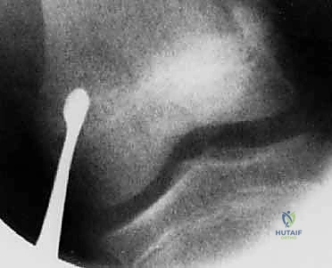
Crucially, the nerve graft must be reversed in its longitudinal orientation prior to inset. Because the sural nerve gives off collateral branches along its course, orienting the nerve in its native direction would allow regenerating axons to escape through these defunct collateral branches into the surrounding soft tissue. Reversing the graft ensures all regenerating axons are directed exclusively toward the distal target. Tensionless coaptation is absolutely critical; the graft must have slight redundancy to prevent traction injury during postoperative mobilization. Epineurial or group fascicular repair is performed using 8-0 or 9-0 monofilament nylon sutures under the operating microscope.
Microvascular Anastomosis and Perfusion Validation
The microvascular anastomosis is the definitive crux of the entire procedure. For both bone and nerve transfers, the arterial and venous anastomoses are typically performed prior to definitive nerve coaptation or final wound closure to minimize ischemia time. End-to-end or end-to-side anastomoses are performed using 8-0 or 9-0 nylon sutures, depending on the caliber match between the donor and recipient vessels. Venous outflow is equally, if not more, critical than arterial inflow; venous congestion is a leading cause of flap failure. Therefore, two veins should be anastomosed for every artery whenever anatomically feasible.
Following the completion of the anastomoses, the microvascular clamps are released. The surgeon must meticulously validate perfusion. In a vascularized fibula transfer, brisk bleeding from the medullary canal and the periosteum should be observed. In a vascularized nerve transfer, immediate return of bleeding from the epineurial vessels of the graft confirms successful revascularization. The anastomotic sites are bathed in warm papaverine to relieve vasospasm, and an implantable venous Doppler probe may be secured around the efferent vein to allow for continuous postoperative monitoring. The wounds are closed meticulously over closed-suction drains, ensuring absolutely no compression occurs over the vascular pedicle.
Complications, Incidence Rates, and Salvage Management
Microvascular Failure and Thrombosis
While the theoretical and physiological benefits of vascularized tissue transfers are immense, the procedures are fraught with profound technical challenges and potential complications. The most devastating complication is microvascular thrombosis, which typically occurs within the first 72 hours postoperatively. If the arterial pedicle thromboses, the massive composite graft is instantly converted into a massive avascular graft. In the case of a vascularized nerve graft, because of its thickness, it will undergo complete central necrosis, resulting in absolutely zero axonal regeneration and complete failure of the reconstruction. In a physeal transfer, thrombosis results in immediate death of the physis and cessation of growth.
The incidence of microvascular thrombosis in experienced high-volume centers ranges from 3% to 7%. Rapid identification is paramount. If clinical monitoring of the skin paddle shows pallor (arterial insufficiency) or violaceous engorgement (venous congestion), or if the implantable Doppler signal is lost, the patient must be returned to the operating room immediately for surgical exploration. Salvage management involves taking down the anastomosis, performing a thorough thrombectomy, utilizing local thrombolytics (e.g., tissue plasminogen activator or heparinized saline), and revising the anastomosis, potentially utilizing interpositional vein grafts if the pedicle is damaged or under tension.
Orthopaedic and Neurologic Complications
Beyond microvascular failure, numerous orthopaedic and neurologic complications can arise. In vascularized physeal transfers, premature physeal closure can occur even if the anastomosis remains patent. This is often due to unrecognized ischemic injury to the epiphyseal vessels during harvest, iatrogenic injury from hardware crossing the physis, or mechanical failure of the fixation leading to shear stress across the growth plate. The incidence of premature closure varies but can be as high as 15-20% in complex reconstructions. Salvage management for premature fusion may require subsequent limb lengthening procedures (distraction osteogenesis) or contralateral epiphysiodesis to manage limb-length discrepancies.
Donor site morbidity is another significant concern. Following proximal fibula harvest, patients may experience common peroneal nerve palsy (transient or permanent), knee instability due to disruption of the lateral collateral ligament complex (which must be meticulously reconstructed during closure), or ankle valgus instability in young children (necessitating a prophylactic distal tibiofibular syndesmotic screw). Following sural nerve harvest, patients universally experience loss of sensation in the lateral aspect of the foot and are at risk for painful neuroma formation at the proximal stump.
| Complication | Estimated Incidence | Prevention Strategy | Salvage Management / Treatment |
|---|---|---|---|
| Microvascular Thrombosis | 3% - 7% | Meticulous intimal repair, tension-free anastomosis, avoidance of pedicle kinking, dual venous outflow. | Immediate surgical re-exploration, thrombectomy, anastomotic revision with vein grafts. |
| Premature Physeal Closure | 15% - 20% | Preservation of capsular/epiphyseal vessels, absolute avoidance of trans-physeal hardware, rigid diaphyseal fixation. | Distraction osteogenesis (Ilizarov/Taylor Spatial Frame), contralateral epiphysiodesis. |
| Common Peroneal Nerve Palsy | 2% - 5% | Meticulous neurolysis and protection during proximal fibula harvest, avoidance of aggressive retraction. | Ankle-foot orthosis (AFO), delayed tendon transfers (e.g., posterior tibial tendon transfer) if permanent. |
| Nonunion at Osteotomy Site | 5% - 10% | Rigid internal fixation, preservation of periosteal blood supply, adequate debridement of recipient bed. | Autologous bone grafting, revision of internal fixation, electrical bone stimulation. |
| Donor Site Neuroma (Sural) | 5% - 15% | Burying the proximal nerve stump deep into the muscle belly (e.g., gastrocnemius) away from the incision line. | Surgical excision of neuroma, targeted muscle reinnervation (TMR), or proximal relocation. |
Phased Post-Operative Rehabilitation Protocols
Immediate Post-Operative Flap Monitoring and Immobilization
The immediate postoperative phase (Days 0 to 7) is defined by strict immobilization and hyper-vigilant flap monitoring. The reconstructed limb is immobilized in a custom, well-padded orthosis or a bivalved cast. It is absolutely critical that the immobilization device is designed with a large window to allow for unencumbered visualization of the skin paddle and to ensure that absolutely no external pressure is applied over the anatomical course of the vascular pedicle. The limb is typically elevated to promote venous drainage and minimize edema, which can secondarily compress the microvascular anastomosis.
Flap monitoring is conducted continuously. Clinical assessment of the skin paddle's color, capillary refill, and turgor is performed every hour for the first 48 hours, and then every 2 to 4 hours subsequently. If an implantable venous Doppler was utilized, the auditory signal is monitored constantly by the nursing staff. The patient is maintained on a specialized microsurgical protocol, which includes strict bed rest, maintenance of normothermia, aggressive hydration to ensure high cardiac output and optimal peripheral perfusion, and pharmacological prophylaxis against vasospasm and thrombosis (typically utilizing low molecular weight dextran, aspirin, or subcutaneous heparin, depending on institutional protocols).
Intermediate Phase and Progressive Mobilization
The intermediate phase (Weeks 2 to 8) marks the transition from strict immobilization to protected, progressive mobilization. For vascularized joint and bone transfers, radiographic evaluation is performed at regular intervals to assess the progression of metaphyseal union at the osteotomy sites. Strict non-weight-bearing or non-loading of the reconstructed extremity is rigidly enforced. However, to prevent devastating joint stiffness and tendon adhesions, protected passive range of motion (ROM) of the adjacent joints is initiated under the direct supervision of a specialized orthopaedic hand or physical therapist.
For vascularized nerve grafts, the intermediate phase involves monitoring for the early signs of axonal regeneration. Because the vascularized graft circumvents the prolonged delay associated with Wallerian degeneration and plasmatic imbibition, the advancing Tinel's sign can often be detected earlier than in conventional grafts. The therapist focuses on maintaining the passive suppleness of the paralyzed target muscles and preventing joint contractures through daily passive stretching and the use of dynamic splinting. Electrical stimulation of the denervated muscles may be employed to delay the onset of irreversible muscle atrophy while awaiting reinnervation.
Long-Term Surveillance and Functional Restoration
The long-term phase (Months 3 to 24+) is focused on functional restoration, strengthening, and longitudinal surveillance. For physeal transfers, once solid radiographic metaphyseal union is achieved (typically between 8 to 12 weeks), progressive weight-bearing or functional loading is permitted. The most critical aspect of long-term follow-up for these patients is the serial radiographic measurement of longitudinal growth. Scanograms or long-leg/long-arm alignment films are obtained every 6 months until skeletal maturity to ensure the transplanted physis remains open and is growing at a predictable rate. Any evidence of premature fusion or angular deformity must be identified early to allow for timely surgical intervention.
For nerve transfers, the long-term phase is a waiting game governed by the biology of nerve regeneration. As the regenerating axons finally reach the target motor endplates, the patient will begin to experience muscle fasciculations followed by trace voluntary contractions. At this stage, the rehabilitation protocol shifts dramatically toward active-assisted ROM, neuromuscular re-education, and progressive resistance training. Biofeedback techniques and mirror therapy are frequently utilized to help the patient integrate the newly reinnervated musculature into functional motor patterns. Maximum neurological recovery following massive vascularized nerve grafting may take up to 2 to 3 years, necessitating immense patience and psychological support for the patient.
Summary of Landmark Literature and Clinical Guidelines
Foundational Microsurgical Literature
The current clinical guidelines and operative techniques for vascularized composite tissue transfers are built upon the foundational, pioneering work of a select group of visionary microsurgeons. The concept of vascularized bone transfer was popularized by Taylor and colleagues in the 1970s, who first described the successful transfer of the vascularized fibula. Subsequently, Weiland et al. and Wray et al. produced landmark clinical reports definitively establishing that longitudinal growth continues after the vascularized transfer of physes in pediatric patients. Their meticulous documentation of long-term outcomes proved that the vascularized physis behaves biologically like a normal growth plate, provided its dual blood supply is maintained.
The experimental and clinical validation of vascularized nerve grafts is inextricably linked to the work of Taylor. In his seminal 1976 publication, Taylor described the first successful clinical application of a vascularized nerve graft, utilizing the radial nerve to bridge a massive median nerve defect. His subsequent experimental work demonstrated the profound physiological advantages
