Introduction & Epidemiology
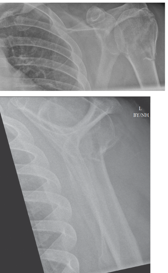
Proximal humerus fractures (PHFs) are common injuries, particularly affecting two distinct demographic groups: younger individuals involved in high-energy trauma and, more frequently, older, osteoporotic patients sustaining low-energy falls. These fractures represent approximately 5% of all fractures and are the third most common fracture in individuals over 65, following hip and distal radius fractures. The incidence is projected to increase with the aging global population.
The Neer classification system, widely adopted for its prognostic and therapeutic implications, categorizes PHFs based on the number of displaced "parts" (humeral head, greater tuberosity, lesser tuberosity, and surgical neck) and displacement criteria (displacement >1 cm or angulation >45 degrees). This system aids in surgical decision-making by correlating fracture morphology with potential vascular compromise and rotator cuff integrity.
*
One-part fractures:
Nondisplaced or minimally displaced.
*
Two-part fractures:
Displacement of one major segment relative to the others (e.g., surgical neck, greater tuberosity).
*
Three-part fractures:
Displacement of two segments relative to the humeral head.
*
Four-part fractures:
Displacement of all three segments (greater tuberosity, lesser tuberosity, surgical neck) relative to the humeral head.
*
Fracture-dislocations:
Any of the above combined with glenohumeral dislocation.
Understanding the epidemiology and classification is fundamental for appropriate diagnosis, prognostication, and the selection of optimal management strategies, ranging from conservative care to complex reconstructive surgery.
Surgical Anatomy & Biomechanics
A thorough understanding of the surgical anatomy and biomechanics of the proximal humerus is paramount for successful management of PHFs.
Surgical Anatomy
-
Proximal Humerus Bony Anatomy:
The proximal humerus comprises the humeral head, anatomical neck, greater tuberosity, lesser tuberosity, and surgical neck.
- Humeral Head: Articulates with the glenoid, covered by articular cartilage. Its blood supply is crucial for preventing avascular necrosis (AVN).
- Greater Tuberosity: Located laterally, serves as the insertion site for the supraspinatus, infraspinatus, and teres minor tendons (posterior cuff). Fractures here can lead to impingement and rotator cuff dysfunction.
- Lesser Tuberosity: Located anteriorly, serves as the insertion site for the subscapularis tendon (anterior cuff). Fractures can result in internal rotation deformity.
- Surgical Neck: The narrow region distal to the tuberosities, a common site for fracture.
- Anatomical Neck: The groove separating the humeral head from the tuberosities.
- Vascularity: The blood supply to the humeral head is predominantly from the ascending branch of the anterior circumflex humeral artery, forming the arcuate artery, which penetrates the head near the bicipital groove. The posterior circumflex humeral artery also contributes. Disruption of these vessels, particularly in multi-part fractures with significant displacement, is a major risk factor for AVN. The integrity of the medial calcar metaphyseal extension and its periosteal attachments is also vital for vascularity.
-
Neurovascular Structures:
- Axillary Nerve: Most commonly injured nerve in PHFs or during surgical approaches. It wraps around the surgical neck, approximately 5-7 cm distal to the acromion, innervating the deltoid and teres minor.
- Musculocutaneous Nerve: Located more medially and anteriorly, supplies the biceps and brachialis.
- Radial Nerve: Located more distally and posteriorly in the spiral groove.
- Brachial Plexus: Vulnerable in high-energy injuries or fracture-dislocations.
- Axillary Artery and Vein: Positioned medially and anteriorly, at risk during medial screw placement or extreme retraction.
- Soft Tissue & Rotator Cuff: The rotator cuff tendons (supraspinatus, infraspinatus, teres minor, subscapularis) insert into the greater and lesser tuberosities. Their integrity and position are critical for shoulder function. Muscle pull from the rotator cuff and deltoid can create significant deforming forces in displaced fractures. The bicipital groove houses the long head of the biceps tendon, which can be an anatomical landmark during surgical approaches.
Biomechanics
The stability of the proximal humerus is derived from its bony architecture, rotator cuff muscle balance, and ligamentous structures.
*
Deforming Forces:
*
Supraspinatus:
Pulls the greater tuberosity superiorly and posteriorly.
*
Subscapularis:
Pulls the lesser tuberosity anteriorly and medially.
*
Deltoid:
Pulls the humeral shaft proximally and laterally.
*
Pectoralis Major/Latissimus Dorsi/Teres Major:
Insert more distally, contributing to adduction and internal rotation of the shaft relative to the head.
*
Medial Calcar:
This dense trabecular bone region acts as a crucial weight-bearing structure, resisting varus collapse and providing critical support for internal fixation. Loss of medial calcar support significantly increases the risk of screw cutout and construct failure.
*
Bone Quality:
In osteoporotic bone, the ability of screws to achieve adequate purchase and withstand physiological loads is compromised, influencing fixation strategies and implant choice.
Indications & Contraindications
The decision-making process for managing proximal humerus fractures involves a careful assessment of patient factors, fracture characteristics, and potential complications. Both non-operative and operative strategies have distinct indications and contraindications.
Non-Operative Indications
Non-operative management is the treatment of choice for the majority of proximal humerus fractures, particularly one-part fractures, and often for two-part surgical neck fractures in elderly, low-demand patients with minimal displacement.
- Minimally Displaced or Nondisplaced Fractures: Generally defined as <1 cm displacement and <45 degrees angulation in any plane. These fractures inherently have good healing potential.
- Stable Fractures: Fractures where deforming forces are balanced, or the fracture pattern provides inherent stability (e.g., valgus-impacted fractures where the head is impacted into the shaft in a valgus position).
- Elderly, Low-Demand Patients: Individuals with significant comorbidities, limited functional expectations, or poor bone quality where the risks of surgery outweigh potential benefits.
- High Surgical Risk: Patients with severe medical comorbidities (cardiac, pulmonary, renal, neurological) that make anesthesia and surgery excessively dangerous.
- Poor Soft Tissue Envelope: Compromised skin or soft tissues that increase the risk of wound complications.
Operative Indications
Surgical intervention is generally reserved for displaced, unstable fractures, fracture-dislocations, and patients who are physiologically amenable to surgery with higher functional demands.
- Displaced Two-Part Surgical Neck Fractures: Significant displacement (>1 cm) or angulation (>45 degrees), especially in younger, active patients.
- Displaced Greater Tuberosity Fractures: Displacement >5 mm, particularly in active patients, due to the high risk of impingement and rotator cuff dysfunction if left unreduced. Can lead to subacromial impingement and weakness.
- Displaced Lesser Tuberosity Fractures: Less common, but can lead to internal rotation contracture and subscapularis dysfunction.
- Three-Part and Four-Part Fractures: Often require surgical intervention due to high displacement, instability, and risk of AVN. The specific strategy (ORIF, hemiarthroplasty, reverse total shoulder arthroplasty) depends on age, bone quality, rotator cuff integrity, and fracture morphology.
- Fracture-Dislocations: Require urgent reduction and often internal fixation to stabilize the fracture and prevent recurrence.
- Head-Splitting Fractures: Fractures involving the articular surface that are significantly displaced.
- Open Fractures: Require urgent surgical debridement and stabilization to prevent infection.
- Neurovascular Compromise: Acute neurovascular injury necessitates immediate surgical exploration and repair, often followed by fracture stabilization.
- Polytrauma Patients: When the proximal humerus fracture is part of a larger injury pattern and needs definitive stabilization to facilitate rehabilitation of other injuries.
- Younger, Active Patients: Higher functional expectations often warrant aggressive management to restore anatomy and function.
Contraindications
Absolute Contraindications:
* Active infection (local or systemic) in the presence of an open fracture.
* Severe soft tissue compromise, precluding safe surgical incision and closure.
* Patient unwillingness or inability to comply with post-operative rehabilitation protocols.
Relative Contraindications:
* Severe osteoporosis that may not hold internal fixation.
* Extreme medical comorbidities where the risk of surgery outweighs potential benefits, even in displaced fractures.
* Pre-existing shoulder arthritis or significant rotator cuff tear (may influence choice towards arthroplasty).
* Non-displaced or minimally displaced fractures that are stable.
Operative vs. Non-Operative Indications
| Feature / Factor | Non-Operative Management | Operative Management |
|---|---|---|
| Fracture Pattern | One-part, minimally displaced (Neer I) | Two-part displaced (surgical neck, tuberosity) |
| Stable, valgus-impacted fractures | Three-part, Four-part fractures | |
| Fracture-dislocations | ||
| Head-splitting fractures with significant displacement | ||
| Displacement/Angulation | <1 cm displacement, <45 degrees angulation | >1 cm displacement, >45 degrees angulation |
| Patient Age/Demand | Elderly, low-demand, sedentary patients | Younger, active, high-demand patients |
| Comorbidities | Significant medical comorbidities, high surgical risk | Physiologically fit, able to tolerate surgery |
| Bone Quality | Severe osteoporosis (may not hold fixation well) | Good to moderate bone quality for fixation |
| Associated Injuries | No significant neurovascular compromise | Open fractures, neurovascular injury |
| Significant soft tissue compromise (requiring debridement) |
Pre-Operative Planning & Patient Positioning
Meticulous pre-operative planning is essential for anticipating challenges and optimizing outcomes in proximal humerus fracture surgery.
Pre-Operative Assessment
-
Clinical Evaluation:
- Thorough history including mechanism of injury, hand dominance, pre-injury function, pain levels, and medical comorbidities.
- Detailed neurovascular examination, documenting sensory and motor function, pulses, and capillary refill. Axillary nerve function (deltoid contraction, sensation over lateral arm) is critical.
- Assessment of soft tissue integrity.
-
Imaging:
- Standard Radiographs: True AP, scapular Y, and axillary lateral views are mandatory. These allow for initial Neer classification, assessment of displacement, angulation, and humeral head position relative to the glenoid.
-
Computed Tomography (CT) Scan:
Indispensable for complex fractures, particularly three- and four-part fractures, fracture-dislocations, and head-splitting injuries.
- Provides detailed information on comminution, articular involvement, glenoid impression fractures, and precise tuberosity displacement.
- 3D reconstructions are invaluable for understanding fracture morphology and planning reduction maneuvers.
- Magnetic Resonance Imaging (MRI): Less commonly used acutely but can be helpful for assessing rotator cuff integrity, associated ligamentous injuries, or brachial plexus pathology in selected cases.
-
Templating:
- Using contralateral shoulder radiographs (if available) or anatomical templates, evaluate appropriate plate length, screw lengths, and number of screws required.
- Plan screw trajectories, especially calcar screws, to maximize purchase and avoid intra-articular penetration.
- Informed Consent: Discuss potential risks and complications including infection, nonunion, malunion, avascular necrosis (AVN), nerve injury (especially axillary), hardware prominence, stiffness, and the need for possible revision surgery (e.g., arthroplasty).
Patient Positioning
The choice of patient position depends on the planned surgical approach, surgeon preference, and the specific fracture pattern.
-
Beach Chair Position (Semi-Fowler's):
- Advantages: Excellent visualization for the deltopectoral approach, allows for easy assessment of shoulder range of motion during the procedure, less blood loss due to reverse Trendelenburg, and ability to assess rotator cuff integrity.
- Setup: Patient is semi-recumbent (45-60 degrees) with the head secured, often in a horseshoe headrest. The torso is angled, and the legs are slightly flexed. The operative arm is draped free, resting on a sterile hand table or supported by a specialized arm holder, allowing full circumduction and manipulation.
- Precautions: Ensure adequate padding to all pressure points, especially the contralateral elbow, sacrum, and heels. Monitor blood pressure closely for potential cerebral hypoperfusion ("beach chair hypotension").
-
Lateral Decubitus Position:
- Advantages: Can be preferred for certain posterior approaches or if shoulder arthroscopy is concurrently planned. May offer improved stability for patients with certain comorbidities.
- Setup: Patient is placed on the non-operative side, secured with axillary roll, hip and knee supports. The operative arm is draped free in an arm suspension system or on a sterile hand table.
- Precautions: Careful padding of the dependent ear, eye, and arm. Ensure adequate perfusion to the dependent limb.
-
 - Image Note: The image above illustrates typical patient positioning for shoulder surgery, likely in a beach chair or lateral decubitus setup, showcasing draped surgical field. This type of setup is critical for successful exposure and manipulation during proximal humerus fracture repair.
- Supine Position: Less common for proximal humerus plating, but can be used for intramedullary nailing via an anterior approach or for specific fracture patterns requiring limited anterior exposure.
Anesthesia & Prophylaxis
- Anesthesia: General anesthesia is typically employed. A regional nerve block (e.g., interscalene block) can be highly beneficial for post-operative pain control, reducing opioid requirements.
- Antibiotics: Prophylactic intravenous antibiotics (e.g., Cefazolin) administered pre-incision are standard.
- DVT Prophylaxis: Routine DVT prophylaxis should be considered based on patient risk factors and surgical duration.
Detailed Surgical Approach / Technique
The most common surgical technique for displaced proximal humerus fractures requiring open reduction and internal fixation (ORIF) is locking plate osteosynthesis via the deltopectoral approach. Other techniques include intramedullary nailing, hemiarthroplasty, and reverse total shoulder arthroplasty for complex cases.
Deltopectoral Approach for Locking Plate Osteosynthesis
This approach is versatile and provides excellent exposure for reduction and fixation of most proximal humerus fractures, while respecting the axillary nerve.
- Incision: A curvilinear incision is made from the coracoid process, extending distally along the deltopectoral groove for approximately 8-10 cm. The incision can be extended proximally towards the acromion or distally as needed.
-
Dissection:
- Skin and Subcutaneous Tissue: Incise down to the deep fascia.
- Deltopectoral Groove: Identify the cephalic vein running in the deltopectoral groove. This is the key internervous plane. The deltoid muscle is lateral (innervated by the axillary nerve), and the pectoralis major muscle is medial (innervated by the medial and lateral pectoral nerves).
- Cephalic Vein Management: The cephalic vein is typically retracted laterally with the deltoid, but can be ligated and divided if necessary for better exposure, particularly in revision cases or when space is limited. Care should be taken to preserve smaller venous tributaries.
- Deep Dissection: Retract the deltoid laterally and the pectoralis major medially. This exposes the clavipectoral fascia. Incise the clavipectoral fascia distal to the coracoacromial ligament, providing access to the subdeltoid space.
- Subdeltoid Space: Blunt dissection is used to enter the subdeltoid bursa. This allows visualization of the proximal humerus and the rotator cuff. Be mindful of the anterior circumflex humeral artery and axillary nerve, which lie close to the surgical neck, approximately 5-7 cm distal to the acromion.
-
Exposure and Fracture Assessment:
- Clean any hematoma and debris to fully visualize the fracture fragments: humeral head, greater tuberosity, lesser tuberosity, and humeral shaft.
- Identify the bicipital groove and the long head of the biceps tendon, which serves as a crucial anatomical landmark for plate positioning.
- Assess the integrity of the rotator cuff attachments to the tuberosities.
-
Reduction Techniques:
- Indirect Reduction: Gentle longitudinal traction on the arm, often combined with external rotation, can help align the shaft with the head fragment.
-
Direct Reduction:
- K-wires (Joy-stick technique): Insert K-wires into the humeral head, tuberosities, and shaft fragments to manipulate them into anatomical position.
- Bone Hooks/Periosteal Elevators: Can be used to mobilize and reduce fragments.
- Suture Passer: Strong non-absorbable sutures can be passed through the rotator cuff tendons attached to the tuberosities to aid in their reduction and temporary stabilization.
- Reduction Forceps: Clamp around the surgical neck to achieve alignment.
- Restoring Anatomy: The primary goals are to restore the anatomical length, alignment, and rotation of the humeral head relative to the shaft. Special attention is paid to the medial calcar for stability. Provisional fixation with K-wires ensures stability during plate application.
-
Locking Plate Application:
- Plate Selection: Choose a precontoured locking plate (e.g., PHILOS plate) that matches the anatomy of the proximal humerus.
- Plate Positioning: The plate is typically positioned laterally on the humerus, aligned with the bicipital groove, 2-3 cm distal to the superior apex of the greater tuberosity. This position avoids impingement with the acromion and rotator cuff.
- Shaft Fixation: Secure the plate to the humeral shaft with at least 3-4 bicortical locking screws. Ensure proper length and avoid nerve/vessel injury.
-
Head Fixation:
Insert multiple locking screws into the humeral head.
- Calcar Screws: Crucial for medial column support. At least one, ideally two, calcar screws should be directed inferiorly and medially towards the calcar region to resist varus collapse.
- Tuberosity Screws: Directed into the greater and lesser tuberosities, often aiming for subchondral bone, but avoiding articular penetration.
- Suture Augmentation: Non-absorbable sutures are commonly passed through the rotator cuff tendons (supraspinatus, infraspinatus, subscapularis) and then through eyelets or holes in the plate. Tying these sutures provides additional compressive force, enhances tuberosity fixation, and helps prevent screw cutout.
- Articular Surface Check: After final screw insertion, perform a fluoroscopic check in multiple planes (AP, Y, axillary) to ensure no screw penetration into the glenohumeral joint. Manually rotate the shoulder and observe for any impingement or catching.
-
Wound Closure:
- Irrigate the wound thoroughly.
- Close the deep fascia (clavipectoral fascia).
- Approximate subcutaneous tissues.
- Close the skin with appropriate sutures or staples. A drain is usually not necessary.
Alternative Surgical Techniques
- Minimally Invasive Plate Osteosynthesis (MIPO): Utilizes smaller incisions and a subdeltoid tunnel to reduce soft tissue dissection. Requires advanced fluoroscopic skills and specialized instruments.
- Intramedullary Nailing: Less common for complex PHFs but can be used for select two-part surgical neck fractures, particularly in osteopenic bone, offering theoretical advantages of load sharing. However, potential for avascular necrosis from reaming and difficulty controlling head rotation are disadvantages.
- Hemiarthroplasty: Indicated for comminuted four-part fractures, head-splitting fractures with irreconstructible articular surface, severe osteopenia preventing stable fixation, or high risk of AVN, especially in older patients. Involves replacing only the humeral head with a prosthetic implant. Requires intact or repairable rotator cuff.
- Reverse Total Shoulder Arthroplasty (RTSA): Increasingly favored for complex four-part fractures in elderly patients, particularly with a compromised or irrepairable rotator cuff, or with severe osteopenia. Offers predictable pain relief and functional improvement by deltoid substitution for the rotator cuff.
Complications & Management
Despite meticulous surgical technique, proximal humerus fracture management carries a significant risk of complications. Proactive recognition and appropriate management are crucial for salvage and optimizing patient outcomes.
Common Complications
| Complication | Incidence (%) | Etiology / Risk Factors | Management / Salvage Strategy |
|---|---|---|---|
| Avascular Necrosis (AVN) | 5-30% (higher in 3- and 4-part fractures) | Disruption of humeral head blood supply (anterior circumflex humeral artery) in complex fractures, significant displacement, medial calcar disruption. |
Conservative:
Pain management, activity modification for mild cases.
Surgical: Core decompression (early stage), Hemiarthroplasty or RTSA (late stage, collapse). |
| Nonunion / Malunion | 5-15% (nonunion), 10-30% (malunion) | Poor reduction, inadequate fixation, severe comminution, patient non-compliance, smoking, infection, poor bone quality, early aggressive ROM. |
Nonunion:
Revision ORIF with bone grafting (autograft/allograft), plate augmentation; Hemiarthroplasty/RTSA.
Malunion: Osteotomy, arthroplasty, or accept if asymptomatic. |
| Screw Cutout / Implant Failure | 5-20% | Osteoporotic bone, inadequate medial support (calcar screws), varus collapse, premature weight-bearing, poor screw purchase, poor reduction. | Revision ORIF with medial calcar strut graft (fibula, allograft), cement augmentation, or conversion to Hemiarthroplasty/RTSA. |
| Nerve Injury (Axillary) | 2-10% (transient), <1% (permanent) | Direct trauma during injury, excessive retraction, iatrogenic injury during screw insertion, prolonged nerve compression. |
Observation:
Most transient neuropraxias recover within 6-12 months.
Surgical: Neurolysis if no recovery, nerve grafting for complete transection (rare). EMG/NCS for monitoring. |
| Infection | 1-5% (superficial), <1% (deep) | Compromised soft tissue, open fracture, prolonged surgery, patient comorbidities (diabetes, smoking). |
Superficial:
Oral antibiotics, wound care.
Deep: Surgical debridement, IV antibiotics, implant retention (if stable) or removal (if unstable/failed). Two-stage revision if necessary. |
| Shoulder Stiffness | 10-40% (common post-immobilization) | Prolonged immobilization, inadequate rehabilitation, pain, capsular contracture, heterotopic ossification, rotator cuff adhesions. |
Early guided rehabilitation, pain control, physical therapy.
Surgical: Manipulation Under Anesthesia (MUA), arthroscopic capsular release. |
| Rotator Cuff Pathology | 5-15% (impingement, tear) | Tuberosity malunion, hardware prominence, persistent tear from injury, iatrogenic damage during surgery. |
Conservative:
Physical therapy, NSAIDs, injections.
Surgical: Hardware removal, rotator cuff repair, acromioplasty. |
| Hardware Prominence / Pain | 10-25% | Subacromial impingement by plate, prominent screw heads/plate edges, bursal irritation. | Hardware removal after fracture union (typically 12-18 months post-op), only if symptomatic. |
General Management Principles
- Early Recognition: High index of suspicion for developing complications based on patient symptoms and imaging.
- Imaging: Repeat radiographs are essential to monitor fracture healing, implant position, and identify signs of complications (e.g., cutout, loss of reduction). CT scans may be needed for detailed assessment of nonunion, malunion, or articular involvement.
- Patient Communication: Open and honest discussion with patients about potential risks and expectations, especially regarding the possibility of reoperation or less-than-perfect functional outcomes.
- Multidisciplinary Approach: Involve pain management specialists, physical therapists, and potentially infectious disease specialists for complex cases.
Post-Operative Rehabilitation Protocols
A structured and progressive post-operative rehabilitation program is critical for restoring function, preventing stiffness, and optimizing outcomes following proximal humerus fracture fixation. The protocol must be tailored to the individual patient, fracture stability, bone quality, and the surgeon's preference, with close communication between the surgeon and therapist.
General Principles
- Protect Fixation: The primary goal in the early phase is to protect the surgical repair.
- Gradual Progression: Movements are introduced progressively, balancing the need for mobility with the requirement for fracture healing.
- Pain Management: Adequate pain control is essential to allow participation in therapy.
- Patient Compliance: Patient education and adherence to the protocol are paramount.
Phase I: Immobilization and Early Passive Motion (0-6 weeks)
- Goals: Protect surgical repair, minimize pain and swelling, initiate early passive range of motion (PROM) to prevent stiffness, maintain distal extremity function.
-
Immobilization:
- Sling immobilization, typically for 4-6 weeks, with an abduction pillow/splint if desired by the surgeon (especially for tuberosity fractures to prevent impingement).
- Remove sling only for hygiene and exercises.
-
Exercises (Daily, supervised by therapist initially, then home program):
- Pendulum Exercises: Gentle, gravity-assisted swings of the arm (flexion/extension, internal/external rotation, circumduction). Initiated early, often within the first week, based on pain and fracture stability.
-
Passive Range of Motion (PROM):
- Forward Elevation (Flexion): Supine patient, therapist assists arm into flexion, staying below 90 degrees initially.
- External Rotation: Supine patient, therapist assists external rotation to tolerance, typically 0-30 degrees initially. Avoid aggressive stretching.
- Internal Rotation/Abduction: Often more restricted initially.
- Elbow, Wrist, Hand AROM: Encourage active range of motion for the elbow, wrist, and hand to prevent stiffness and promote circulation.
- Scapular Mobility: Gentle scapular glides and retraction exercises to maintain scapulothoracic rhythm.
-
Precautions:
- No active shoulder movement.
- No lifting, pushing, or pulling.
- Avoid sudden or uncontrolled movements.
- No weight-bearing on the affected arm.
Phase II: Active-Assisted Range of Motion & Gentle Strengthening (6-12 weeks)
- Goals: Restore active range of motion (AROM), initiate gentle strengthening, improve neuromuscular control.
- Progression Criteria: Clinically and radiographically stable fracture, minimal pain, patient understanding of precautions.
-
Exercises:
-
Active-Assisted Range of Motion (AAROM):
- Wall climbs/finger walks.
- Pulley exercises (assisted flexion and abduction).
- Cane/stick exercises (assisted external/internal rotation, flexion).
- Active Range of Motion (AROM): Gradually progress from AAROM to AROM as pain allows.
-
Gentle Isometric Strengthening:
- Isometric external/internal rotation, abduction, adduction (against resistance without movement).
- Shoulder shrugs and scapular stabilization exercises.
- Proprioception: Begin with light proprioceptive exercises (e.g., weight shifts).
-
Active-Assisted Range of Motion (AAROM):
-
Precautions:
- Continue to avoid heavy lifting or sudden, forceful movements.
- No resistive strengthening until fracture union is confirmed.
- Avoid excessive external rotation or abduction, which can stress the healing tuberosities.
Phase III: Progressive Strengthening & Advanced Function (12+ weeks)
- Goals: Restore full strength, endurance, and functional use of the arm; return to sport/work-specific activities.
- Progression Criteria: Full, pain-free AROM, evidence of radiographic fracture union, good strength in initial resistance exercises.
-
Exercises:
-
Progressive Resistive Exercises (PREs):
- Light weights, resistance bands (initially light, increasing gradually).
- Concentric and eccentric strengthening for all planes of shoulder motion.
- Focus on rotator cuff and deltoid strengthening.
-
Advanced Proprioception & Neuromuscular Control:
- Plyometric exercises (if appropriate for sport).
- Dynamic stabilization exercises.
-
Functional Training:
- Simulate work or sport-specific movements.
- Gradual return to activities, starting with light tasks.
-
Progressive Resistive Exercises (PREs):
-
Return to Activity:
- Light recreational activities: 4-6 months.
- Contact sports or heavy manual labor: 6-12 months, pending full strength and surgeon approval.
- Long-Term: Continue home exercise program to maintain strength and mobility.
Summary of Key Literature / Guidelines
The management of proximal humerus fractures has evolved significantly, driven by advancements in implant technology, surgical techniques, and a deeper understanding of fracture biomechanics and vascularity. Current literature emphasizes a patient-centered approach, considering fracture morphology, bone quality, age, functional demands, and surgeon experience.
-
Non-Operative vs. Operative Management:
- Traditionally, a majority of PHFs have been treated non-operatively, with good outcomes for minimally displaced or stable fractures.
- The PROXIMAL trial (2015) , a randomized controlled trial, compared locking plate fixation with non-operative treatment for displaced proximal humerus fractures in adults. It found no significant difference in the Oxford Shoulder Score or EQ-5D score at 2 years, suggesting that for many displaced fractures, operative fixation may not offer superior outcomes compared to careful non-operative management, especially in an older population. This study has sparked considerable debate and encouraged more critical selection of surgical candidates.
- However, the trial has been criticized for methodological limitations, and many surgeons still advocate for ORIF in younger, active patients or specific fracture patterns (e.g., displaced greater tuberosity fractures or valgus-impacted fractures where reduction is stable) to optimize anatomical reduction and functional recovery.
- Current consensus supports non-operative treatment for most one-part fractures and many two-part surgical neck fractures, particularly in the elderly.
-
Locking Plate Osteosynthesis (LPO):
- LPO has become the gold standard for ORIF of complex PHFs due to its angular stability, which provides better fixation in osteoporotic bone compared to conventional plates.
- The importance of medial calcar support is a consistent theme in the literature. Studies by Gardner et al. and others have shown that adequate medial support (through anatomical reduction, calcar screws, or medial comminution reduction) significantly reduces the risk of varus collapse and screw cutout.
- Suture augmentation of tuberosities to the plate is also widely recommended to enhance rotator cuff fixation and stability.
- Proper plate positioning (2-3 cm distal to the greater tuberosity apex, aligned with the bicipital groove) is crucial to avoid impingement and AVN.
-
Arthroplasty for Complex Fractures:
- For highly comminuted four-part fractures, head-splitting fractures, or cases with severe osteopenia and high risk of AVN (e.g., high-risk Neer 4-part fracture with compromised blood supply), hemiarthroplasty has historically been a viable option, particularly in older patients. However, outcomes can be variable, often limited by tuberosity healing and rotator cuff function.
- Reverse Total Shoulder Arthroplasty (RTSA) has gained significant traction, especially in elderly patients with complex PHFs, pre-existing rotator cuff dysfunction, or severe osteopenia. Multiple studies demonstrate more predictable pain relief and functional outcomes with RTSA compared to hemiarthroplasty or ORIF in this specific demographic, as it bypasses the need for tuberosity healing and relies on the deltoid for elevation. This represents a significant paradigm shift in recent years.
- The decision between ORIF, hemiarthroplasty, or RTSA is a complex one, influenced by fracture type, patient age, bone quality, rotator cuff status, and functional demands.
-
Complications and Prevention:
- Literature consistently highlights AVN, nonunion, malunion, and hardware-related complications as major concerns.
- Strategies to mitigate AVN include careful soft tissue handling, avoiding stripping of the medial calcar, and anatomical reduction.
- Prevention of screw cutout relies on proper medial support, appropriate screw length, and meticulous surgical technique.
- Early, structured rehabilitation is universally recommended to prevent stiffness, but progression must respect fracture healing.
In conclusion, the management of proximal humerus fractures is nuanced. While non-operative treatment remains the cornerstone for many stable fractures, surgical intervention, particularly with locking plate fixation or arthroplasty, offers reliable solutions for unstable, displaced, or complex patterns. The increasing role of reverse total shoulder arthroplasty in the elderly, coupled with meticulous surgical technique and structured rehabilitation, underscores the ongoing pursuit of optimal patient outcomes in this challenging domain of orthopedic trauma. Future research will likely focus on further refining patient selection criteria, improving implant longevity, and advancing rehabilitation strategies.
