Combined Plantar Fascia & Tarsal Tunnel Release: An Intraoperative Masterclass
Key Takeaway
This masterclass guides fellows through combined plantar fascia and tarsal tunnel release for chronic heel pain with neurogenic symptoms. We cover meticulous patient selection, detailed surgical anatomy, and a granular, real-time intraoperative execution, emphasizing critical dissection planes and nerve decompression. Pearls for avoiding pitfalls and comprehensive postoperative management are provided, ensuring optimal patient outcomes in this complex foot and ankle pathology.
Welcome, colleagues and fellows, to the operating theater. Today, we are undertaking a comprehensive exploration of a challenging yet incredibly rewarding pathology: chronic plantar heel pain stemming from recalcitrant plantar fasciitis compounded by distal tarsal tunnel syndrome. This is a frequently underrecognized and misdiagnosed disorder wherein the typical mechanical enthesopathy of the plantar fascia develops secondary neurogenic symptoms, rendering it entirely unresponsive to standard nonoperative management. Our objective in this masterclass is to detail a definitive, comprehensive surgical approach: a focused, measured release of the plantar fascia combined with a meticulous proximal and distal tarsal tunnel decompression.
Comprehensive Introduction and Patho-Epidemiology
Chronic plantar heel pain is one of the most ubiquitous complaints encountered in the orthopedic foot and ankle clinic, affecting roughly ten percent of the general population over their lifetime. While the vast majority of these patients suffer from isolated, self-limiting mechanical plantar fasciitis that resolves with conservative measures, a distinct subset presents with a recalcitrant clinical picture. We postulate that chronic plantar heel pain unresponsive to rigorous, standard nonoperative protocols exceeding nine to twelve months frequently results from an attenuated, fibrotic plantar fascia coupled with secondary neuritis or frank nerve entrapment. This "double crush" or adjacent-tissue traction phenomenon transforms a straightforward mechanical enthesopathy into a complex neuro-mechanical syndrome.
The pathophysiology of this combined disorder is rooted in the intimate anatomical relationship between the medial calcaneal tuberosity, the origin of the plantar fascia, and the arborization of the tibial nerve. Chronic microtearing and subsequent fibrotic healing of the plantar fascia lead to localized hypertrophy, edema, and a localized inflammatory cascade. This inflammatory milieu, combined with the mechanical tethering of the hypertrophied fascia and the deep fascia of the abductor hallucis, creates a rigid, unyielding compartment. The adjacent neural structures, particularly the lateral plantar nerve and its first branch, are subjected to abnormal compression and dynamic traction during the gait cycle, leading to demyelination, axonal damage, and chronic neuropathic pain.
Specifically, our focus must remain on the most common manifestation of distal tarsal tunnel syndrome in this demographic: the entrapment of the lateral plantar nerve and, critically, its first branch, eponymously known as Baxter’s nerve. The entrapment typically occurs as the nerve courses between the deep fascia of the abductor hallucis muscle and the medial margin of the quadratus plantae. As the plantar fascia thickens and the abductor hallucis hypertrophies—often seen in athletes or laborers—the spatial constraints on Baxter’s nerve become critical. Recognizing this dual pathology is the cornerstone of successful surgical intervention; addressing only the fascia while ignoring the nerve, or vice versa, guarantees suboptimal outcomes and continued patient suffering.
Epidemiologically, this combined presentation is most frequently observed in middle-aged adults, though it is increasingly recognized in high-level endurance athletes. Risk factors include obesity, prolonged occupational standing, pes planus or rigid pes cavus deformities, and a history of explosive, repetitive lower extremity loading. The chronicity of the disease is a defining feature; by the time these patients reach the operating theater, they have typically endured months or years of debilitating pain, failed numerous conservative modalities, and exhibit profound functional limitations.
Detailed Surgical Anatomy and Biomechanics
Before a scalpel ever touches the skin, a profound, three-dimensional understanding of the regional anatomy is absolutely paramount. The medial ankle and hindfoot represent a complex, highly congested confluence of tendinous, neurovascular, and fascial structures. In this anatomical watershed, surgical precision is non-negotiable, and a millimeter of deviation can result in catastrophic iatrogenic injury.
The Proximal Tarsal Tunnel
The classic proximal tarsal tunnel, initially detailed in the seminal works of Koppell and Thompson, involves the entrapment of the main trunk of the tibial nerve as it courses beneath the flexor retinaculum, posterior and inferior to the medial malleolus. The roof of this tunnel is formed by the flexor retinaculum (laciniate ligament), a robust structure created by the coalescence of the deep and superficial aponeuroses of the leg. This retinaculum is intimately tethered to the sheaths of the posterior tibial, flexor digitorum longus, and flexor hallucis tendons, creating distinct fibro-osseous compartments.
The contents of the proximal tarsal tunnel are arranged in a specific, predictable order from anterior to posterior, lying deep to the retinaculum. Most anteriorly lies the tibialis posterior tendon, followed immediately by the flexor digitorum longus tendon. Posterior to these tendinous structures lies the neurovascular bundle, consisting of the posterior tibial artery and its accompanying venae comitantes, which flank the tibial nerve. The tibial nerve itself is our primary target for proximal decompression; it typically resides between the vascular leash and the more posteriorly situated flexor hallucis longus tendon. The classic mnemonic "Tom, Dick, And Harry" (Tibialis posterior, flexor Digitorum longus, posterior tibial Artery, tibial Nerve, flexor Hallucis longus) remains an invaluable tool for recalling this spatial orientation.
The biomechanics of the proximal tunnel are dynamic. During ankle dorsiflexion and eversion, the cross-sectional area of the tunnel decreases, while tension on the tibial nerve increases. In the presence of tenosynovitis, venous engorgement, or a space-occupying lesion (such as a ganglion cyst or lipoma), this dynamic compression becomes pathological, leading to the classic symptoms of proximal tarsal tunnel syndrome. Surgical release must therefore ensure complete transection of the flexor retinaculum well proximal to the medial malleolus to guarantee absolute neural freedom.
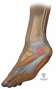
The Distal Tarsal Tunnel and Neural Arborization
As the tibial nerve navigates the distal extent of the proximal tunnel, it typically trifurcates into its terminal branches: the medial plantar nerve, the lateral plantar nerve, and the medial calcaneal nerve(s). The medial calcaneal nerves typically emerge from the main trunk of the tibial nerve proximal to, or exactly at, the superior margin of the abductor hallucis muscle. These branches can be singular or multiple and provide critical sensory innervation to the posteromedial and plantar heel pad.
The medial plantar nerve diverges from the tibial nerve just proximal to or beneath the abductor hallucis. It courses anteriorly under the muscle belly, providing motor innervation to it, before continuing distally to form the intermetatarsal nerves supplying the first, second, and third interspaces. The lateral plantar nerve follows a more tortuous and complex trajectory. It initially dives deep to the fascia of the abductor hallucis and the medial margin of the plantar fascia, traversing over the fascia of the quadratus plantae. It then hooks distally beneath the flexor digitorum brevis, emerging to supply the fourth and fifth interspaces.
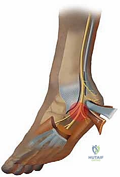
The first branch of the lateral plantar nerve (Baxter's nerve) is the critical epicenter of our distal decompression today. This nerve typically branches immediately after the lateral plantar nerve originates from the posterior tibial trunk. It descends vertically between the deep fascia of the abductor hallucis and the medial fascia of the quadratus plantae. It then sharply changes direction, coursing laterally in a horizontal plane between the quadratus plantae and the flexor digitorum brevis. It provides sensory fibers to the calcaneal periosteum and terminates as the primary motor branch to the abductor digiti quinti. Entrapment here is a mechanical vise, caught between the hypertrophied abductor hallucis fascia, the medial band of the plantar fascia, and the medial caudal margin of the quadratus plantae.
The Plantar Fascia and Windlass Mechanism
The plantar fascia, or plantar aponeurosis, is a dense band of connective tissue originating from the medial tuberosity of the os calcis. It is anatomically divided into three distinct segments: the central, medial, and lateral bands. Clinically and biomechanically, the central portion is the most significant, originating from the medial calcaneal tuberosity and fanning out to insert into the base of all five proximal phalanges via the plantar plates.
This structure is the primary engine of the "windlass mechanism," first described by Hicks. During the terminal stance phase of gait, dorsiflexion of the metatarsophalangeal (MTP) joints winds the plantar fascia around the metatarsal heads. This action dynamically shortens the distance between the calcaneus and the metatarsals, elevating the longitudinal arch, locking the midtarsal joints, and converting the foot into a rigid lever for propulsion. Pathological attenuation or chronic contracture of this fascia disrupts this vital biomechanical sequence.
Surgical intervention in this region carries significant neurovascular risks that must be respected. Superficially, the saphenous vein and nerve lie anterior to the standard incision line and must be meticulously identified and retracted. Deep within the tunnel, the posterior tibial artery and its delicate venous plexus surround the tibial nerve. Iatrogenic injury to these vessels causes immediate, obscuring hemorrhage, while damage to the tibial nerve or its branches results in devastating, often irreversible sensory and motor deficits. Identification must always precede resection.
Exhaustive Indications and Contraindications
The decision to proceed with a combined plantar fascia and tarsal tunnel release is not taken lightly and must be based on a rigorous, evidence-based assessment of the patient's clinical presentation and response to conservative care. The primary indication is chronic, recalcitrant plantar heel pain that exhibits clear clinical signs of both mechanical enthesopathy and compressive neuropathy, having failed exhaustive nonoperative management for a minimum of nine to twelve months.
Surgeons must carefully differentiate between isolated plantar fasciitis and the combined neuro-mechanical syndrome. Patients with pure mechanical fasciitis typically experience "first-step" morning pain that improves with activity, whereas those with concomitant nerve entrapment report a burning, radiating pain (often extending proximally up the medial leg or distally into the toes) that worsens with prolonged standing and persists as a throbbing ache at rest. The presence of a positive Tinel's sign over the tarsal tunnel or localized tenderness over Baxter's nerve strongly supports the indication for combined release.
Contraindications must be strictly observed to prevent catastrophic outcomes. Active local or systemic infection is an absolute contraindication. Severe peripheral vascular disease that compromises wound healing must be addressed prior to any elective foot surgery. Furthermore, patients with diffuse, metabolically driven peripheral neuropathy (such as advanced diabetic sensorimotor polyneuropathy) are generally poor candidates, as surgical decompression cannot reverse the underlying metabolic axonal degeneration, though it may be considered in highly selected cases of superimposed focal entrapment.
| Category | Specific Clinical Scenarios |
|---|---|
| Absolute Indications | - Chronic plantar heel pain > 9-12 months failing all conservative care. - Clinical evidence of distal tarsal tunnel/Baxter's nerve entrapment (positive Tinel's, radiating pain). - MRI confirmation of severe fascial thickening with adjacent neural edema. - Intractable pain significantly limiting activities of daily living. |
| Relative Indications | - Recurrent heel pain after previous isolated plantar fasciotomy. - Heel pain associated with space-occupying lesions (ganglion, lipoma) in the tarsal tunnel. - High-level athletes requiring definitive resolution after failed extended rest. |
| Absolute Contraindications | - Active local soft tissue infection or osteomyelitis. - Severe, unoptimized peripheral vascular disease (ABI < 0.4). - Purely psychogenic pain disorders. - Inability to comply with strict non-weight-bearing postoperative protocols. |
| Relative Contraindications | - Poorly controlled diabetes mellitus (HbA1c > 8.0%). - Advanced, diffuse diabetic peripheral neuropathy without focal entrapment signs. - Inflammatory arthropathies (e.g., Rheumatoid Arthritis) causing systemic enthesitis without focal mechanical compression. |
Pre-Operative Planning, Templating, and Patient Positioning
Meticulous preoperative planning is the bedrock of a successful surgical outcome. This begins with a comprehensive, granular history and physical examination designed to isolate the dual nature of the pathology. We must determine the exact character, timing, and radiation of the pain to differentiate it from lumbar radiculopathy, isolated metatarsalgia, or a calcaneal stress fracture.
Clinical Evaluation and Diagnostic Modalities
The physical examination must be exhaustive. For the plantar fasciitis component, we look for focal, reproducible tenderness directly over the medial calcaneal tubercle. We assess the integrity of the fascia by recreating the windlass mechanism (passively dorsiflexing the ankle and all MTP joints) and palpating the medial cord. Asymmetry in tension suggests a chronic, partial rupture. For the neuritic component, we map the radiation of pain. A positive Valleix phenomenon (proximal radiation up the medial leg) or distal radiation along the lateral plantar aspect strongly suggests nerve irritation.
We specifically palpate the "soft spot" at the medial border of the heel, corresponding to the exit point of the lateral plantar nerve beneath the abductor hallucis fascia. Tenderness here is a hallmark of Baxter's nerve entrapment. Diagnostic imaging is critical for ruling out confounding pathologies. Weight-bearing radiographs evaluate for calcaneal stress fractures, degenerative joint disease, or significant osseous deformity. While MRI is not universally mandated, it is highly sensitive for confirming fascial thickening (typically >4mm), interstitial tearing, marrow edema in the calcaneus, and occult space-occupying lesions within the tarsal tunnel. Electrodiagnostic studies (EMG/NCS) are frequently obtained; however, surgeons must recognize that a negative EMG does not rule out traction neuritis, which is often dynamic and position-dependent.
Optimization and Failed Conservative Care
By definition, patients indicated for this procedure have failed an exhaustive regimen of nonoperative management. This typically includes relative rest, Achilles and plantar fascia specific stretching protocols, NSAIDs, and customized orthoses designed with a deep heel cup and a "nerve relief channel" to offload the lateral plantar nerve.
It is critical to note that certain conservative modalities may actually exacerbate the neurogenic component. Physical therapy utilizing deep heat (ultrasound, diathermy) can increase local neural inflammation. Furthermore, we strongly discourage repeated corticosteroid injections. Corticosteroids placed into the fat pad or near the nerve can cause fat pad atrophy, precipitate a complete iatrogenic rupture of the plantar fascia, and paradoxically increase neuritic symptoms through direct crystal irritation or secondary scarring. Extracorporeal shock wave therapy (ESWL), while effective for isolated mechanical fasciitis, is generally contraindicated in cases with clear neurogenic symptoms, as the acoustic waves can further traumatize the inflamed nerve.
Operating Room Setup and Patient Positioning
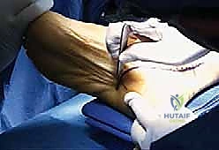
Proper positioning is essential for optimal visualization of the medial hindfoot. The patient is positioned supine on the operating table. A high thigh tourniquet is applied over abundant padding; a bloodless field is an absolute requirement for the safe, microscopic dissection of the delicate neural branches.
A substantial bump is placed beneath the ipsilateral hip. This internally rotates the entire lower extremity, bringing the medial aspect of the ankle and foot perfectly parallel to the floor and directly into the surgeon's line of sight. The entire lower leg, from the tibial tubercle down, is prepped and draped to allow full manipulation of the foot and ankle during the procedure. While routine fluoroscopy is rarely required for the soft tissue decompression itself, a C-arm should be readily available in the suite to assess for and potentially resect any large, impinging plantar calcaneal osteophytes if preoperative imaging dictates.
Step-by-Step Surgical Approach and Fixation Technique
We now transition to the operative execution. This procedure demands patience, magnification (loupes are mandatory), and a deep respect for the surrounding neurovascular structures. Our goal is absolute, unhindered decompression of the neural elements followed by a measured release of the mechanical tether.
Incision and Superficial Dissection
Our approach utilizes a generous curvilinear incision designed to expose both the proximal tunnel and the distal arborization zones. I initiate the incision approximately 6 to 8 centimeters proximal to the tip of the medial malleolus, situated exactly halfway between the posterior border of the medial malleolus and the anterior border of the Achilles tendon.
The incision curves gently distally, paralleling the course of the tibial nerve, passing inferior to the medial malleolus, and extending into the medial arch of the foot, terminating just dorsal to the plantar skin transition line. This distal extension is critical for accessing the abductor hallucis and the insertion of the plantar fascia.
Superficial dissection is carried out with sharp scalpel work and blunt spreading with tenotomy scissors. We must immediately identify and protect the superficial venous network and branches of the saphenous nerve lying anteriorly. Hemostasis of the subcutaneous vessels must be meticulous using bipolar electrocautery to prevent postoperative hematoma, which can cause secondary compression.
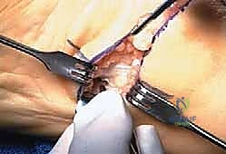
Proximal Tarsal Tunnel Decompression
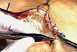
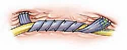
Once the subcutaneous fat is parted, the glistening, transverse fibers of the flexor retinaculum (laciniate ligament) come into view. Using a #15 blade, I carefully incise the proximal margin of the retinaculum. I then introduce a grooved director or the tips of a fine Metzenbaum scissor beneath the retinaculum to protect the underlying neurovascular bundle.
The retinaculum is divided entirely along its length. It is imperative to extend this release proximally into the deep fascia of the leg to ensure no proximal tethering remains. Upon opening the tunnel, the posterior tibial artery, its venae comitantes, and the tibial nerve are exposed. The venous plexus here can be incredibly friable and tortuous; any bleeding must be controlled immediately with bipolar cautery to maintain the pristine visualization required for nerve tracing.
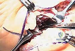
Distal Decompression and Nerve Tracing
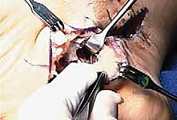
With the proximal trunk identified, we trace the tibial nerve distally to its trifurcation. Vessel loops can be gently placed around the main trunk and its primary branches (medial and lateral plantar nerves) for ease of identification, taking care not to apply excessive traction.
The dissection proceeds distally along the course of the lateral plantar nerve. This requires mobilizing the superior margin of the abductor hallucis muscle belly. The deep fascia of the abductor hallucis is a thick, unyielding structure that forms the roof of the distal tarsal tunnel.
Using sharp dissection, the deep investing fascia of the abductor hallucis is completely transected. We must trace the lateral plantar nerve until the origin of its first branch (Baxter's nerve) is clearly visualized. We follow Baxter's nerve as it dives deep, ensuring it is entirely free from any fascial bands connecting the abductor hallucis to the quadratus plantae.
Plantar Fasciotomy
Once the neural structures are completely decompressed and protected dorsally, we address the mechanical component. Retractors are placed to expose the medial and central bands of the plantar fascia at their origin on the calcaneal tuberosity.
Using a #15 blade, a measured, partial plantar fasciotomy is performed. We typically release the medial band and the medial one-third to one-half of the central band. It is absolutely critical to preserve the lateral band and the lateral portion of the central band. A complete release severely compromises the windlass mechanism, leading to devastating lateral column overload, cuboid syndrome, and a catastrophic collapse of the longitudinal arch.
Following the release, the surgical field is copiously irrigated. The tourniquet is deflated prior to closure to ensure absolute hemostasis, particularly addressing the venae comitantes. Only the subcutaneous tissue and skin are closed; the flexor retinaculum and abductor fascia are left wide open to prevent recurrent compression.
Complications, Incidence Rates, and Salvage Management
Despite meticulous surgical technique, the combined plantar fascia and tarsal tunnel release carries a distinct profile of potential complications. The most devastating, though fortunately rare, is iatrogenic nerve injury. Direct laceration or excessive traction neuropraxia of the tibial nerve or its branches can result in permanent anesthesia of the plantar foot, intrinsic muscle paralysis, and the formation of painful terminal neuromas.
Another significant biomechanical complication is lateral column overload, often termed "cuboid syndrome." This occurs if the plantar fasciotomy is overly aggressive (releasing >50% of the fascia). The loss of the medial tension band shifts the weight-bearing forces laterally, resulting in severe pain over the cuboid, fifth metatarsal base, and lateral midfoot. This complication is notoriously difficult to treat and often requires prolonged orthotic management, custom bracing, or, in severe cases, lateral column stabilizing osteotomies.
Wound healing complications, including dehiscence and superficial infection, are more common in this region due to the tenuous vascular supply of the medial ankle skin and the dynamic forces applied during early mobilization. Furthermore, Complex Regional Pain Syndrome (CRPS) is a known risk following any nerve decompression in the distal extremity, characterized by disproportionate pain, allodynia, and autonomic dysregulation.
| Complication | Estimated Incidence | Salvage Management / Mitigation Strategy |
|---|---|---|
| Iatrogenic Nerve Injury (Neuroma/Laceration) | < 1 - 2% | Immediate intraoperative microsurgical repair if identified. Postoperative neuromas may require excision and burying into deep muscle (e.g., abductor hallucis). Prevention via meticulous microsurgical technique is paramount. |
| Lateral Column Overload (Cuboid Syndrome) | 5 - 10% (Higher with complete fascial release) | Custom orthotics with lateral posting, stiff-soled shoes, prolonged taping. Surgical salvage requires lateral column fusion or calcaneal osteotomy in refractory cases. Strict adherence to releasing only the medial 1/3 to 1/2 of the fascia prevents this. |
| Wound Dehiscence / Infection | 2 - 5% | Local wound care, oral or IV antibiotics. Avoid early aggressive range of motion. Ensure tension-free skin closure and meticulous hemostasis prior to closure. |
| Recurrent/Persistent Neuralgia | 5 - 8% | Often due to incomplete release of the abductor hallucis fascia or perineural scarring. Managed with gabapentinoids, targeted nerve blocks. Revision neurolysis is technically demanding and yields diminishing returns. |
Phased Post-Operative Rehabilitation Protocols
The postoperative rehabilitation protocol must strike a delicate balance between protecting the surgical soft tissue envelope and preventing restrictive perineural scarring. The protocol is divided into three distinct phases, tailored to the patient's biological healing response.
Phase I: Immediate Post-Operative Protection (Weeks 0-2)
Immediately following closure, the patient is placed in a bulky, well-padded posterior splint with the ankle maintained in a neutral (90-degree) position. The patient is instructed to remain strictly non-weight-bearing on the operative extremity using crutches or a knee
