Acute Patellar Dislocation: Evaluation & Surgical Management
Key Takeaway
Acute lateral patellar dislocations require meticulous clinical and radiographic evaluation to identify osteochondral fractures and medial patellofemoral ligament (MPFL) tears. While primary management is often non-operative, involving closed reduction and immobilization, the presence of intra-articular loose bodies or massive retinacular disruption necessitates surgical intervention. This guide details the biomechanics, diagnostic algorithms, and step-by-step surgical techniques for managing acute patellar instability and associated osteochondral defects.
Comprehensive Introduction and Patho-Epidemiology
Acute dislocation of the patella represents a significant traumatic event that primarily afflicts the young, active, and athletic patient populations, accounting for approximately two to three percent of all knee injuries evaluated in emergency departments. The patella almost exclusively dislocates in a lateral direction, a phenomenon driven by the inherent biomechanical vectors of the extensor mechanism, the physiologic Q-angle, and the dominant lateral pull of the vastus lateralis relative to the medial stabilizing structures. Understanding the intricate pathoanatomy, particularly the cascading disruption of the medial soft-tissue restraints, is paramount for orthopedic surgeons when formulating both conservative and operative management strategies. The initial dislocation event is rarely an isolated soft-tissue stretch; rather, it is a catastrophic failure of the medial patellofemoral complex that permanently alters the kinematics of the patellofemoral joint.
The epidemiological profile of acute patellar dislocations demonstrates a distinct bimodal distribution, with the primary peak occurring in adolescents and young adults between the ages of ten and twenty years, often with a slight female predominance due to baseline ligamentous laxity and wider pelvic anatomy increasing the Q-angle. A secondary peak is frequently observed in older patients engaged in high-impact or pivoting sports. The mechanism of injury typically involves a non-contact twisting motion—specifically, internal rotation of the femur on a planted, externally rotated tibia with the knee in early flexion. This position maximizes the lateralizing vector forces on the extensor mechanism just before the patella fully engages the osseous stability of the trochlear groove. Direct trauma to the medial aspect of the patella can also precipitate a dislocation, though this is significantly less common than the non-contact, torsional mechanism.
While the majority of primary acute dislocations historically have been managed successfully with closed reduction and structured rehabilitation, contemporary orthopedic literature highlights an unacceptably high rate of recurrent instability and subsequent chondral degradation following non-operative care in specific high-risk cohorts. The natural history of a first-time dislocator reveals a recurrence rate approaching fifteen to forty-four percent, which exponentially increases with subsequent dislocation events. Furthermore, the high incidence of associated osteochondral shear fractures and profound medial patellofemoral ligament (MPFL) avulsions necessitates a remarkably high index of suspicion and rigorous diagnostic evaluation. The classic "kissing contusion" pattern—bone bruising on the medial patellar facet and the lateral femoral condyle—is the hallmark of a transient lateral patellar dislocation and subsequent relocation, serving as the primary mechanism for these debilitating osteochondral shear injuries that mandate acute surgical intervention.
Detailed Surgical Anatomy and Biomechanics
The stability of the patellofemoral joint relies on a highly complex, synergistic interplay between active muscular restraints, passive ligamentous structures, and the osseous geometry of the trochlear groove. The osseous architecture provides static stability primarily in deeper degrees of knee flexion. As the knee flexes beyond thirty degrees, the patella engages the trochlear groove, and the bony conformity becomes the dominant restraint to lateral translation. Anomalies in this bony architecture, such as trochlear dysplasia (characterized by a shallow or convex trochlear groove) or patella alta (an abnormally high-riding patella), delay or completely negate this osseous engagement, leaving the joint dangerously dependent on soft-tissue restraints during the vulnerable early arcs of motion. The Dejour classification system remains the gold standard for quantifying trochlear dysplasia, categorizing the morphology from a shallow sulcus (Type A) to a convex, cliff-like lateral facet (Type D), each representing progressively higher risks for recurrent instability.
The Medial Patellofemoral Ligament (MPFL) is unequivocally the primary passive restraint to lateral patellar translation, providing approximately fifty to sixty percent of the total restraining force from zero to thirty degrees of knee flexion. Anatomically, the MPFL is a distinct condensation of capsular fibers within layer two of the medial knee. Its femoral origin is meticulously defined at Schöttle’s point—a radiographic landmark located anterior to the posterior femoral cortical line, proximal to the posterior extension of Blumensaat's line, and distal to the medial epicondyle. From this isometric femoral footprint, the ligament fans out anteriorly to insert broadly onto the proximal two-thirds of the medial patellar margin and the deep fascia of the vastus medialis obliquus (VMO). During an acute lateral dislocation, the MPFL is universally injured, undergoing plastic deformation or complete macroscopic rupture.
Beyond the MPFL, the secondary medial restraints include the medial patellotibial ligament (MPTL) and the medial patellomeniscal ligament (MPML), which collectively contribute approximately twenty to thirty percent of the restraining force, primarily resisting lateral translation in deeper flexion angles. The dynamic stability of the patellofemoral joint is orchestrated by the quadriceps musculature, with the VMO playing a critical role. The VMO fibers insert at a fifty-to-fifty-five-degree angle onto the superomedial pole of the patella, providing a dynamic medializing vector during active extension. In the setting of an acute dislocation, the VMO is frequently injured, either via interstitial tearing or direct avulsion from the patella, leading to rapid atrophy and a profound loss of dynamic medial tracking. Understanding the precise anatomical footprints and the biomechanical tensioning of these structures is absolutely critical when attempting anatomical repair or reconstruction, as non-anatomic graft placement or over-tensioning will inevitably lead to iatrogenic medial subluxation, severe patellofemoral arthrosis, and disastrous clinical outcomes.
Exhaustive Indications and Contraindications
The decision-making process following an acute patellar dislocation requires a nuanced evaluation of the patient's pathoanatomy, the presence of intra-articular derangement, and the individual's baseline risk factors for recurrence. Historically, almost all first-time dislocations were treated non-operatively. However, the modern paradigm has shifted toward aggressive early surgical intervention in the presence of specific structural injuries. The absolute primary indication for acute surgical intervention is the identification of a displaced osteochondral fracture originating from either the medial patellar facet or the lateral femoral condyle. These shear fractures, if left untreated, not only create symptomatic loose bodies that cause mechanical locking and catching, but also leave critical weight-bearing chondral defects that rapidly progress to early-onset patellofemoral osteoarthritis.
Another compelling indication for acute surgical management is an irreducible patellar dislocation. While rare, irreducibility is typically caused by the interposition of the torn medial retinaculum, the MPFL, or a flipped osteochondral fragment into the patellofemoral joint space, physically blocking concentric reduction. Additionally, massive avulsion of the medial soft tissues—particularly when the VMO is visibly stripped from the adductor tubercle or the medial patella, resulting in gross, multidirectional patellar instability even in extension—warrants acute open or arthroscopically assisted repair. In the pediatric and adolescent populations, the presence of a large, displaced sleeve fracture of the medial patella also dictates urgent open reduction and internal fixation to restore the continuity of the extensor mechanism and prevent catastrophic growth disturbances.
Conversely, absolute contraindications to acute surgical intervention include a first-time dislocation with an uncompromised articular surface, no evidence of osteochondral shear fractures, and a concentrically reduced patella on post-reduction imaging. In these scenarios, the biological healing capacity of the medial structures, combined with rigorous physical therapy focusing on VMO strengthening and core/gluteal stabilization, yields excellent functional outcomes. Relative contraindications encompass patients with profound, uncorrected underlying anatomical abnormalities (such as severe patella alta or a TT-TG distance exceeding 20 millimeters) who present with an acute-on-chronic dislocation. In these complex cases, isolated acute soft-tissue repair is doomed to fail; these patients are better served by initial conservative management, followed by a comprehensive, staged reconstructive procedure (e.g., tibial tubercle osteotomy combined with MPFL reconstruction) once the acute inflammatory phase has subsided.
| Variable | Indications for Operative Management | Contraindications for Operative Management |
|---|---|---|
| Intra-articular Pathology | Displaced osteochondral fracture (>1 cm); symptomatic loose body. | No osteochondral fracture; purely cartilaginous fragments <5 mm in non-weight-bearing zones. |
| Reduction Status | Irreducible dislocation (soft tissue or bony interposition). | Spontaneously reduced or easily reduced closed with concentric tracking. |
| Soft Tissue Integrity | Massive, palpable VMO/retinacular avulsion with gross lateral instability in extension. | First-time dislocation with stable tracking post-reduction; intact VMO tone. |
| Concomitant Injuries | Associated major ligamentous rupture (e.g., ACL, MCL requiring acute intervention). | Isolated MPFL sprain/partial tear without secondary ligamentous involvement. |
| Patient Factors | High-demand athlete with recurrent instability and structural damage. | Poor surgical candidate; non-compliant patient; chronic, painless habitual dislocator. |
| Anatomical Variants | Normal anatomy with traumatic structural failure. | Severe, unaddressed bony dysplasia (relative contraindication for isolated acute soft tissue repair). |
Pre-Operative Planning, Templating, and Patient Positioning
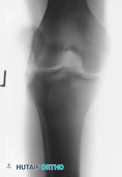
Thorough pre-operative planning is the cornerstone of successful surgical management for acute patellar dislocations. Following the initial clinical evaluation and closed reduction, a comprehensive radiographic series is mandatory to confirm concentric reduction and to rule out gross osteochondral pathology. A standard trauma knee series must include a true Anteroposterior (AP) view, a true Lateral view, and a Sunrise or Merchant view. The AP view evaluates overall tibiofemoral alignment and may reveal large osteochondral fragments or avulsion fractures from the medial patella (the MPFL insertion site). The Lateral view is critical for assessing patellar height using the Insall-Salvati or Caton-Deschamps ratios, and for identifying signs of trochlear dysplasia, such as the crossing sign, a supratrochlear spur, or a double contour sign.
The Sunrise or Merchant view is particularly invaluable for evaluating patellofemoral congruence, static patellar tilt, and the presence of intra-articular fragments within the dependent portions of the joint space. As demonstrated in the advanced imaging protocols, plain radiographs alone possess low sensitivity for purely cartilaginous or small osteochondral shear injuries. Therefore, if a tense hemarthrosis is present following a dislocation, Magnetic Resonance Imaging (MRI) is strictly warranted. MRI serves a dual purpose: it meticulously maps the exact location and severity of the MPFL tear (femoral origin, midsubstance, or patellar insertion) and identifies occult articular cartilage injuries. Furthermore, Computed Tomography (CT) scans may be utilized in complex cases to accurately measure the Tibial Tubercle-Trochlear Groove (TT-TG) distance; a TT-TG distance greater than 20 millimeters indicates severe lateralization of the extensor mechanism, which may necessitate an osseous realignment procedure rather than an isolated soft-tissue repair.

Patient positioning and preparation in the operating theater must be meticulously executed to allow for dynamic intra-operative assessment. The patient is placed supine on the operating table. General anesthesia is typically employed, often supplemented with a regional block, such as an adductor canal block, to provide robust postoperative analgesia without compromising quadriceps motor function—a critical factor for early rehabilitation. A well-padded tourniquet is applied to the proximal thigh. The operative limb is prepped and draped free, utilizing a lateral post or a specialized leg holder that permits unhindered, full range of motion from zero to one hundred and forty degrees of flexion. Prior to the initial incision, a thorough Examination Under Anesthesia (EUA) is mandatory. The surgeon must meticulously assess patellar tracking, quantify the degree of lateral translation in quadrants at zero, thirty, and ninety degrees of flexion, and compare the competence of the medial restraints directly to the contralateral, uninjured knee.
Step-by-Step Surgical Approach and Fixation Technique
When surgical intervention is mandated—most frequently due to a displaced osteochondral fragment or a massive soft-tissue avulsion—a formal open exploration combined with arthroscopic assessment remains the gold standard for comprehensive joint management. The procedure commences with a diagnostic arthroscopy through standard anterolateral and anteromedial portals. Copious irrigation is utilized to evacuate the tense hemarthrosis and flush out micro-cartilaginous debris. A systematic diagnostic sweep is performed, scrutinizing the medial patellar facet and the lateral femoral condyle for osteochondral shear fractures. If a fixable fragment is identified (typically defined as greater than 1 to 1.5 centimeters with sufficient subchondral bone), the arthroscopic approach is transitioned to an open arthrotomy to facilitate precise, anatomical reduction and rigid internal fixation.
The Open Approach and Arthrotomy
A longitudinal medial parapatellar incision is meticulously planned, centered over the medial border of the patella and extending from the distal pole proximally toward the VMO insertion. Dissection is carried sharply through the subcutaneous tissues to expose the superficial fascia and the underlying medial patellar retinaculum. The surgeon must carefully identify the traumatic rent in the retinacular tissues. This disruption is frequently accompanied by significant ecchymosis and hematoma, which must be evacuated to visualize the torn edges of the MPFL and the VMO. The traumatic tear is then longitudinally extended to create a formal medial arthrotomy, providing excellent visualization of the patellofemoral articulation.
Osteochondral Fragment Management
Once the joint is fully exposed, the osteochondral fragment is retrieved and assessed on the back table. The donor crater on the lateral femoral condyle or medial patella is meticulously debrided of loose, non-viable cartilage to create stable, vertical margins, exposing healthy, bleeding subchondral bone to promote healing. The fragment is then anatomically reduced into the crater. Provisional fixation is achieved using smooth Kirschner wires. Definitive fixation is highly dependent on the fragment's characteristics. For fragments with robust subchondral bone, headless titanium compression screws or bioabsorbable screws are utilized. It is absolutely critical that the heads of these implants are countersunk at least 1 to 2 millimeters below the surrounding articular cartilage surface to prevent catastrophic abrasion of the opposing articular facet during knee flexion. For purely cartilaginous or highly comminuted fragments that cannot hold rigid hardware, modern chondral darts or fibrin glue may be employed, or the fragment may be excised entirely, followed by microfracture of the defect if it lies in a critical weight-bearing zone.
Anatomical Retinacular and MPFL Repair
Following the management of intra-articular pathology, attention is directed toward the medial soft-tissue repair. The torn edges of the MPFL and the medial retinaculum are identified and mobilized. If the MPFL is avulsed directly from the medial border of the patella, anatomical repair is achieved using two or three small-diameter suture anchors (e.g., 2.5mm or 3.0mm) placed along the articular margin of the proximal two-thirds of the patella. If the tear is located at the femoral origin (Schöttle’s point), suture anchors are placed at the anatomic footprint between the adductor tubercle and the medial epicondyle. For midsubstance ruptures, a direct end-to-end repair is performed using heavy, non-absorbable sutures (e.g., #2 FiberWire) utilizing a running locking Krackow or whipstitch technique.
Tensioning and Closure
The most critical step of the soft-tissue repair is the tensioning phase. The repair must be tensioned with the knee flexed to approximately thirty to forty-five degrees, the arc of motion where the patella naturally engages the trochlea. The surgeon must avoid the severe pitfall of over-tensioning the medial structures. An over-constrained repair will result in iatrogenic medial patellar subluxation, drastically increased patellofemoral contact pressures, and rapid, painful arthrosis. Post-repair, the surgeon must confirm that the patella can still be manually translated laterally by one to two quadrants. Once satisfactory tracking and tension are confirmed, the tourniquet is deflated, meticulous hemostasis is achieved to prevent postoperative hematoma, and the wound is closed in a layered fashion. The limb is subsequently placed in a hinged knee brace locked in full extension.
Complications, Incidence Rates, and Salvage Management
Surgical management of acute patellar dislocations, while highly effective for addressing intra-articular pathology and restoring gross stability, carries a distinct profile of postoperative complications. The surgeon must be acutely aware of these risks to counsel patients appropriately and to implement meticulous surgical techniques that mitigate their occurrence. The most frequent and arguably most debilitating complication following open medial retinacular repair is arthrofibrosis, characterized by a profound loss of knee flexion and painful, restricted patellar glide. This stiffness arises from excessive scar tissue formation within the medial gutters and the suprapatellar pouch, often exacerbated by prolonged postoperative immobilization or inadequate hemostasis leading to a massive, organized hemarthrosis.
Another severe complication is iatrogenic medial patellar instability or medial overload syndrome, which directly results from over-tensioning the MPFL repair or placing the femoral fixation non-anatomically (typically too proximal or too anterior). This non-anatomic tensioning forces the patella to track excessively medially, drastically increasing contact pressures on the medial patellar facet. Patients present with severe anterior knee pain, inability to ascend or descend stairs, and a positive medial apprehension sign. If conservative management with targeted physical therapy fails, salvage management requires surgical release of the over-tightened structures and a formal MPFL reconstruction using an autograft or allograft to restore physiologic kinematics.
Recurrent lateral instability remains a persistent risk, particularly in patients with unrecognized or unaddressed underlying anatomical risk factors, such as severe trochlear dysplasia, patella alta, or an excessive TT-TG distance. In these scenarios, an isolated soft-tissue repair is biomechanically insufficient to overcome the massive lateralizing vectors. When recurrent dislocation occurs after a primary repair, salvage management mandates a comprehensive realignment strategy. This typically involves a tibial tubercle osteotomy (anteromedialization, such as the Fulkerson osteotomy) to correct the TT-TG distance and patellar height, combined with a robust MPFL reconstruction. In cases of extreme, high-grade trochlear dysplasia (Dejour Types B, C, or D), a sulcus-deepening trochleoplasty may be indicated to surgically construct a competent bony groove, though this remains a technically demanding procedure reserved for specialized patellofemoral centers.
| Complication | Estimated Incidence | Etiology / Risk Factors | Salvage Management / Treatment |
|---|---|---|---|
| Arthrofibrosis (Stiffness) | 5% - 15% | Prolonged immobilization; massive postoperative hematoma; over-tensioned repair. | Aggressive physical therapy; intra-articular corticosteroid injection; arthroscopic lysis of adhesions and manipulation under anesthesia (MUA). |
| Recurrent Lateral Instability | 10% - 30% | Unaddressed bony dysplasia (patella alta, high TT-TG); failure of soft tissue repair; non-compliant rehab. | Comprehensive realignment: Tibial tubercle osteotomy (TTO) +/- MPFL reconstruction +/- Trochleoplasty. |
| Iatrogenic Medial Subluxation | 2% - 5% | Over-tensioning of the MPFL repair; non-anatomic femoral anchor placement (too proximal/anterior). | Surgical release of the tight medial structures; revision anatomical MPFL reconstruction. |
| Hardware Failure / Migration | 1% - 3% | Poor bone quality; inadequate countersinking of osteochondral screws; premature weight-bearing. | Arthroscopic hardware removal; revision fixation if fragment is viable; chondroplasty/microfracture if fragment is lost. |
| Patellofemoral Osteoarthritis | 15% - 25% (Long-term) | Missed osteochondral fractures; chronic altered kinematics; severe initial chondral impact ("kissing contusion"). | Activity modification; NSAIDs; visco-supplementation; eventually patellofemoral arthroplasty (PFA) or total knee arthroplasty (TKA) in older patients. |
Phased Post-Operative Rehabilitation Protocols
The postoperative rehabilitation following either conservative management or surgical repair of an acute patellar dislocation is a critical determinant of long-term functional success. The rehabilitation protocol must be carefully phased to protect the healing medial soft tissues from excessive lateralizing forces while simultaneously preventing arthrofibrosis and restoring the dynamic stabilizing capacity of the quadriceps musculature. The biological timeline of ligamentous healing dictates the progression, requiring a delicate balance between immobilization for tissue approximation and early motion for collagen organization.
Phase I (Weeks 0-3): Maximal Protection and Muscle Activation.
Immediately following surgery or closed reduction, the primary goals are to minimize effusion, control pain, and prevent profound quadriceps inhibition. The knee is immobilized in a hinged knee brace locked in full extension during all weight-bearing activities. Weight-bearing status is generally dictated by the presence of osteochondral fixation; isolated soft-tissue repairs may bear weight as tolerated in the locked brace, whereas osteochondral fracture fixations typically require strict non-weight-bearing or touch-down weight-bearing for four to six weeks to protect the articular cartilage repair. Rehabilitation focuses on isometric quadriceps activation, specifically targeting the VMO through biofeedback, multiple-angle isometrics, and straight leg raises in the brace. Patellar mobilization in the superior and inferior planes is initiated to prevent capsular contracture, but lateral glides are strictly prohibited to protect the medial repair.
Phase II (Weeks 3-6): Controlled Range of Motion and Early Strengthening.
During this phase, the biological repair begins to consolidate, allowing for the introduction of controlled mechanical stress to organize the healing collagen fibers. The hinged brace is unlocked to permit progressive, active-assisted, and passive range of motion. The clinical goal is to achieve ninety degrees of knee flexion by the end of week four, and full, symmetric range of motion by week six. Strengthening exercises transition from strict isometrics to closed-kinetic-chain (CKC) exercises, such as shallow mini-squats (zero to forty-five degrees) and bilateral leg presses. CKC exercises are preferred over open-kinetic-chain (OKC) knee extensions, as CKC movements increase joint compressive forces, thereby enhancing patellofemoral stability and minimizing shear stress across the healing articular surfaces.
Phase III (Weeks 6-12): Advanced Strengthening and Proprioception.
By week six, the hinged brace is typically discontinued, provided the patient demonstrates a normal gait pattern and sufficient quadriceps control (absence of an extension lag). The focus shifts toward heavy resistance training, targeting not only the quadriceps but the entire lower extremity kinetic chain. Gluteal and core strengthening are paramount, as proximal hip weakness leads to dynamic femoral internal rotation and subsequent valgus collapse at the knee—a primary biomechanical driver of lateral patellar instability. Single-leg balance exercises, proprioceptive neuromuscular facilitation (PNF), and progressive step-up/step-down programs are integrated to restore neuromuscular control.
Phase IV (Months 3-6): Return to Sport and High-Level Function.
The final phase of rehabilitation bridges the gap between clinical recovery and athletic performance. Plyometric training, agility drills, and sport-specific cutting maneuvers are gradually introduced under the supervision of a physical therapist. The criteria for return to unrestricted athletic play are rigorous and objective. Patients must demonstrate a complete absence of pain and effusion, full symmetric range of motion, and no apprehension during dynamic valgus stress testing. Furthermore, functional testing—including single-leg hop tests, crossover hops, and isokinetic dynamometer testing—must demonstrate limb symmetry indices (LSI) of greater than ninety percent compared to the uninjured contralateral limb before the surgeon clears the patient for full collision or pivoting sports.
Summary of Landmark Literature and Clinical Guidelines
The evolution of treatment algorithms for acute patellar dislocations is deeply rooted in several landmark orthopedic studies that have systematically defined the natural history, the precise pathoanatomy, and the long-term outcomes of various interventions. Historically, the management of first-time dislocations was overwhelmingly conservative. However, the seminal natural history study by Fithian et al. fundamentally altered this perspective by demonstrating that patients with a history of patellar dislocation have a dramatically higher risk of recurrent instability compared to first-time dislocators, and that conservative management in patients with underlying anatomical risk factors yields unacceptably high failure rates. This study underscored the necessity of stratifying patients based on their individual risk profiles rather than applying a universal non-operative protocol.
The anatomical and radiological understanding of the injury was revolutionized by the comprehensive MRI studies conducted by Balcarek et al. and Nomura et al. Balcarek’s research definitively proved that the MPFL is injured in nearly ninety-eight percent of acute lateral patellar dislocations, mapping the specific injury patterns (femoral, midsubstance, and patellar insertions) and highlighting the high incidence of multifocal retinacular failure. Nomura’s work further correlated these MRI findings with arthroscopic observations, bringing widespread attention to the alarming prevalence of osteochondral shear fractures—identifying articular cartilage injury in over ninety percent of acute dislocation events. These imaging studies established the current clinical guideline that a tense hemarthrosis following a dislocation mandates an MRI to rule out catastrophic chondral damage.
In the realm of surgical intervention, long-term outcome studies by Sillanpää et al. have provided critical insights into the efficacy of primary MPFL repair versus reconstruction. Their data suggests that while primary end-to-end repair of the MPFL can be successful in acute settings with bony avulsions, midsubstance repairs often fail due to the inherent plastic deformation and attenuation of the ligamentous tissue sustained during the initial traumatic event. Consequently, modern clinical guidelines increasingly advocate for formal MPFL reconstruction using autograft or allograft—even in the acute or subacute setting—for high-demand athletes or patients with significant tissue attenuation, ensuring a more robust and biomechanically sound restoration of the medial restraints. These landmark contributions collectively form the evidence-based foundation upon which contemporary orthopedic surgeons evaluate, image, and surgically reconstruct the acutely dislocated patellofemoral joint.
