Score: 0%
ORTHOPEDIC MCQS ONLINE 014 ANATOMY IMAGING
QUESTION 1
of 100
Figures 1a through 1c
Figures 1a through 1c
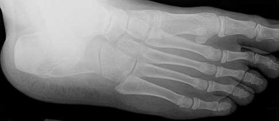
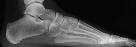
1
Normal foot
2
Calcaneonavicular (CN) coalition
3
Talocalcaneal (TC) middle facet coalition


4
TC posterior facet coalition

A
1
B C
_
- Calcaneonavicular (CN) coalition**_
QUESTION 2
of 100
Figures 2a through 2h
Figures 2a through 2h
1
Normal foot
2
Calcaneonavicular (CN) coalition
3
Talocalcaneal (TC) middle facet coalition
4
TC posterior facet coalition


A B


C


2

D E F
G

H
- Calcaneonavicular (CN) coalition
QUESTION 3
of 100
Figures 3a through 3h
A B
C
3
D E F
G
H
Figures 3a through 3h
A B
C
3
D E F
G
H
1
Normal foot
2
Calcaneonavicular (CN) coalition
3
Talocalcaneal (TC) middle facet coalition
4
TC posterior facet coalition
- TC posterior facet coalition
QUESTION 4
of 100
Figures 4a through 4j
A B 4
D C .
E
F G H
5
I J
Figures 4a through 4j
A B 4
D C .
E
F G H
5
I J
1
Normal foot
2
Calcaneonavicular (CN) coalition
3
Talocalcaneal (TC) middle facet coalition
4
TC posterior facet coalition
- Normal foot
QUESTION 5
of 100
Figures 5a through 5h
A B
C
6
D E F
G
H
Figures 5a through 5h
A B
C
6
D E F
G
H
1
Normal foot
2
Calcaneonavicular (CN) coalition
3
Talocalcaneal (TC) middle facet coalition
4
TC posterior facet coalition
- Calcaneonavicular (CN) coalition
QUESTION 6
of 100
Figures 6a through 6j
A B 7
C F
D
E
G H
Figures 6a through 6j
A B 7
C F
D
E
G H
1
Normal foot
2
Calcaneonavicular (CN) coalition
3
Talocalcaneal (TC) middle facet coalition
4
TC posterior facet coalition
- Calcaneonavicular (CN) coalition
QUESTION 7
of 100
Figures 7a through 7h
8
A B D …
C
E F G
Figures 7a through 7h
8
A B D …
C
E F G
1
Normal foot
2
Calcaneonavicular (CN) coalition
3
Talocalcaneal (TC) middle facet coalition
4
TC posterior facet coalition
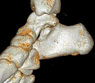
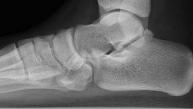
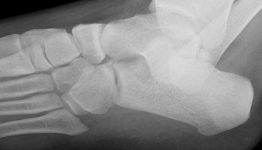
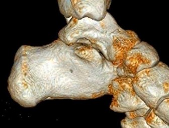
9
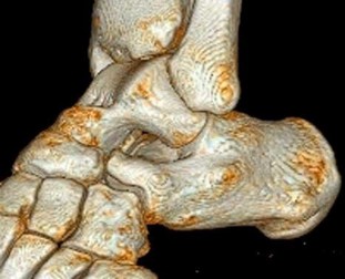
Tarsal coalitions occur when primitive mesenchymal cells fail to differentiate and form the
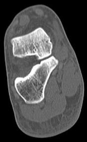
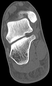
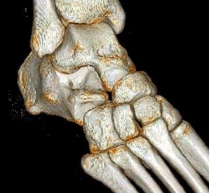
normal articular separations between the tarsal bones of the hindfoot. Overall incidence is difficult to determine because many affected people are minimally symptomatic or asymptomatic. Symptomatic tarsal coalitions typically present in adolescents as a painful flatfoot; however, there are a number of possible presentations, and occasionally symptoms do not appear until adulthood. Most tarsal coalitions are between the calcaneus and the navicular (CN) and the talus and the calcaneus (TC). Although most TC coalitions are across the middle facet, posterior facet coalitions do occur. Plain radiographic evaluation of suspected tarsal coalition is the mainstay for diagnosis. However, coalitions can be bony or fibrous, and making the diagnosis can be difficult. The addition of CT images to distinguish bony definition and MR images to decipher soft tissue can aid in diagnostics. Bony coalitions appear as definite bony bridging between the bones, while fibrous coalitions are suspected when distortion of the bony anatomy is seen. Bony coalitions are best seen on the oblique view (CN) and Harris axial view (TC). There are a number of secondary signs such as the anteater (AE) sign (elongation of the anterior process of the calcaneus as it extends to the navicular as seen on the lateral view [CN]). talar beaking (traction spur of the talar neck thought to result from abnormal stresses as seen on the lateral view [both CN and TN]), and the “C” sign (a continuous cortical contour from the medial talus to the sustentaculum tali [ST]) as seen on the lateral view (TC). A number of newer signs are not as well known, such as a broad mediolateral dimension of the navicular on the anteroposterior (AP) view (the
navicular is wider than the talar head [CN]), nonvisualization of the middle facet on the lateral view (TC), the brick sign (a normal ST is flat, but a distorted ST is enlarged and curved [CN]), and a tapered lateral navicular bone as seen on the AP view (the medial navicular [CN] is much thicker than the lateral navicular).
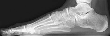
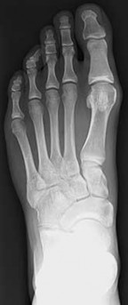
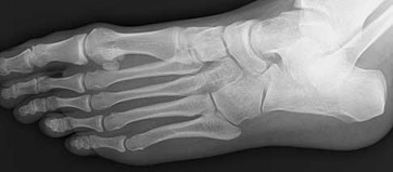
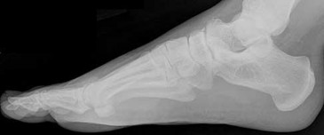
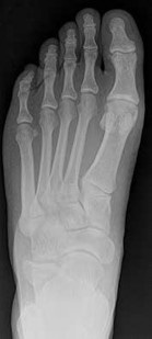
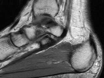
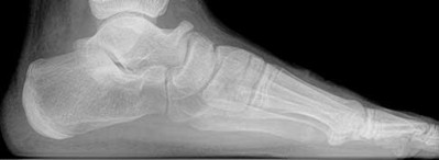
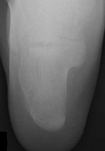
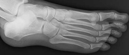
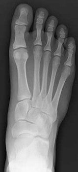
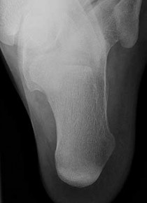
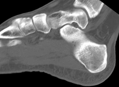
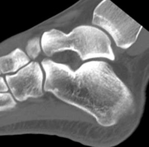
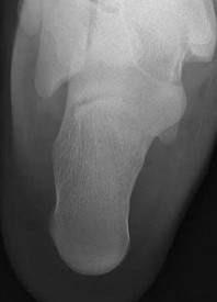
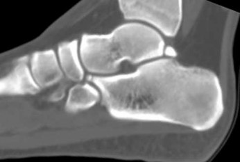
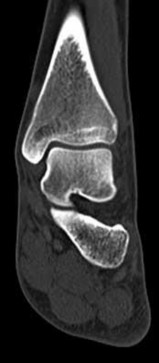
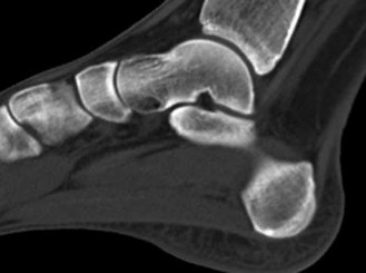
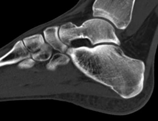
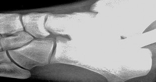
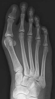
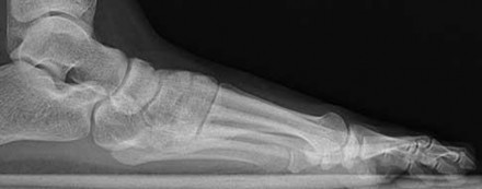
Figure 1a shows talar beaking (TB), an AE, and an open middle facet (MF). Figure 1b shows a wide navicular (WN), and Figure 1c shows an abnormal articulation between the calcaneus and the navicular, all consistent with a CN coalition.
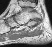
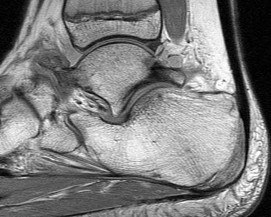
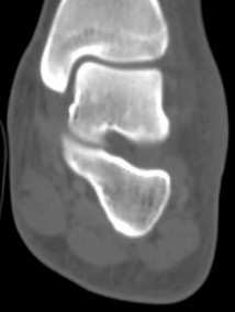
Figure 2a shows an irregularity of the anterior calcaneus. Figure 2b shows TB, AE, and MF. Figure 2c is an oblique view and shows nothing specific. Figure 2d shows an MF. Figure 2e shows an AE. Figures 2f, 2g, and 2h show edema and an abnormal connection between the calcaneus and the navicular, all consistent with a CN coalition.
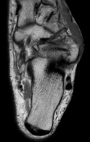
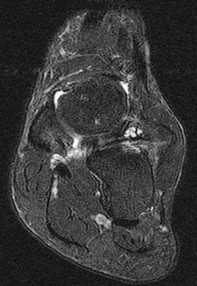
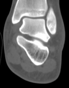
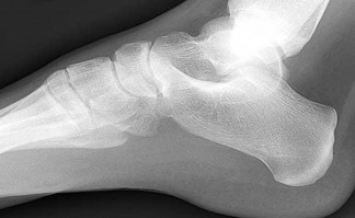
Figure 3a shows a flatfoot. Figure 3b shows an MF and TB, but not a C sign. Figure 3c shows a bony irregularity between the calcaneus and the navicular and a WN. Figure 3d shows an MF. Figure 3e shows an MF, but narrowing or loss of the posterior facet. Figures 3f through 3h show medial edema and joint irregularities consistent with a posterior facet coalition.
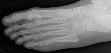
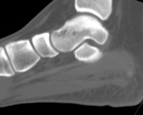
Figures 4a through 4j do not show any signs of a coalition.
11
Figure 5a shows a WN and tapering of the lateral navicular. Figure 5b shows TB and MF, but no definite AE. Figure 5c shows an abnormal articulation between the calcaneus and the navicular with fragmentation. Figures 5d and 5e show an MF. Figure 5f shows TB and fragmentation of the articulation between the calcaneus and the navicular. Figures 5g and 5h show an MF consistent with a CN coalition.
Figure 6a shows a WN and tapering of the lateral navicular. Figure 6b shows AE and TB. Figure 6c shows an abnormal articulation between the calcaneus and the navicular. Figures 6d, 6e, 6g, 6h, and 6j show MF. Figures 6f and 6i show an abnormal articulation between the calcaneus and the navicular, all consistent with a CN coalition.
Figure 7a shows a mild flatfoot with lateral peritalar subluxation of the navicular. Figure 7b does not show an open MF and has a questionable C sign. Figure 7c shows that the opening between the calcaneus and the navicular appears normal without distortion. Figures 7d, 7e, 7g, and 7h show a lateral sloping distorted middle facet consistent with a middle facet coalition, and Figure 7f shows a normal posterior facet.
RECOMMENDED READINGS
1. [Crim JR, Kjeldsberg KM. Radiographic diagnosis of tarsal coalition. AJR Am J Roentgenol. 2004 Feb;182(2):323-8. PubMed PMID: 14736655. ](http://www.ncbi.nlm.nih.gov/pubmed/14736655)[View Abstract at](http://www.ncbi.nlm.nih.gov/pubmed/14736655)[ ](http://www.ncbi.nlm.nih.gov/pubmed/14736655)[PubMed](http://www.ncbi.nlm.nih.gov/pubmed/14736655)
2. [Swiontkowski MF, Scranton PE, Hansen S. Tarsal coalitions: long-term results of surgical treatment. J Pediatr Orthop. 1983 Jul;3(3):287-92. PubMed PMID: 6874924. ](http://www.ncbi.nlm.nih.gov/pubmed/6874924)[View Abstract at PubMed](http://www.ncbi.nlm.nih.gov/pubmed/6874924)
3. [Morgan RC Jr, Crawford AH. Surgical management of tarsal coalition in adolescent athletes. Foot Ankle. 1986 Dec;7(3):183-93. PubMed PMID: 3804141. ](http://www.ncbi.nlm.nih.gov/pubmed/3804141)[View Abstract](http://www.ncbi.nlm.nih.gov/pubmed/3804141)[ ](http://www.ncbi.nlm.nih.gov/pubmed/3804141)[at PubMed](http://www.ncbi.nlm.nih.gov/pubmed/3804141)
Video 8
Tarsal coalitions occur when primitive mesenchymal cells fail to differentiate and form the
normal articular separations between the tarsal bones of the hindfoot. Overall incidence is difficult to determine because many affected people are minimally symptomatic or asymptomatic. Symptomatic tarsal coalitions typically present in adolescents as a painful flatfoot; however, there are a number of possible presentations, and occasionally symptoms do not appear until adulthood. Most tarsal coalitions are between the calcaneus and the navicular (CN) and the talus and the calcaneus (TC). Although most TC coalitions are across the middle facet, posterior facet coalitions do occur. Plain radiographic evaluation of suspected tarsal coalition is the mainstay for diagnosis. However, coalitions can be bony or fibrous, and making the diagnosis can be difficult. The addition of CT images to distinguish bony definition and MR images to decipher soft tissue can aid in diagnostics. Bony coalitions appear as definite bony bridging between the bones, while fibrous coalitions are suspected when distortion of the bony anatomy is seen. Bony coalitions are best seen on the oblique view (CN) and Harris axial view (TC). There are a number of secondary signs such as the anteater (AE) sign (elongation of the anterior process of the calcaneus as it extends to the navicular as seen on the lateral view [CN]). talar beaking (traction spur of the talar neck thought to result from abnormal stresses as seen on the lateral view [both CN and TN]), and the “C” sign (a continuous cortical contour from the medial talus to the sustentaculum tali [ST]) as seen on the lateral view (TC). A number of newer signs are not as well known, such as a broad mediolateral dimension of the navicular on the anteroposterior (AP) view (the
navicular is wider than the talar head [CN]), nonvisualization of the middle facet on the lateral view (TC), the brick sign (a normal ST is flat, but a distorted ST is enlarged and curved [CN]), and a tapered lateral navicular bone as seen on the AP view (the medial navicular [CN] is much thicker than the lateral navicular).
Figure 1a shows talar beaking (TB), an AE, and an open middle facet (MF). Figure 1b shows a wide navicular (WN), and Figure 1c shows an abnormal articulation between the calcaneus and the navicular, all consistent with a CN coalition.
Figure 2a shows an irregularity of the anterior calcaneus. Figure 2b shows TB, AE, and MF. Figure 2c is an oblique view and shows nothing specific. Figure 2d shows an MF. Figure 2e shows an AE. Figures 2f, 2g, and 2h show edema and an abnormal connection between the calcaneus and the navicular, all consistent with a CN coalition.
Figure 3a shows a flatfoot. Figure 3b shows an MF and TB, but not a C sign. Figure 3c shows a bony irregularity between the calcaneus and the navicular and a WN. Figure 3d shows an MF. Figure 3e shows an MF, but narrowing or loss of the posterior facet. Figures 3f through 3h show medial edema and joint irregularities consistent with a posterior facet coalition.
Figures 4a through 4j do not show any signs of a coalition.
11
Figure 5a shows a WN and tapering of the lateral navicular. Figure 5b shows TB and MF, but no definite AE. Figure 5c shows an abnormal articulation between the calcaneus and the navicular with fragmentation. Figures 5d and 5e show an MF. Figure 5f shows TB and fragmentation of the articulation between the calcaneus and the navicular. Figures 5g and 5h show an MF consistent with a CN coalition.
Figure 6a shows a WN and tapering of the lateral navicular. Figure 6b shows AE and TB. Figure 6c shows an abnormal articulation between the calcaneus and the navicular. Figures 6d, 6e, 6g, 6h, and 6j show MF. Figures 6f and 6i show an abnormal articulation between the calcaneus and the navicular, all consistent with a CN coalition.
Figure 7a shows a mild flatfoot with lateral peritalar subluxation of the navicular. Figure 7b does not show an open MF and has a questionable C sign. Figure 7c shows that the opening between the calcaneus and the navicular appears normal without distortion. Figures 7d, 7e, 7g, and 7h show a lateral sloping distorted middle facet consistent with a middle facet coalition, and Figure 7f shows a normal posterior facet.
RECOMMENDED READINGS
1. [Crim JR, Kjeldsberg KM. Radiographic diagnosis of tarsal coalition. AJR Am J Roentgenol. 2004 Feb;182(2):323-8. PubMed PMID: 14736655. ](http://www.ncbi.nlm.nih.gov/pubmed/14736655)[View Abstract at](http://www.ncbi.nlm.nih.gov/pubmed/14736655)[ ](http://www.ncbi.nlm.nih.gov/pubmed/14736655)[PubMed](http://www.ncbi.nlm.nih.gov/pubmed/14736655)
2. [Swiontkowski MF, Scranton PE, Hansen S. Tarsal coalitions: long-term results of surgical treatment. J Pediatr Orthop. 1983 Jul;3(3):287-92. PubMed PMID: 6874924. ](http://www.ncbi.nlm.nih.gov/pubmed/6874924)[View Abstract at PubMed](http://www.ncbi.nlm.nih.gov/pubmed/6874924)
3. [Morgan RC Jr, Crawford AH. Surgical management of tarsal coalition in adolescent athletes. Foot Ankle. 1986 Dec;7(3):183-93. PubMed PMID: 3804141. ](http://www.ncbi.nlm.nih.gov/pubmed/3804141)[View Abstract](http://www.ncbi.nlm.nih.gov/pubmed/3804141)[ ](http://www.ncbi.nlm.nih.gov/pubmed/3804141)[at PubMed](http://www.ncbi.nlm.nih.gov/pubmed/3804141)
Video 8
QUESTION 8
of 100
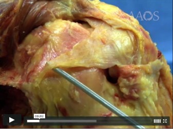
Video 8 features a total knee arthroplasty while trialing; which structure is too tight?
Video 8 features a total knee arthroplasty while trialing; which structure is too tight?
1
Medial collateral ligament.
2
Posterior cruciate ligament 11
3
Posterior capsule of the knee
4
Patellar tendon
The video demonstration shows a trial range of motion of a cruciate-retaining total knee arthroplasty. The video shows lift off of the trial surface while the knee is flexed. This is indicative of a tight posterior cruciate ligament. If asymmetric tightness were present on the medial side, this could signify a tight medial collateral ligament. The posterior capsule is involved in tightness of the knee in extension. The patellar tendon does not play a role in this scenario.
RECOMMENDED READINGS
1. Villanueva M, Chana F, Pereiro J, Ríos-Luna A, Rojo-Manaute J, Benito Del Carmen F, Fahandez-Saddi H, Perez-Caballer A. AAOS Orthopaedic Video Theater: Instability after Total Knee Arthroplasty: Limits of Constraint. Rosemont, IL: American Academy of Orthopaedic Surgeons; 2013.
2. Hoppenfeld S, deBoer P. Surgical Exposures in Orthopedics. 3rd ed. Philadelphia, PA: Lippincott Williams & Wilkins; 2003:493-568.
QUESTION 9
of 100
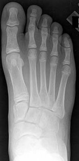
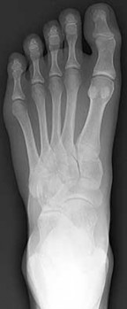
Figures 9a and 9b are the radiographs of a 19-year-old woman with a painful juvenile bunion. The pathologic findings associated with this deformity
include a
Figures 9a and 9b are the radiographs of a 19-year-old woman with a painful juvenile bunion. The pathologic findings associated with this deformity
include a
1
Laterally deviated distal metatarsal articular surface, a
lax or disrupted distal 1-2 transverse intermetatarsal 12
ligament, and a contracted lateral collateral (lateral first
metatarsophalangeal) ligament.
2
Laterally deviated distal metatarsal articular surface, a contracted lateral collateral (lateral first metatarsophalangeal) ligament, and a medially deviated or hypermobile first metatarsocuneiform joint.
3
Medially deviated or hypermobile first metatarsocuneiform joint, a lax or disrupted distal 1-2 transverse intermetatarsal ligament, and a contracted lateral collateral (lateral first metatarsophalangeal) ligament.
4
Lax or disrupted distal 1-2 transverse intermetatarsal ligament, laterally deviated distal metatarsal articular surface, and a medially deviated or hypermobile first metatarsocuneiform joint.
The radiographs show a hallux valgus deformity with a laterally deviated distal metatarsal articular surface, a large intermetatarsal angle with medial deviation at the first metatarsocuneiform joint, an elongated medial collateral ligament, and a contracted lateral collateral ligament. There is no distal 1-2 transverse intermetatarsal ligament. The distal transverse ligament in the first interspace extends from the second metatarsal to the lateral (fibular) sesamoid, remains intact, and keeps the sesamoids in a lateral position as the first metatarsal head migrates medially.
RECOMMENDED READINGS
1. Coughlin MJ. Roger A. Mann Award. Juvenile hallux valgus: etiology and treatment. Foot Ankle Int. 1995 Nov;16(11):682-97. PubMed PMID: 8589807.
[View Abstract at PubMed](http://www.ncbi.nlm.nih.gov/pubmed/8589807)
2. Coughlin MJ, Mann RA. Hallux valgus. In: Coughlin MJ, Mann RA, Saltzman CL, eds. Surgery of the Foot and Ankle. 8th ed. Philadelphia, PA: Mosby Elsevier; 2007:183-226.
QUESTION 10
of 100
13
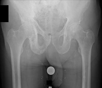
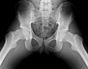
Figure 10 is an anteroposterior pelvis radiograph of an 82-year-old man who had right hip pain that began 2 weeks ago but has since resolved with use of over-the-counter nonsteroidal anti-inflammatory drugs (NSAIDs). Currently he has no pain. Examination of his hip shows decreased internal rotation and minimal pain at the extremes of motion. What is the most appropriate treatment at this point?
13
Figure 10 is an anteroposterior pelvis radiograph of an 82-year-old man who had right hip pain that began 2 weeks ago but has since resolved with use of over-the-counter nonsteroidal anti-inflammatory drugs (NSAIDs). Currently he has no pain. Examination of his hip shows decreased internal rotation and minimal pain at the extremes of motion. What is the most appropriate treatment at this point?
1
Observation and NSAID use as needed
2
MRI scan
3
Bone biopsy
4
Serum protein electrophoresis (SPEP) and urine protein electrophoresis (UPEP).
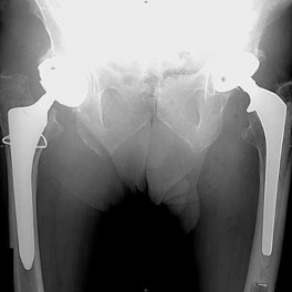
The radiograph shown is consistent with Paget disease of the bone. It demonstrates classic findings of widened lamellae and disorganized sclerotic and lytic areas. The cause is not clearly defined, but may be linked to a viral infection and subsequent alterations of osteoblastic and osteoclastic activity. Most patients are asymptomatic, and Paget disease is often found incidentally on radiographs. In this case, the patient’s symptoms likely were caused by hip arthritis, but Paget disease can cause diffuse bone pain in some cases. Considering the patient’s mild and short-term symptoms, observation and NSAID use is most appropriate. An MRI scan or biopsy is indicated if sarcomatous transformation is suspected, but this condition is rare and is associated with a substantial, unrelenting increase in pain. SPEP and UPEP are tests for multiple myeloma, of which the radiographs show no signs.
RECOMMENDED READINGS
1. [Ralston SH. Pathogenesis of Paget's disease of bone. Bone. 2008 Nov;43(5):819-25. doi: 10.1016/j.bone.2008.06.015. Epub 2008 Jul 11. Review. PubMed PMID: 18672105.](http://www.ncbi.nlm.nih.gov/pubmed/18672105)[View Abstract at PubMed](http://www.ncbi.nlm.nih.gov/pubmed/18672105)
2. Bonenberger E, Einhorn T. Metabolic bone diseases. In: Callaghan JJ, Rosenberg
AG, Rubash HE, eds. The Adult Hip. 2nd ed. Philadelphia, PA: Lippincott Williams 14
& Wilkins; 2007:514-533.
QUESTION 11
of 100
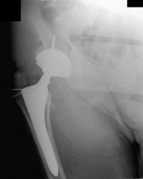
What is the most likely explanation for the change between the initial intraoperative radiograph (Figure 11a) and the radiograph taken 4 weeks after surgery (Figure 11b) in an 87-year-old man who underwent primary hip replacement for osteoarthritis?
What is the most likely explanation for the change between the initial intraoperative radiograph (Figure 11a) and the radiograph taken 4 weeks after surgery (Figure 11b) in an 87-year-old man who underwent primary hip replacement for osteoarthritis?
1
Deep-wound infection.
2
Aseptic loosening
3
Osteoporosis
4
Initial implant stability
The image demonstrates subsidence of the femoral implant. The implant subsided because it did not have good initial stability. The tapered femoral implant was placed after initial preparation for an anatomic femoral stem. A limited, nondisplaced femoral neck fracture was encountered during the procedure and treated. Two advantages of tapered stems are the efficient transfer of stress to the proximal femur and the ability to accommodate some subsidence to achieve enhanced stability. Although subsidence of a tapered stem to a more stable position can produce a good result, quality of metaphyseal bone should be considered. Attention to surgical technique remains important to optimize component stability for biological fixation.
RECOMMENDED READINGS
1. Savory CG, Hamilton WG, Engh CA Sr, Della Valle CJ, Rosenberg AG, Galante JO. 15 Hip designs. In: Barrack RL, Booth RE Jr, Lonner JH, McCarthy JC, Mont MA, Rubash HE, eds. Orthopaedic Knowledge Update: Hip and Knee Reconstruction 3.
Rosemont, IL: American Academy of Orthopaedic Surgeons; 2006:345-368.
2. Blaha JD, Borus TA. Press-fit femoral components. In: Callaghan J, Rosenberg A, and Rubash H, eds The Adult Hip. 2nd ed. Philadelphia, PA: Lippincott Williams & Wilkins; 2007:1036-1043.
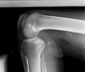
QUESTION 12
of 100
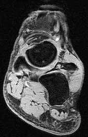
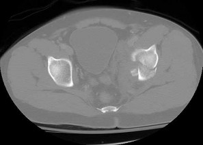
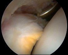
Figure 12 shows an arthroscopic view from an inferolateral portal of a right knee. The asterisk indicates which structure?
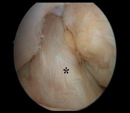
Figure 12 shows an arthroscopic view from an inferolateral portal of a right knee. The asterisk indicates which structure?
1
Anterior cruciate ligament, anteromedial bundle
2
Anterior cruciate ligament, anterolateral bundle
3
Anterior cruciate ligament, posteromedial bundle
4
Anterior cruciate ligament, posterolateral bundle
The asterisk indicates the anteromedial bundle of the anterior cruciate ligament. The anterior cruciate ligament consists of 2 functional bundles: anteromedial and posterolateral. During extension of the knee, the posterolateral bundle becomes taut. In flexion, the anteromedial bundle is tight and the posterolateral bundle relaxes. Recently, techniques for double-bundle reconstruction have been described to recreate the normal anatomic relationship of the 2 bundles.
RECOMMENDED READINGS
1. [Chhabra A, Zelle BA, Feng MT, Fu FH. The arthroscopic appearance of a normal anterior cruciate ligament in a posterior cruciate ligament-deficient knee: the posterolateral bundle (PLB) sign. Arthroscopy. 2005 Oct;21(10):1267. PubMed PMID: 16226658. ](http://www.ncbi.nlm.nih.gov/pubmed/16226658)[View Abstract at PubMed](http://www.ncbi.nlm.nih.gov/pubmed/16226658)
2. [Cha PS, Brucker PU, West RV, Zelle BA, Yagi M, Kurosaka M, Fu FH. Arthroscopic double-bundle anterior cruciate ligament reconstruction: an anatomic approach. Arthroscopy. 2005 Oct;21(10):1275. PubMed PMID: 16226666. ](http://www.ncbi.nlm.nih.gov/pubmed/16226666)[View Abstract ](http://www.ncbi.nlm.nih.gov/pubmed/16226666)[at](http://www.ncbi.nlm.nih.gov/pubmed/16226666)
[PubMed](http://www.ncbi.nlm.nih.gov/pubmed/16226666)
16
3. Clarke HD, Scott WN, Insall JN, et al. Anatomy. In: Insall JN, Scott WN, eds. Surgery
of the Knee. Vol 1. 4th ed. Philadelphia, PA: Churchill Livingstone; 2006:3-66.
QUESTION 13
of 100
Gerdy tubercle
Gerdy tubercle
1
Figure 13a.
2
Figure 13b
3
Figure 13c
4
Figure 13d
5
Figure 13e
- Figure 13a
QUESTION 14
of 100
Site of tendon insertion that is proximal to its muscular origin
Site of tendon insertion that is proximal to its muscular origin
1
Figure 13a.
2
Figure 13b
3
Figure 13c
4
Figure 13d
5
Figure 13e
- Figure 13d
QUESTION 15
of 100
Stieda fracture
Stieda fracture
1
Figure 13a.
2
Figure 13b
3
Figure 13c
4
Figure 13d
5
Figure 13e
- Figure 13e
QUESTION 16
of 100
Structure responsible for the pivot shift phenomenon as it transitions to become a knee flexor from being a knee extensor, thereby causing tibial reduction
Structure responsible for the pivot shift phenomenon as it transitions to become a knee flexor from being a knee extensor, thereby causing tibial reduction
1
Figure 13a.
2
Figure 13b
3
Figure 13c
4
Figure 13d
5
Figure 13e
- Figure 13b
QUESTION 17
of 100
Primary stabilizer of the knee to valgus stress in approximately 30 degrees of flexion 1- Figure 13a.
Primary stabilizer of the knee to valgus stress in approximately 30 degrees of flexion 1- Figure 13a.
1
Figure 13b
2
Figure 13c
3
Figure 13d
4
Figure 13e
5
Figure 13f
The Gerdy tubercle is located on the anterolateral portion of the proximal tibia and is the insertion for the iliotibial (IT) tract and the origin for fibers of the tibialis anterior muscle. The popliteus muscle originates from the posteromedial aspect of the proximal tibia and inserts into the popliteal groove on the distal lateral femur, deep to and distal to the origin of the fibular collateral ligament. Avulsion fracture of the medial distal femur at the origin of the medial collateral ligament is known as a Stieda fracture and may later develop into a Pelligrini-Stieda lesion. With the knee in extension and the tibia internally rotated, the lateral plateau subluxes anteriorly to the lateral femoral condyle in the anterior cruciate ligament-deficient knee. As the knee is moved from an extended to a flexed position, the IT band switches from being a knee extensor, and, as the forces of the IT band move posterior to the
center of rotation of the lateral femur, the IT band contributes to the posterior reduction of 19
the lateral tibial plateau and the palpable clunk that is felt. The medial collateral ligament is the primary stabilizer when valgus stress is placed on the knee. This is most pronounced when the knee is flexed at 25 to 30 degrees.
RECOMMENDED READINGS
1. [Haims AH, Medvecky MJ, Pavlovich R Jr, Katz LD. MR imaging of the anatomy of and injuries to the lateral and posterolateral aspects of the knee. AJR Am J Roentgenol. 2003 Mar;180(3):647-53. PubMed PMID: 12591668.](http://www.ncbi.nlm.nih.gov/pubmed/12591668)[View Abstract at](http://www.ncbi.nlm.nih.gov/pubmed/12591668)[ ](http://www.ncbi.nlm.nih.gov/pubmed/12591668)[PubMed](http://www.ncbi.nlm.nih.gov/pubmed/12591668)
2. [Recondo JA, Salvador E, Villanúa JA, Barrera MC, Gervás C, Alústiza JM. Lateral stabilizing structures of the knee: functional anatomy and injuries assessed with MR imaging. Radiographics. 2000 Oct;20 Spec No:S91-S102. PubMed PMID: 11046165. ](http://www.ncbi.nlm.nih.gov/pubmed/11046165)[View Abstract at PubMed](http://www.ncbi.nlm.nih.gov/pubmed/11046165)
3. [Hunter TB, Peltier LF, Lund PJ. Radiologic history exhibit. Musculoskeletal eponyms: who are those guys? Radiographics. 2000 May-Jun;20(3):819-36. PubMed PMID: 10835130.](http://www.ncbi.nlm.nih.gov/pubmed/10835130)[View Abstract at PubMed](http://www.ncbi.nlm.nih.gov/pubmed/10835130)
4. [Lane CG, Warren R, Pearle AD. The pivot shift. J Am Acad Orthop Surg. 2008 Dec;16(12):679-88. Review. PubMed PMID: 19056917.](http://www.ncbi.nlm.nih.gov/pubmed/19056917)[View Abstract at PubMed](http://www.ncbi.nlm.nih.gov/pubmed/19056917)
5. [Phisitkul P, James SL, Wolf BR, Amendola A. MCL injuries of the knee: current concepts review. Iowa Orthop J. 2006;26:77-90. Review. PubMed PMID: 16789454. ](http://www.ncbi.nlm.nih.gov/pubmed/16789454)View Abstract at PubMed .
QUESTION 18
of 100
A 56-year-old woman fell off a stepladder and sustained the injury shown in Figures 18a and 18b. In addition to the pain from her injury, she has numbness and weakness in her foot. Upon examination, the findings most consistent with her radiographs are decreased sensation
21
A B
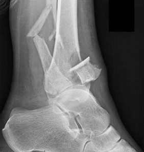
A 56-year-old woman fell off a stepladder and sustained the injury shown in Figures 18a and 18b. In addition to the pain from her injury, she has numbness and weakness in her foot. Upon examination, the findings most consistent with her radiographs are decreased sensation
21
A B
1
in her first interspace and an inability to dorsiflex her toes.
2
over her lateral forefoot and an inability to evert her foot.
3
over her medial forefoot and an inability to invert her foot.
4
over her lateral forefoot and an inability to plantar flex her first metatarsal.
The radiographs reveal a tibial pilon fracture with an extruded and rotated anterior tibial fragment that lies deep to the anterior compartment neurovascular bundle, which contains the deep peroneal nerve. This nerve innervates the anterior compartment muscles and the extensor digitorum brevis and extensor hallucis brevis muscles and provides sensation to the dorsal aspect of the first interspace. An injury to the deep peroneal nerve at this level will only affect the innervation to the extensor digitorum brevis and extensor hallucis brevis muscles and the innervation of the first interspace. The superficial peroneal nerve innervates
the lateral compartment muscles above the level of this injury and innervates the dorsum of the foot. The medial forefoot is innervated by the saphenous nerve and the posterior tibial nerve innervates the posterior compartment muscles above the level of the injury. The sural nerve innervates the lateral foot and has no motor component, and the superficial peroneal nerve innervates the peroneus longus, which plantar flexes the first metatarsal above the level of the injury.
RECOMMENDED READINGS
1. Agur AM, Dalley AF, eds. Grant’s Atlas of Anatomy. 13th ed. Philadelphia, PA: Wolters Kluwer/Lippincott Williams & Wilkins; 2013:362-370.
2. Hoppenfeld S, de Boer P, Buckley R, eds. Surgical Exposures in Orthopaedics: The Anatomic Approach. 4th ed. Philadelphia, PA: Lippincott Williams & Wilkins; 2009:625-673.
QUESTION 19
of 100
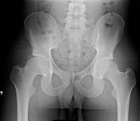
Figure 19 is an anteroposterior pelvis radiograph of a 60-year-old woman who has a 4-month history of right hip pain. She has a
medical history of lupus and has used
21
prednisone in the past, but not currently.
Her pain is persistent despite use of over-the-counter pain medications and activity modifications. What is the most appropriate treatment at this point?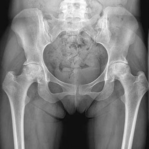
1
Weight loss and protected weight bearing
2
Bisphosphonate therapy
3
Total hip resurfacing arthroplasty
4
Total hip arthroplasty
This scenario describes osteonecrosis of the hip, likely attributable to chronic corticosteroid use. Her radiographs show bilateral hip involvement with whole-head involvement and collapse of the femoral head. Observation, protected weight bearing, and bisphosphonates have been advocated during the precollapse stage, but, considering the advanced
involvement of the femoral heads, these treatments are not indicated. Hip arthroplasty is the most reliable treatment option at this point to resolve her symptoms. Total hip resurfacing is not indicated for multiple reasons, including poor bone stock from corticosteroid use and her age. Large-head involvement and concerns about metal-on-metal articulation in someone with potential for renal impairment (such as a patient with lupus) also are contraindications to total hip resurfacing. Total hip arthroplasty will offer the best chance for success.
RECOMMENDED READINGS
1. Mont M, Bezwasa H. Osteonecrosis: Strategies for treatment. In: Callaghan JJ, Rosenberg AG, Rubash HE, eds. The Adult Hip. 2nd ed. Philadelphia, PA: Lippincott Williams & Wilkins; 2007:477-499.
2. [Lieberman JR, Berry DJ, Mont MA, Aaron RK, Callaghan JJ, Rajadhyaksha AD, Urbaniak JR. Osteonecrosis of the hip: management in the 21st century. Instr Course Lect. 2003;52:337-55. Review. PubMed PMID: 12690862.](http://www.ncbi.nlm.nih.gov/pubmed/12690862)[View Abstract at PubMed](http://www.ncbi.nlm.nih.gov/pubmed/12690862)
QUESTION 20
of 100
22
The lesser or small saphenous vein passes along the sural nerve in the mid calf
22
The lesser or small saphenous vein passes along the sural nerve in the mid calf
1
medially.
2
laterally.
3
anteriorly.
4
posteriorly.
The sural nerve is at risk when incisions are placed near the Achilles tendon musculotendinous junction of the posterior calf. The nerve, which can be easily identified when the vein is visualized, is at risk during Achilles tendon recession procedures, and the vein provides a landmark with which to identify the nerve.
RECOMMENDED READINGS
1. [Eid EM, Hegazy AM. Anatomical variations of the human sural nerve and its role in clinical and surgical procedures .Clin Anat.2011Mar;24(2):237-45.doi: 10.1002/ ca.21068. Epub 2010 Oct 14.PubMed PMID: 20949489.](http://www.ncbi.nlm.nih.gov/pubmed/20949489)[View Abstract at PubMed](http://www.ncbi.nlm.nih.gov/pubmed/20949489)
2. Hoppenfeld S, deBoer P, Buckley R. Surgical Exposures in Orthopaedics. The Anatomic Approach. Philadelphia, PA: Lippincott Williams &Wilkins; 2009:585-622.
3. [Aktan Ikiz ZA, Uçerler H, Bilge O. The anatomic features of the sural nerve with an emphasis on its clinical importance. Foot Ankle Int. 2005 Jul;26(7):560-7. PubMed PMID: 16045849.](http://www.ncbi.nlm.nih.gov/pubmed/16045849)[View Abstract at PubMed](http://www.ncbi.nlm.nih.gov/pubmed/16045849)
QUESTION 21
of 100
Release of which structure results in the largest hip internal rotation increase in both flexion and extension ?
Release of which structure results in the largest hip internal rotation increase in both flexion and extension ?
1
Medial arm of the iliofemoral ligament.
2
Lateral arm of the iliofemoral ligament
3
Pubofemoral ligament
4
Ischiofemoral ligament
Hip stability is augmented by thickened portions of the articular capsule. A sectioning study of the hip capsular ligaments identified the ischiofemoral ligament to have the most significant effect in limiting hip internal rotation in both extension and flexion. The strongest of the capsular ligaments is the iliofemoral ligament. The medial arm of the iliofemoral ligament provides the most significant restraint against anterior hip translation with hip extension and external rotation. The lateral arm of the iliofemoral ligament provides restriction to both internal and external rotation with the hip in extension. The pubofemoral ligament augments stability of the hip against external rotation in extension.
RECOMMENDED READINGS
1. Martin HD, Savage A, Braly BA, Palmer IJ, Beall DP, Kelly B. The function of the hip capsular ligaments: a quantitative report. Arthroscopy. 2008 Feb;24(2):188-95. doi: 10.1016/j.arthro.2007.08.024. Epub 2007 Nov 26. PubMed PMID: 18237703.
[View Abstract at PubMed](http://www.ncbi.nlm.nih.gov/pubmed/18237703)
2. Wasielewski RC.The Hip. In: Callaghan J, Rosenberg A, Rubash H, The Adult Hip. 2nd ed. Philadelphia, PA: Lippincott Williams & Wilkins; 2007:53.
CLINICAL SITUATION FOR QUESTIONS 22 THROUGH 25
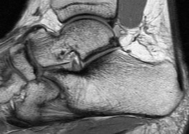
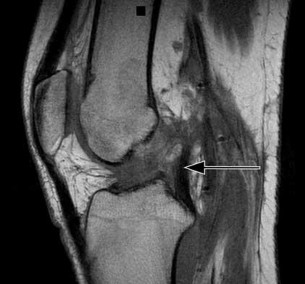
A 22-year-old man sustains an injury to his right knee in a motor vehicle collision. Figure 22a is the posterior stress radiograph of the involved knee, and Figure 22b is a selected MR image that identifies the injured structure.
A
B
RECOMMENDED READINGS
1. Martin HD, Savage A, Braly BA, Palmer IJ, Beall DP, Kelly B. The function of the hip capsular ligaments: a quantitative report. Arthroscopy. 2008 Feb;24(2):188-95. doi: 10.1016/j.arthro.2007.08.024. Epub 2007 Nov 26. PubMed PMID: 18237703.
[View Abstract at PubMed](http://www.ncbi.nlm.nih.gov/pubmed/18237703)
2. Wasielewski RC.The Hip. In: Callaghan J, Rosenberg A, Rubash H, The Adult Hip. 2nd ed. Philadelphia, PA: Lippincott Williams & Wilkins; 2007:53.
CLINICAL SITUATION FOR QUESTIONS 22 THROUGH 25
A 22-year-old man sustains an injury to his right knee in a motor vehicle collision. Figure 22a is the posterior stress radiograph of the involved knee, and Figure 22b is a selected MR image that identifies the injured structure.
A
B
QUESTION 22
of 100
Which relationship is noted for the structure identified by the arrow in Figure 22b? 24
Which relationship is noted for the structure identified by the arrow in Figure 22b? 24
1
The anterolateral and posteromedial bundles are relaxed in mid flexion and tensioned in high flexion.
2
The anterolateral and posteromedial bundles are tensioned in mid flexion and tensioned in high flexion.
3
The anterolateral bundle is tensioned in mid flexion, and the posteromedial bundle is tensioned in both extension and high flexion.
4
The posteromedial bundle is tensioned in mid flexion, and the anterolateral bundle is tensioned in both flexion and extension.
- The anterolateral bundle is tensioned in mid flexion, and the posteromedial bundle is tensioned in both extension and high flexion.
QUESTION 23
of 100
Which relationship is noted for the fibers of the structure injured in Figure 22b?
Which relationship is noted for the fibers of the structure injured in Figure 22b?
1
The anterolateral bundle is longer, thicker, and stronger than the posteromedial bundle.
2
The anterolateral bundle is shorter, thicker, and stronger than the posteromedial bundle.
3
The posterolateral bundle is longer, thicker, and stronger than the anterolateral bundle.
4
The posteromedial bundle is shorter, thicker, and stronger than the anterolateral bundle.
- The anterolateral bundle is shorter, thicker, and stronger than the posteromedial bundle.
QUESTION 24
of 100
When surgical reconstruction is accomplished using a 2-bundled reconstruction technique, recreating the function of the posteromedial bundle contributes uniquely to the recovery of stability against which direction of force?
When surgical reconstruction is accomplished using a 2-bundled reconstruction technique, recreating the function of the posteromedial bundle contributes uniquely to the recovery of stability against which direction of force?
1
Posterior tibial translation in extension
2
Anterior tibial translation in extension
3
External rotation of the tibia in extension
4
Hyperextension
- Hyperextension
QUESTION 25
of 100
With respect to the structure identified by the arrow in Figure 22b, the meniscofemoral 25
ligaments are
With respect to the structure identified by the arrow in Figure 22b, the meniscofemoral 25
ligaments are
1
uniformly present, and are positioned posterior to the injured ligament.
2
uniformly present, with one positioned anterior and the other positioned posterior to the injured ligament.
3
variably present, and are positioned posterior to the injured ligament.
4
variably present, with one positioned anterior and the other positioned posterior to the injured ligament.
The stress radiographs demonstrate posterior instability of the right knee in flexion. The MR images demonstrate injury to both the anterior and posterior cruciate ligament (PCL), with the stump identified with the arrow on the MR image (Figure 22b). The PCL has 2 functional bands. The anterolateral bundle originates from the roof of the intercondylar notch. It runs in a posterolateral direction onto the tibial crest between the posterior attachment of the medial and lateral menisci. During a double-bundled posterior ligament reconstruction, the
anterolateral bundle is tensioned with the knee in a position of mid flexion. The posteromedial bundle has a variable pattern of tension both in extension and in high flexion. Tensioning of the posteromedial bundle in extension may contribute to resistance against knee hyperextension.
The meniscofemoral ligaments are variably present. Although 93% of knees have been reported to have at least 1 meniscofemoral ligament present, both ligaments are simultaneously present in approximately 50% of knees. The ligament of Humphrey (anterior meniscofemoral ligament) and ligament of Wrisberg (posterior meniscofemoral ligament) are delineated by their anatomic relationship to the posterior cruciate.
RECOMMENDED READINGS
1. [Amis AA, Bull AM, Gupte CM, Hijazi I, Race A, Robinson JR. Biomechanics of the PCL and related structures: posterolateral, posteromedial and meniscofemoral ligaments. Knee Surg Sports Traumatol Arthrosc. 2003 Sep;11(5):271-81. Epub 2003 Sep 5. Review. PubMed PMID: 12961064.](http://www.ncbi.nlm.nih.gov/pubmed/12961064)[View Abstract at PubMed](http://www.ncbi.nlm.nih.gov/pubmed/12961064)
2. Amis AA, Gupte CM, Bull AM, Edwards A. Anatomy of the posterior cruciate ligament and the meniscofemoral ligaments. Knee Surg Sports Traumatol Arthrosc. 2006 Mar;14(3):257-63. Epub 2005 Oct 14. Review. PubMed PMID: 16228178.
View Abstract at PubMed . 26
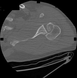
QUESTION 26
of 100
The injury pattern shown in the CT image in Figure 26 is most commonly associated with which mechanism?
The injury pattern shown in the CT image in Figure 26 is most commonly associated with which mechanism?
1
Traction injury.
2
Seizures
3
Collision athletic events
4
Postpolio syndrome
Posterior shoulder dislocations are most commonly the result of seizures and electrical shock. Collision athletic events, postpolio syndrome, and traction injury are rarely associated with posterior shoulder dislocations. The bony defect caused by impaction of the anterior superior humeral head on the posterior glenoid has been referred to as a “reverse Hill-Sachs lesion.”
RECOMMENDED READINGS
1. McLaughlin HL: Posterior dislocation of the shoulder. J Bone Joint Surg Am 1952;64:1584-1590.
2. [Kowalsky MS, Levine WN. Traumatic posterior glenohumeral dislocation: classification, pathoanatomy, diagnosis, and treatment. Orthop Clin North Am. 2008 Oct;39(4):519-33, viii. doi: 10.1016/j.ocl.2008.05.008. Review. PubMed PMID: 18803981.](http://www.ncbi.nlm.nih.gov/pubmed/18803981)[View Abstract at PubMed](http://www.ncbi.nlm.nih.gov/pubmed/18803981)
QUESTION 27
of 100
Which structure is shown in Video 27? 27
Which structure is shown in Video 27? 27
1
Superficial medial collateral ligament
2
Semimembranosus tendon
3
Posterior oblique ligament
4
Medial patellofemoral ligament
Video 27 shows the medial patellofemoral ligament running from the medial epicondyle of the femur to the medial portion of the patella. The posterior oblique ligament and the superficial medial collateral ligament run from medial epicondyle to the tibia.
RECOMMENDED READINGS
1. Babb JR, Detterline AJ, Noyes FR. AAOS Orthopaedic Video Theater. The Key to the Knee: A Layer-by-Layer Video Demonstration of Medial and Anterior Aatomy. Rosemont, IL: American Academy of Orthopaedic Surgeons; 2009.
2. Hoppenfeld S, deBoer P. Surgical Exposures in Orthopedics. 3rd ed. Philadelphia, PA: Lippincott Williams & Wilkins; 2003:493-568.
QUESTION 28
of 100
Which structure, indicated at the tip of the arrow in Figure 28, is at risk for anterior cortical penetration during placement of C1 lateral mass screws?
Which structure, indicated at the tip of the arrow in Figure 28, is at risk for anterior cortical penetration during placement of C1 lateral mass screws?
1
Hypoglossal nerve.
2
Jugular vein
3
Lingual artery
28
4
Internal carotid artery
The internal carotid artery can run in close proximity to the anterior surface of C1 in many patients; consequently, a drill bit or screw tip poses risk. This anatomy always must be considered when placing bicortical C1 screws.
RECOMMENDED READINGS
1. [Currier BL, Maus TP, Eck JC, Larson DR, Yaszemski MJ. Relationship of the internal carotid artery to the anterior aspect of the C1 vertebra: implications for C1-C2 transarticular and C1 lateral mass fixation. Spine (Phila Pa 1976). 2008 Mar 15;33(6):635-9. PubMed PMID: 18344857. ](http://www.ncbi.nlm.nih.gov/pubmed/18344857)[View Abstract at PubMed](http://www.ncbi.nlm.nih.gov/pubmed/18344857)
2. [Hoh DJ, Maya M, Jung A, Ponrartana S, Lauryssen CL. Anatomical relationship of the internal carotid artery to C-1: clinical implications for screw fixation of the atlas. J Neurosurg Spine. 2008 Apr;8(4):335-40. PubMed PMID: 18377318. ](http://www.ncbi.nlm.nih.gov/pubmed/18377318)[View Abstract at](http://www.ncbi.nlm.nih.gov/pubmed/18377318)[ ](http://www.ncbi.nlm.nih.gov/pubmed/18377318)[PubMed](http://www.ncbi.nlm.nih.gov/pubmed/18377318)
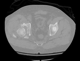
Match the description with the corresponding response. Question 29 of 100
Neural structure most likely damaged as a result of this injury (Figure 29)
1. Corona mortis
2. Tibial division, sciatic nerve
3. Sciatic nerve, peroneal division
4. Fifth lumbar nerve root
5. Kocher-Langenbeck approach
6. Stoppa approach
7. Obturator vessels
8. L4 nerve root
PREFERRED RESPONSE: 3- Sciatic nerve, peroneal division
RECOMMENDED READINGS
1. [Currier BL, Maus TP, Eck JC, Larson DR, Yaszemski MJ. Relationship of the internal carotid artery to the anterior aspect of the C1 vertebra: implications for C1-C2 transarticular and C1 lateral mass fixation. Spine (Phila Pa 1976). 2008 Mar 15;33(6):635-9. PubMed PMID: 18344857. ](http://www.ncbi.nlm.nih.gov/pubmed/18344857)[View Abstract at PubMed](http://www.ncbi.nlm.nih.gov/pubmed/18344857)
2. [Hoh DJ, Maya M, Jung A, Ponrartana S, Lauryssen CL. Anatomical relationship of the internal carotid artery to C-1: clinical implications for screw fixation of the atlas. J Neurosurg Spine. 2008 Apr;8(4):335-40. PubMed PMID: 18377318. ](http://www.ncbi.nlm.nih.gov/pubmed/18377318)[View Abstract at](http://www.ncbi.nlm.nih.gov/pubmed/18377318)[ ](http://www.ncbi.nlm.nih.gov/pubmed/18377318)[PubMed](http://www.ncbi.nlm.nih.gov/pubmed/18377318)
Match the description with the corresponding response. Question 29 of 100
Neural structure most likely damaged as a result of this injury (Figure 29)
1. Corona mortis
2. Tibial division, sciatic nerve
3. Sciatic nerve, peroneal division
4. Fifth lumbar nerve root
5. Kocher-Langenbeck approach
6. Stoppa approach
7. Obturator vessels
8. L4 nerve root
PREFERRED RESPONSE: 3- Sciatic nerve, peroneal division
QUESTION 29
of 100
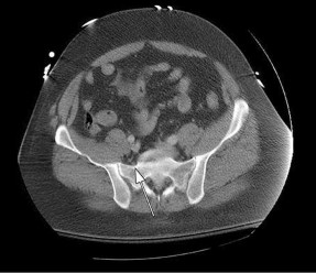
Which structure is most at risk when exposing the most lateral aspect of the medial window (identified by the arrows in Figure 30)? 29
Which structure is most at risk when exposing the most lateral aspect of the medial window (identified by the arrows in Figure 30)? 29
1
Corona mortis
2
Tibial division, sciatic nerve
3
Sciatic nerve, peroneal division
4
Fifth lumbar nerve root
5
Kocher-Langenbeck approach
- Corona mortis
QUESTION 30
of 100
Structures at risk from traction when visualizing the quadrilateral plate from the medial window of the Stoppa approach in this fracture (Figure 31)
Structures at risk from traction when visualizing the quadrilateral plate from the medial window of the Stoppa approach in this fracture (Figure 31)
1
Corona mortis
2
Tibial division, sciatic nerve
3
Sciatic nerve, peroneal division
4
Fifth lumbar nerve root
5
Kocher-Langenbeck approach
- Obturator vessels
QUESTION 31
of 100
Which surgical approach is most commonly used for this fracture (Figure 32)?
Which surgical approach is most commonly used for this fracture (Figure 32)?
1
Corona mortis
2
Tibial division, sciatic nerve
3
Sciatic nerve, peroneal division
4
Fifth lumbar nerve root
5
Kocher-Langenbeck approach
- Kocher-Langenbeck approach
QUESTION 32
of 100
Which structure is indicated by the arrow in Figure 33?
Which structure is indicated by the arrow in Figure 33?
1
Corona mortis
2
Tibial division, sciatic nerve
3
Sciatic nerve, peroneal division
4
Fifth lumbar nerve root
5
Kocher-Langenbeck approach
The posterior position of the sciatic nerve in relation to the acetabulum and the lateral peroneal division makes the peroneal division of the sciatic nerve the portion of the nerve that is most likely to be injured in a posterior traumatic hip dislocation, accounting for up to 10% of concomitant nerve injuries with posterior hip dislocation. The corona mortis is an anatomic variant that results in vascular anastomosis between the obturator and either the external iliac or inferior epigastric arteries. This variant occurs in approximately 80% of patients and varies in its position, being located 4 cm to 9 cm lateral to the symphysis pubis. The obturator vascular bundle is situated in the fat medial to the obturator internus muscle and must be mobilized to access the quadrilateral plate. Dissection may be carried out both above and below this vascular leash. The Kocher-Langenbeck approach is indicated for fractures involving the posterior wall and/or posterior column of the acetabulum and for both column fractures that require direct posterior visualization. This approach is not indicated for direct reduction of the anterior wall or column when direct visualization is required anteriorly. The L5 nerve root is located on the anterior sacrum and is indicated by the arrow.
The position of this neural structure must be considered whether the surgeon is stabilizing 31 the sacroiliac (SI) joint with percutaneous iliosacral screws or with anterior SI plating through the lateral window of the ilioinguinal approach.
RECOMMENDED READINGS
1. [Cornwall R, Radomisli TE. Nerve injury in traumatic dislocation of the hip. Clin Orthop Relat Res. 2000 Aug;(377):84-91. Review. PubMed PMID: 10943188. ](http://www.ncbi.nlm.nih.gov/pubmed/10943188)[View](http://www.ncbi.nlm.nih.gov/pubmed/10943188)[ ](http://www.ncbi.nlm.nih.gov/pubmed/10943188)[Abstract at PubMed](http://www.ncbi.nlm.nih.gov/pubmed/10943188)
2. [Darmanis S, Lewis A, Mansoor A, Bircher M. Corona mortis: an anatomical study with clinical implications in approaches to the pelvis and acetabulum. Clin Anat. 2007 May;20(4):433-9. PubMed PMID: 16944498. ](http://www.ncbi.nlm.nih.gov/pubmed/16944498)[View Abstract at PubMed](http://www.ncbi.nlm.nih.gov/pubmed/16944498)
3. [Archdeacon MT, Kazemi N, Guy P, Sagi HC. The modified Stoppa approach for acetabular fracture. J Am Acad Orthop Surg. 2011 Mar;19(3):170-5. PubMed PMID: 21368098. ](http://www.ncbi.nlm.nih.gov/pubmed/21368098)[View Abstract at PubMed](http://www.ncbi.nlm.nih.gov/pubmed/21368098)
4. Rommens P. The Kocher-Langenbeck approach for the treatment of acetabular fractures. Operat Orthop Traumatol 2004; 16:59-74.
5. [Langford JR, Burgess AR, Liporace FA, Haidukewych GJ. Pelvic fractures: part 2. Contemporary indications and techniques for definitive surgical management. J Am Acad Orthop Surg. 2013 Aug;21(8):458-68. doi: 10.5435/JAAOS-21-08-458. Review. PubMed PMID: 23908252.](http://www.ncbi.nlm.nih.gov/pubmed/23908252)[View Abstract at PubMed](http://www.ncbi.nlm.nih.gov/pubmed/23908252)
QUESTION 33
of 100
Which nerve root contributes to both the sciatic and femoral nerves?
Which nerve root contributes to both the sciatic and femoral nerves?
1
L2
2
L3
3
L4
4
L5
The lumbosacral plexus is formed from the lumbar and sacral roots that are redistributed into the obturator, femoral, and sciatic nerves. The obturator nerve is composed of the L1, L2, and L3 roots. The femoral nerve has contributions from the L3 and L4 roots. The sciatic nerve contains the L4, L5, S1, and lower sacral roots. Therefore, only the L4 root contributes to the femoral and sciatic (via the lumbosacral trunk) nerves, which allows it to innervate the quadriceps and the anterior tibialis muscles.
RECOMMENDED READINGS
32
1. Netter FH. The Ciba Collection of Medical Illustrations: The Musculoskeletal System, Part 1: Anatomy, Physiology and Metabolic Disorders. Summit, NJ: Ciba-Geigy; 1991:77-82.
2. [Samudrala S Department Of Neurosurgery University Of Southern California Medical School Los Angeles California And Department Of Neurosurgery University Of Florida Medical School Gainesville Florida, Khoo LT, Rhim SC, Fessler RG. Complications during anterior surgery of the lumbar spine: an anatomically based study and review. Neurosurg Focus. 1999 Dec 15;7(6):e9. PubMed PMID: 16918208. ](http://www.ncbi.nlm.nih.gov/pubmed/16918208)[View Abstract at PubMed](http://www.ncbi.nlm.nih.gov/pubmed/16918208)
QUESTION 34
of 100
The structure that runs just beneath the peroneal tubercle of the calcaneus is the
The structure that runs just beneath the peroneal tubercle of the calcaneus is the
1
flexor hallucis longus tendon.
2
peroneus brevis tendon.
3
peroneus longus tendon.
4
calcaneal fibular ligament.
The peroneal tubercle is often a good landmark at which to identify the peroneus longus tendon surgically, and a hypertrophic tubercle has been associated with peroneus longus tendinopathy. Both peroneal tendons curve anteriorly around the tip of the fibula, with the peroneal tubercle separating the 2 tendons at the level of the calcaneus. The peroneus brevis runs in front of the tubercle and the longus behind. The flexor hallucis longus runs through a fibro-osseus tunnel posterior to the hindfoot formed by the posterolateral (os trigonum) and posteromedial tubercle of the talus. The calcaneal fibular ligament attaches to the calcaneus below the posterior facet of the subtalar joint and deep to the peroneal tendons.
RECOMMENDED READINGS
1. [Hyer CF, Dawson JM, Philbin TM, Berlet GC, Lee TH. The peroneal tubercle: description, classification, and relevance to peroneus longus tendon pathology. Foot Ankle Int. 2005 Nov;26(11):947-50. Pub PMID: 16309609.](http://www.ncbi.nlm.nih.gov/pubmed/16309609)[View Abstract at PubMed](http://www.ncbi.nlm.nih.gov/pubmed/16309609)
2. Bruce WD, Christofersen MR, Phillips DL. Stenosing tenosynovitis and impingement
[of the peroneal tendons associated with hypertrophy of the peroneal tubercle. Foot Ankle Int. 1999 Jul;20(7):464-7. PubMed PMID: 10437932.](http://www.ncbi.nlm.nih.gov/pubmed/10437932)[View Abstract at PubMed](http://www.ncbi.nlm.nih.gov/pubmed/10437932)
33
3. Lee S, Lin J. Tendon disorders. In: Lieberman JR, ed. AAOS Comprehensive
Orthopaedic Review. Vol 2. Rosemont, IL: American Academy of Orthopaedic Surgeons; 2009:1193-1204.
4. Hollingshead WH. Anatomy for Surgeons: The Back and Limbs. Vol 3. 3rd ed.
Philadelphia, PA: Harper and Row; 1982:792-793.
QUESTION 35
of 100
The process at C5-6 shown in Figures 36a and 36b is from radiographs taken in 2006 and 2009, and can occur over time following an anterior cervical discectomy and fusion. At what rate per year is this thought to occur?
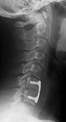
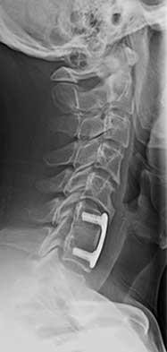

The process at C5-6 shown in Figures 36a and 36b is from radiographs taken in 2006 and 2009, and can occur over time following an anterior cervical discectomy and fusion. At what rate per year is this thought to occur?
1
Less than 1%
2
3%
3
7%
4
15%


A B
34
The process shown in the figures is that of degenerative change adjacent to an anterior cervical discectomy and fusion. The observed rate of degenerative adjacent changes is estimated at 2% to 3% per year following a single-level fusion. These changes are partly related to the natural aging process or degenerative process and can occur regardless of an adjacent fusion, but the influence of a solid adjacent fusion with the increased stress at the next level is thought to be a contributor.
RECOMMENDED READINGS
1. [Rihn JA, Lawrence J, Gates C, Harris E, Hilibrand AS. Adjacent segment disease after cervical spine fusion. Instr Course Lect. 2009;58:747-56. PubMed PMID: 19385583. ](http://www.ncbi.nlm.nih.gov/pubmed/19385583)[View Abstract at PubMed](http://www.ncbi.nlm.nih.gov/pubmed/19385583)
2. Hilibrand AS, Carlson GD, Palumbo MA, Jones PK, Bohlman HH. Radiculopathy and myelopathy at segments adjacent to the site of a previous anterior cervical arthrodesis. J Bone Joint Surg Am. 1999 Apr;81(4):519-28. PubMed PMID: 10225797.
[View Abstract at PubMed](http://www.ncbi.nlm.nih.gov/pubmed/10225797)
CLINICAL SITUATION FOR QUESTIONS 37 THROUGH 39
A 22-year-old woman has had right hip pain for 12 months. Her symptoms have not improved with nonsurgical treatment involving physical therapy and intra-articular injections.
A
B
35
C D E
QUESTION 36
of 100
The plain radiographs and MR image shown in Figures 37a through 37c indicate which condition?
The plain radiographs and MR image shown in Figures 37a through 37c indicate which condition?
1
Pincer-type femoroacetabular impingement with disruption of the ligamentum teres.
2
Pincer-type femoroacetabular impingement with an acetabular labral tear.
3
Cam-type femoroacetabular impingement with disruption of the ligamentum teres.
4
Cam-type femoroacetabular impingement with an acetabular labral tear.
- Cam-type femoroacetabular impingement with an acetabular labral tear
QUESTION 37
of 100
Which condition would you expect to identify during a hip arthroscopy procedure for this patient based on the radiographic findings in Figures 37a through 37c?
Which condition would you expect to identify during a hip arthroscopy procedure for this patient based on the radiographic findings in Figures 37a through 37c?
1
Articular cartilage delamination.
2
Ligamentum teres rupture
3
Osteochondral loose body
4
Paralabral cyst
- Articular cartilage delamination
QUESTION 38
of 100
The patient undergoes hip arthroscopy and the image of the right hip is shown in Figure 39. Repair of the injured structure would be expected to improve
The patient undergoes hip arthroscopy and the image of the right hip is shown in Figure 39. Repair of the injured structure would be expected to improve
1
hip joint survival.
2
hip joint lubrication.
3
hip joint motion. 36
4
hip joint stability.
The radiographic studies reveal both acetabular dysplasia and cam-type femoroacetabular impingement. The MR image shows an acetabular labral tear. Structural abnormalities of the hip, including femoroacetabular impingement, have commonly been identified in association with labral tears. Disruption of the ligamentum teres is not associated with impingement conditions in the absence of trauma.
The patient has acetabular dysplasia with a decreased lateral center-edge angle and also has visible cam-type femoroacetabular impingement. The common pathway for joint degeneration in hips with cam-type femoral head anatomy includes the development of cartilage damage in the anterior or superolateral aspects of the acetabular cartilage. Paralabral cysts may be seen more commonly in association with acetabular dysplasia, although the patient’s radiographs did not demonstrate substantial cystic changes. Osteochondral loose bodies and ligamentum teres ruptures can be seen at arthroscopy in a small number of cases.
There are several proposed roles of the acetabular labrum. It can increase the depth of the acetabular socket by as much as 21% to 28%. Roles of the acetabular labrum include joint lubrication, shock absorption, and pressure distribution. Recent studies assessing the effects of loading on joint stability for both normal and dysplastic hips did not demonstrate a substantial role of the labrum in differences in loading. Although joint stability might be improved following surgical repair, acetabular dysplasia is not likely to be resolved with acetabular labral repair alone.
RECOMMENDED READINGS
1. [Tibor LM, Leunig M. The pathoanatomy and arthroscopic management of femoroacetabular impingement. Bone Joint Res. 2012 Oct 1;1(10):245-57. doi: 10.1302/2046-3758.110.2000105.PubMed: 23610655. ](http://www.ncbi.nlm.nih.gov/pubmed/23610655)[View Abstract at PubMed](http://www.ncbi.nlm.nih.gov/pubmed/23610655)
2. [Peelle MW, Della Rocca GJ, Maloney WJ, Curry MC, Clohisy JC. Acetabular and femoral radiographic abnormalities associated with labral tears. Clin Orthop Relat Res. 2005 Dec;441:327-33. PubMed PMID: 16331022. ](http://www.ncbi.nlm.nih.gov/pubmed/16331022)[View Abstract at PubMed](http://www.ncbi.nlm.nih.gov/pubmed/16331022)
3. Ross JR, Zaltz I, Nepple JJ, Schoenecker PL, Clohisy JC. Arthroscopic disease classification and interventions as an adjunct in the treatment of acetabular dysplasia. Am J Sports Med. 2011 Jul;39 Suppl:72S-8S. doi: 10.1177/0363546511412320.
[PubMed PMID: 21709035. ](http://www.ncbi.nlm.nih.gov/pubmed/21709035)[View Abstract at PubMed](http://www.ncbi.nlm.nih.gov/pubmed/21709035)
4. James SL, Ali K, Malara F, Young D, O'Donnell J, Connell DA. MRI findings of 37
[femoroacetabular impingement. AJR Am J Roentgenol. 2006 Dec;187(6):1412-9. PubMed PMID: 17114529. ](http://www.ncbi.nlm.nih.gov/pubmed/17114529)[View Abstract at PubMed](http://www.ncbi.nlm.nih.gov/pubmed/17114529)
5. [Groh MM, Herrera J. A comprehensive review of hip labral tears. Curr Rev Musculoskelet Med. 2009 Jun;2(2):105-17. doi: 10.1007/s12178-009-9052-9. Epub 2009 Apr 7. PubMed PMID: 19468871. ](http://www.ncbi.nlm.nih.gov/pubmed/19468871)[View Abstract at PubMed](http://www.ncbi.nlm.nih.gov/pubmed/19468871)
6. [Henak CR, Ellis BJ, Harris MD, Anderson AE, Peters CL, Weiss JA. Role of the acetabular labrum in load support across the hip joint. J Biomech. 2011 Aug 11;44(12):2201-6. doi: 10.1016/j.jbiomech.2011.06.011. Epub 2011 Jul 14. PubMed PMID: 21757198. ](http://www.ncbi.nlm.nih.gov/pubmed/21757198)[View Abstract at PubMed](http://www.ncbi.nlm.nih.gov/pubmed/21757198)
QUESTION 39
of 100
What condition favors pollicization in hypoplasia of the thumb?
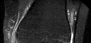
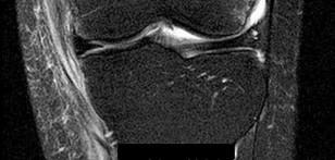
What condition favors pollicization in hypoplasia of the thumb?
1
Extrinsic tendon abnormalities involving the flexor and extensor
2
Narrowing of the first web space
3
Insufficiency of the ulnar collateral ligament of the metaphalangeal joint
4
Unstable carpometacarpal joint
The decision to ablate and pollicize vs preserve and reconstruct is based on the stability of the carpometacarpal joint. A stable thumb is more easily used in prehension activities of grasping and pinching. All other responses are associated with surgical options for reconstruction of the thumb. The carpometacarpal joint is the “keystone” for thumb-to-hand attachment. Without a stable carpometacarpal joint, pollicization may be required. No other response necessitates pollicization.
RECOMMENDED READINGS
1. [McCarroll HR. Congenital anomalies: a 25-year overview. J Hand Surg Am. 2000 Nov;25(6):1007-37. Review. PubMed PMID: 11119659. ](http://www.ncbi.nlm.nih.gov/pubmed/11119659)[View Abstract at PubMed](http://www.ncbi.nlm.nih.gov/pubmed/11119659)
2. [Manske PR, Goldfarb CA. Congenital failure of formation of the upper limb. Hand Clin. 2009 May;25(2):157-70. doi: 10.1016/j.hcl.2008.10.005. Review. PubMed PMID: 19380058.](http://www.ncbi.nlm.nih.gov/pubmed/19380058)[View Abstract at PubMed](http://www.ncbi.nlm.nih.gov/pubmed/19380058)
38
RESPONSES FOR QUESTIONS 41 THROUGH 44
Match the patient and image with the treatment listed.
QUESTION 40
of 100
A 22-year-old man who plays recreational soccer (Figure 41)
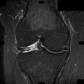
A 22-year-old man who plays recreational soccer (Figure 41)
1
Ligamentous reconstruction
2
Meniscal repair
3
Meniscectomy
4
Immobilization
- Immobilization
QUESTION 41
of 100
A 34-year-old woman who is a professional skier (Figure 42)
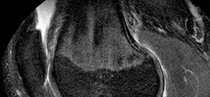
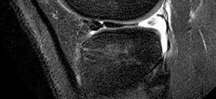
A 34-year-old woman who is a professional skier (Figure 42)
1
Ligamentous reconstruction
2
Meniscal repair
3
Meniscectomy
4
Immobilization
- Meniscectomy
QUESTION 42
of 100
A 16-year-old boy who is a competitive basketball player (Figure 43)
A 16-year-old boy who is a competitive basketball player (Figure 43)
1
Ligamentous reconstruction
2
Meniscal repair
3
Meniscectomy 39
4
Immobilization
- Meniscal repair
QUESTION 43
of 100
An 18-year-old woman who plays high school volleyball (Figure 44)
An 18-year-old woman who plays high school volleyball (Figure 44)
1
Ligamentous reconstruction
2
Meniscal repair
3
Meniscectomy
4
Immobilization
Figure 41 is a coronal MR image demonstrating a medial collateral ligament (MCL) tear. MCL injuries usually are treated with immobilization for 4 to 6 weeks. Surgical treatment is recommended only in cases of persistent valgus laxity or rotatory instability after bracing. Subacute or chronic medial instability associated with cruciate injury is also an indication for surgery.
Figures 42 and 43 are MR images demonstrating lateral meniscal tears. In Figure 42, the coronal view shows a tear through the white-white zone in a discoid lateral meniscus. Because of the location of the tear, this patient is treated with arthroscopic partial meniscectomy. In Figure 43, the lateral view reveals a bucket handle tear of the lateral meniscus that is treated with repair. Figure 44 is an axial MR image demonstrating characteristic bony edema over the medial patella and lateral femoral condyle after an acute lateral patellar dislocation. Immobilization is also recommended for first-time acute patellar dislocations. Surgical treatment for a first-time patellar dislocation is recommended only when a displaced osteochondral fragment is present.
RECOMMENDED READINGS
1. Asik M, Sen C, Taser OF, Alturfan AK, Sozen YV. Discoid lateral meniscus:
41
diagnosis and results of arthroscopic treatment. Knee Surg Sports Traumatol Arthrosc.
[2003 Mar;11(2):99-104. Epub 2003 Jan 25. PubMed PMID: 12664202. ](http://www.ncbi.nlm.nih.gov/pubmed/12664202)[View Abstract](http://www.ncbi.nlm.nih.gov/pubmed/12664202)[ ](http://www.ncbi.nlm.nih.gov/pubmed/12664202)[at PubMed](http://www.ncbi.nlm.nih.gov/pubmed/12664202)
2. [Jacobson KE, Chi FS. Evaluation and treatment of medial collateral ligament and medial-sided injuries of the knee. Sports Med Arthrosc. 2006 Jun;14(2):58-66. Review. PubMed PMID: 17135948. ](http://www.ncbi.nlm.nih.gov/pubmed/17135948)[View Abstract at PubMed](http://www.ncbi.nlm.nih.gov/pubmed/17135948)
3. Laible C, Stein DA, Kiridly DN. Meniscal repair. J Am Acad Orthop Surg. 2013 Apr;21(4):204-13. doi: 10.5435/JAAOS-21-04-204. Review. PubMed PMID:
[23545726.](http://www.ncbi.nlm.nih.gov/pubmed/23545726)[View Abstract at PubMed](http://www.ncbi.nlm.nih.gov/pubmed/23545726)
4. [Palmu S, Kallio PE, Donell ST, Helenius I, Nietosvaara Y. Acute patellar dislocation in children and adolescents: a randomized clinical trial. J Bone Joint Surg Am. 2008 Mar;90(3):463-70. doi: 10.2106/JBJS.G.00072. PubMed PMID: 18310694. ](http://www.ncbi.nlm.nih.gov/pubmed/18310694)[View](http://www.ncbi.nlm.nih.gov/pubmed/18310694)
[Abstract at PubMed](http://www.ncbi.nlm.nih.gov/pubmed/18310694)
QUESTION 44
of 100
The subcutaneous nerve most at risk for transection during an anterior surgical exposure of the ankle is the
The subcutaneous nerve most at risk for transection during an anterior surgical exposure of the ankle is the
1
dorsal cutaneous branch of the sural nerve.
2
deep peroneal nerve.
3
medial plantar nerve.
4
medial branch of the superficial peroneal nerve.
The dorsal medial cutaneous nerve arising from the superficial peroneal nerve crosses the inferior extent of a routine extensile surgical exposure to the ankle joint. The extensile anterior incision develops the interval between the tibialis anterior and extensor hallucis tendons, and, although the deep peroneal nerve is lateral and posterior to the extensor hallucis longus, the nerve most at risk during this exposure is the cutaneous branch supplying the dorsal medial foot to the great toe. Ankle replacement surgery is becoming more common; consequently, surgeons must be familiar with this anatomic landmark and risks related to its transection during surgery.
The medial plantar nerve is at risk when medial hindfoot incisions are made through the
abductor hallucis muscle during tarsal tunnel release and harvest of the flexor hallucis tendon for transfer. The dorsal cutaneous branch of the sural nerve supplies the lateral dorsal
41
midfoot and is at risk when incisions are made for distal peroneal tendon exposure and
internal fixation of fifth metatarsal Jones fractures. RECOMMENDED READINGS
1. Santi MD, Botte MJ. Nerve injury and repair in the foot and ankle. Foot Ankle Int.
[1996 Jul;17(7):425-39. Review. PubMed PMID: 8832252.](http://www.ncbi.nlm.nih.gov/pubmed/8832252)[View Abstract at PubMed](http://www.ncbi.nlm.nih.gov/pubmed/8832252) 2- Sammarco VJ, Taylor R. Anatomy and biomechanics of the foot and ankle. In:
Lieberman JR, ed. AAOS Comprehensive Orthopaedic Review. Vol 2. Rosemont, IL: American Academy of Orthopaedic Surgeons; 2009:1147-1155.
[3- Adkison DP, Bosse MJ, Gaccione DR, Gabriel KR. Anatomical variations in the course of the superficial peroneal nerve. J Bone Joint Surg Am. 1991 Jan;73(1):112-4. PubMed PMID: 1985980.](http://www.ncbi.nlm.nih.gov/pubmed/1985980)[View Abstract at PubMed](http://www.ncbi.nlm.nih.gov/pubmed/1985980)
QUESTION 45
of 100
The cystic lesion shown on the MR images in Figures 46a through 46c should cause denervation changes in which muscle?
A B
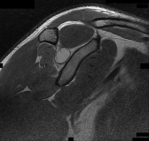
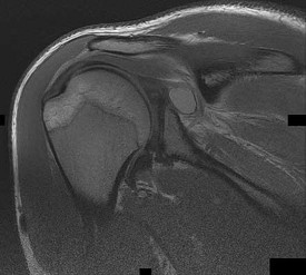
The cystic lesion shown on the MR images in Figures 46a through 46c should cause denervation changes in which muscle?
A B
1
Subscapularis
2
Teres major
3
Infraspinatus
4
Supraspinatus
42
The cystic lesion is shown in an area adjacent to the spinoglenoid notch. A space-occupying lesion in this area has the potential to compress the suprascapular nerve as it passes through the spinoglenoid notch to innervate the infraspinatus. Compression of the nerve can cause denervation and atrophy of the infraspinatus muscle. The supraspinatus is innervated by the suprascapular nerve proximal to the compressive lesions and will not undergo atrophy. The subscapularis and the teres major are innervated by the subscapular nerve.
RECOMMENDED READINGS
1. Getz CL, Ramsey ML, Williams GR. Paralabral cysts of the shoulder. In: Galatz LM, ed. Orthopaedic Knowledge Update: Shoulder and Elbow 3. Rosemont, IL: American Academy of Orthopaedic Surgeons; 2008:339-344.
2. [Takagishi K, Saitoh A, Tonegawa M, Ikeda T, Itoman M. Isolated paralysis of the infraspinatus muscle. J Bone Joint Surg Br. 1994 Jul;76(4):584-7. PubMed PMID: 8027145.](http://www.ncbi.nlm.nih.gov/pubmed/8027145)[View Abstract at PubMed](http://www.ncbi.nlm.nih.gov/pubmed/8027145)
QUESTION 46
of 100
The middle glenohumeral ligament (MGHL) most commonly arises from the
The middle glenohumeral ligament (MGHL) most commonly arises from the
1
coracoacromial ligament at its insertion to the coracoid.
2
inferior aspect of the glenoid as a continuation of the anterior inferior glenohumeral ligament.
3
posterior aspect of the glenoid as a continuation of the posterior superior labrum.
4
labrum and glenoid immediately below the superior glenohumeral ligament.
When present, the MGHL (identified in approximately two-thirds of shoulders) has been shown to arise from the labrum and glenoid immediately below the superior glenohumeral ligament. The MGHL does not arise from the coracohumeral ligament, is not a continuation of the anteroinferior glenohumeral ligament, is located anteriorly, and does not arise from the tip of the coracoid.
RECOMMENDED READINGS 43
1. Ide J, Maeda S, Takagi K. Normal variations of the glenohumeral ligament complex: an anatomic study for arthroscopic Bankart repair. Arthroscopy. 2004 Feb;20(2):164-
[8/. PubMed PMID: 14760349.](http://www.ncbi.nlm.nih.gov/pubmed/14760349)[View Abstract at PubMed](http://www.ncbi.nlm.nih.gov/pubmed/14760349)
2. [O'Brien SJ, Neves MC, Arnoczky SP, Rozbruck SR, Dicarlo EF, Warren RF, Schwartz R, Wickiewicz TL. The anatomy and histology of the inferior glenohumeral ligament complex of the shoulder. Am J Sports Med. 1990 Sep-Oct;18(5):449-56. PubMed PMID: 2252083.](http://www.ncbi.nlm.nih.gov/pubmed/2252083)[View Abstract at PubMed](http://www.ncbi.nlm.nih.gov/pubmed/2252083)
QUESTION 47
of 100
Figures 48a and 48b are the axial and sagittal T1-weighted MR images of the L4-5 disc level of a 38-year-old man. He is symptomatic from the pathology shown. A surgeon would expect the neurological findings to include
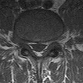
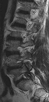
Figures 48a and 48b are the axial and sagittal T1-weighted MR images of the L4-5 disc level of a 38-year-old man. He is symptomatic from the pathology shown. A surgeon would expect the neurological findings to include
1
Right extensor hallucis longus (EHL) weakness, anterior shin numbness, and diminished patella reflex
2
Right quadriceps weakness, anterior shin pain, and diminished patella reflex
3
Right dorsal foot first web numbness, EHL weakness, and normal reflexes
4
Left quadriceps weakness, anterior shin pain, and diminished patella reflex
The pathology shown in the MR images is a right-sided far lateral herniated nucleus pulposus at L4-5, which, if symptomatic, would cause a radiculopathy of the exiting root, L4. A more common posterolateral herniation at L4-5 would cause irritation of the traversing root, L5. The L4 root has a sensory distribution over the anterior thigh that extends along the anterior shin but does not tend to reach the toes. The motor distribution is to the quadriceps and anterior tibialis muscles, with the predominant reflex effect being the patella reflex. The L5 root has a sensory distribution to the first dorsal web space of the foot, motor distribution to the EHL, and no specific reflex.
RECOMMENDED READINGS
1. Standaert CJ, Herring SA, Sinclair JD. The patient history and physical examination: Cervical, thoracic, and lumbar. In: Herkowitz HN, Garfin SR, Eismont FJ, Bell GR, Balderston RA, eds. Rothman-Simeone The Spine. Vol 1. 5th ed. Philadelphia, PA: Saunders Elsevier; 2006:171-186.
2. Bono CM, Wisneski R, Garfin SR: Lumbar disc herniations. In: Herkowitz HN, Garfin SR, Eismont FJ, Bell GR, Balderston RA, eds. Rothman-Simeone The Spine. Vol 1. 5th ed. Philadelphia, PA: Saunders Elsevier; 2006:967-991.
QUESTION 48
of 100
Figure 49 is the chest radiograph of a 47-year-old man who underwent right shoulder arthroscopy under general anesthesia and regional blockade (interscalene). The finding in the radiographic image likely is attributable to which mechanism?
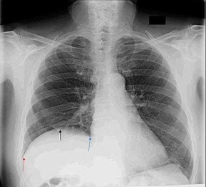
Figure 49 is the chest radiograph of a 47-year-old man who underwent right shoulder arthroscopy under general anesthesia and regional blockade (interscalene). The finding in the radiographic image likely is attributable to which mechanism?
1
Positive end-pressure ventilation
2
Iatrogenic nerve injury during the surgical approach
3
Nerve palsy related to regional anesthetic block
4
Traction injury to the brachial plexus at the time of surgery
45
Positive end-pressure ventilation can cause alveolar rupture and pneumothorax, not elevation of the hemidiaphragm on the surgical side. The phrenic nerve, which controls the right hemidiaphragm, is not encountered during routine shoulder surgery because it courses medial to the scalene musculature near the midline of the neck. A traction injury is unlikely to cause injury to the phrenic nerve because it is part of the cervical plexus, which is not normally subject to traction during shoulder surgery. Hemidiaphragm paralysis via the phrenic nerve has been reported to occur as often as 100% of the time with use of interscalene regional anesthesia.
RECOMMENDED READINGS
1. [Urmey WF, Talts KH, Sharrock NE. One hundred percent incidence of hemidiaphragmatic paresis associated with interscalene brachial plexus anesthesia as diagnosed by ultrasonography. Anesth Analg. 1991 Apr;72(4):498-503. PubMed PMID: 2006740.](http://www.ncbi.nlm.nih.gov/pubmed/2006740)[View Abstract at PubMed](http://www.ncbi.nlm.nih.gov/pubmed/2006740)
2. [Lenters TR, Davies J, Matsen FA 3rd. The types and severity of complications associated with interscalene brachial plexus block anesthesia: local and national evidence. J Shoulder Elbow Surg. 2007 Jul-Aug;16(4):379-87. Epub 2007 Apr 19. PubMed PMID: 17448698.](http://www.ncbi.nlm.nih.gov/pubmed/17448698)[View Abstract at PubMed](http://www.ncbi.nlm.nih.gov/pubmed/17448698)
QUESTION 49
of 100
Figure 50 is the radiograph of a 68-year-old man who has anterior knee pain 6 months after total knee arthroplasty. His knee is flexed 45 degrees. Anteroposterior and lateral radiographs show satisfactory size and alignment of the components. This patient has knee range of motion from 0 to 75 degrees. What is the most appropriate next step in his evaluation?
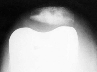
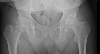
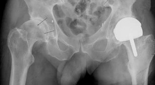
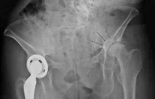
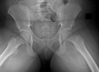
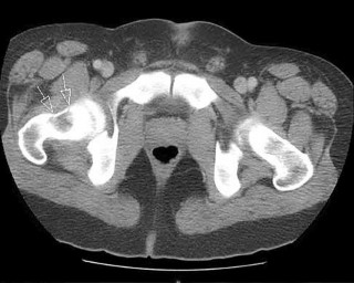
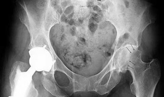
Figure 50 is the radiograph of a 68-year-old man who has anterior knee pain 6 months after total knee arthroplasty. His knee is flexed 45 degrees. Anteroposterior and lateral radiographs show satisfactory size and alignment of the components. This patient has knee range of motion from 0 to 75 degrees. What is the most appropriate next step in his evaluation?
1
MR images with metal artifact reduction
2
Dynamic fluoroscopy
3
CT images of the femoral and tibial components
4
30-degree, 60-degree, and 90-degree patellar radiographs
46
The patient’s radiograph demonstrates patellar instability. He has both pain and suboptimal range of motion following knee arthroplasty. Several considerations may contribute to loss of range of motion, but rotational malalignment of the components is a primary consideration for patients with otherwise well-aligned, well-fixed knee arthroplasties complicated by patellar instability. CT images have demonstrated value in evaluation of femoral and tibial component rotational malalignment. Radiographs in variable degrees of flexion are not necessary because the patient has documented patellar instability on his 45-degree flexion radiograph. Dynamic fluoroscopy may demonstrate ligamentous instability, but does not readily demonstrate patellofemoral tracking. Even with use of a metal-suppression technique, MR image artifact limits its usefulness in evaluating this clinical concern.
RECOMMENDED READINGS
1. Peters CL, Crofoot CD. Knee reconstruction and replacement. In: Fischgrund JS, ed. Orthopaedic Knowledge Update 9. Rosemont, IL: American Academy of Orthopaedic Surgeons; 2008:457-471.
2. [Berger RA, Crossett LS, Jacobs JJ, Rubash HE. Malrotation causing patellofemoral complications after total knee arthroplasty. Clin Orthop Relat Res. 1998 Nov;(356):144-53. PubMed PMID: 9917679.](http://www.ncbi.nlm.nih.gov/pubmed/9917679)[View Abstract at PubMed](http://www.ncbi.nlm.nih.gov/pubmed/9917679)
3. [Jazrawi LM, Birdzell L, Kummer FJ, Di Cesare PE. The accuracy of computed tomography for determining femoral and tibial total knee arthroplasty component rotation. J Arthroplasty. 2000 Sep;15(6):761-6. PubMed PMID: 11021452.](http://www.ncbi.nlm.nih.gov/pubmed/11021452)[View](http://www.ncbi.nlm.nih.gov/pubmed/11021452)[ ](http://www.ncbi.nlm.nih.gov/pubmed/11021452)[Abstract at PubMed](http://www.ncbi.nlm.nih.gov/pubmed/11021452)
4. [Kelly MA. Extensor mechanism complications in total knee arthroplasty. Instr Course Lect. 2004;53:193-9. Review. PubMed PMID: 15116613.](http://www.ncbi.nlm.nih.gov/pubmed/15116613)[View Abstract at PubMed](http://www.ncbi.nlm.nih.gov/pubmed/15116613)
RESPONSES FOR QUESTION 51 THROUGH 55
Match the image above with the description below.
47
A B
C
D
48
E F
QUESTION 50
of 100
Radiograph of a man with knee pain, thyroid abnormalities, and marked decreased hip internal rotation with flexion at 90 degrees
Radiograph of a man with knee pain, thyroid abnormalities, and marked decreased hip internal rotation with flexion at 90 degrees
1
Figure 51a
2
Figure 51b
3
Figure 51c
4
Figure 51d
5
Figure 51e
- Figure 51e
QUESTION 51
of 100
Cross-over sign
Cross-over sign
1
Figure 51a
2
Figure 51b
3
Figure 51c
4
Figure 51d
5
Figure 51e
- Figure 51c
QUESTION 52
of 100
Posterior wall
Posterior wall
1
Figure 51a
49
2
Figure 51b
3
Figure 51c
4
Figure 51d
5
Figure 51e
- Figure 51b
QUESTION 53
of 100
History of high-dose steroid treatment after renal transplantation
History of high-dose steroid treatment after renal transplantation
1
Figure 51a
2
Figure 51b
3
Figure 51c
4
Figure 51d
5
Figure 51e
- Figure 51a
QUESTION 54
of 100
Before proceeding with total hip replacement, consideration should be given to imaging the cervical spine with flexion/extension films
Before proceeding with total hip replacement, consideration should be given to imaging the cervical spine with flexion/extension films
1
Figure 51a
2
Figure 51b
3
Figure 51c
4
Figure 51d
5
Figure 51e
A slipped capital femoral epiphysis is most common in adolescent boys who are overweight. Examination must include evaluation of the hip, thigh, and knee and usually reveals limited internal rotation of the hip. At times, the condition is associated with concomitant renal or endocrine abnormalities or a history of radiation therapy to the region. The cross-over sign is 51 a radiographic finding indicative of acetabular retroversion and is seen in pincer-type femoroacetabular impingement. The posterior wall sign is seen in pincer-type femoroacetabular impingement when the center of the femoral head is lateral to the lateral border of the posterior wall of the acetabulum. It is indicative of acetabulum posterior wall deficiency. Patients who undergo solid organ transplantation frequently are treated with steroid immunosuppression. This treatment is a well-known risk factor for osteonecrosis of
the femoral head. Protrusio acetabula are frequently seen in patients with systemic inflammatory arthropathy. In patients with rheumatoid arthritis involving the hip, the status of the cervical spine must be considered before proceeding with surgery.
RECOMMENDED READINGS
1. [Aronsson DD, Loder RT, Breur GJ, Weinstein SL. Slipped capital femoral epiphysis: current concepts. J Am Acad Orthop Surg. 2006 Nov;14(12):666-79. Review. PubMed PMID: 17077339.](http://www.ncbi.nlm.nih.gov/pubmed/17077339)[View Abstract at PubMed](http://www.ncbi.nlm.nih.gov/pubmed/17077339)
2. Nepple JJ, Prather H, Trousdale RT, Clohisy JC, Beaulé PE, Glyn-Jones S, Rakhra K, Kim YJ. Diagnostic imaging of femoroacetabular impingement. J Am Acad Orthop Surg. 2013;21 Suppl 1:S20-6. doi: 10.5435/JAAOS-21-07-S20. PubMed PMID:
[23818187.](http://www.ncbi.nlm.nih.gov/pubmed/23818187)[View Abstract at PubMed](http://www.ncbi.nlm.nih.gov/pubmed/23818187)
3. [Lavernia CJ, Sierra RJ, Grieco FR. Osteonecrosis of the femoral head. J Am Acad Orthop Surg. 1999 Jul-Aug;7(4):250-61. Review. PubMed PMID: 10434079. ](http://www.ncbi.nlm.nih.gov/pubmed/10434079)[View](http://www.ncbi.nlm.nih.gov/pubmed/10434079)[ ](http://www.ncbi.nlm.nih.gov/pubmed/10434079)[Abstract at PubMed](http://www.ncbi.nlm.nih.gov/pubmed/10434079)
4. McBride MT, Muldoon MP, Santore RF, Trousdale RT, Wenger DR. Protrusio acetabuli: diagnosis and treatment. J Am Acad Orthop Surg. 2001 Mar-Apr;9(2):79-
[88/. Review. PubMed PMID: 11281632.](http://www.ncbi.nlm.nih.gov/pubmed/11281632)[View Abstract at PubMed](http://www.ncbi.nlm.nih.gov/pubmed/11281632)
QUESTION 55
of 100
A 73-year-old man presents to your clinic many years after undergoing total shoulder arthroplasty with pain and the radiographic findings demonstrated in Figure 56. The most likely cause of this patient’s pain is
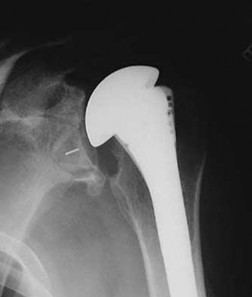
A 73-year-old man presents to your clinic many years after undergoing total shoulder arthroplasty with pain and the radiographic findings demonstrated in Figure 56. The most likely cause of this patient’s pain is
1
stress shielding.
2
humeral osteolysis.
3
glenoid loosening. 51
4
progressive glenoid arthritis.
The radiograph shows proximal humeral migration and loosening of the glenoid component. Proximal migration of the humeral head may represent rotator cuff dysfunction and can lead to progressive failure of the glenoid component. Stress shielding will not cause lysis or loosening of the glenoid component. Humeral osteolysis is an uncommon finding and is not shown. Progressive glenoid arthrosis is not possible with a resurfaced glenoid.
RECOMMENDED READINGS
1. [Hill JM, Norris TR. Long-term results of total shoulder arthroplasty following bone-grafting of the glenoid. J Bone Joint Surg Am. 2001 Jun;83-A(6):877-83. PubMed PMID: 11407796.](http://www.ncbi.nlm.nih.gov/pubmed/11407796)[View Abstract at PubMed](http://www.ncbi.nlm.nih.gov/pubmed/11407796)
2. Fox TJ, Cil A, Sperling JW, Sanchez-Sotelo J, Schleck CD, Cofield RH. Survival of the glenoid component in shoulder arthroplasty. J Shoulder Elbow Surg. 2009 Nov-Dec;18(6):859-63. doi: 10.1016/j.jse.2008.11.020. Epub 2009 Mar 17. PubMed
[PMID: 19297199.](http://www.ncbi.nlm.nih.gov/pubmed/19297199)[View Abstract at PubMed](http://www.ncbi.nlm.nih.gov/pubmed/19297199)
QUESTION 56
of 100
Which nerve is most commonly injured after total knee arthroplasty?
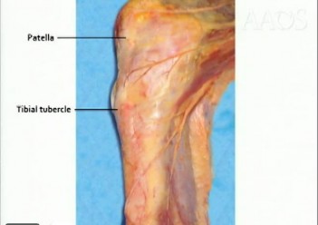
Which nerve is most commonly injured after total knee arthroplasty?
1
Tibial nerve
2
Superficial peroneal nerve
3
Infrapatellar branch of the saphenous
nerve 52
4
Sartorial branch of the saphenous nerve
The tibial or peroneal nerves usually are not injured during total knee arthroplasty. Incidence of peroneal nerve damage is highest in knees with a valgus deformity and an associated flexion contracture attributable to nerve stretch. This nerve injury occurs in as many as 9% of patients undergoing knee arthroplasty. Tibial nerve injury is a rare occurrence and usually an iatrogenic transection injury. The infrapatellar branch of the saphenous nerve and its nerve plexus is commonly injured after the medial parapatellar approach, and altered sensation attributable to injury is reported in up to 70% of cases. Injury typically manifests as numbness inferior to the patella. The sartorial branch of the saphenous nerve provides sensation distal to the knee and is uncommonly injured with a medial parapatellar approach. These concepts are illustrated in video 57, “Selective Exposures in Orthopaedic Surgery: The Knee, 2nd Edition.”
RECOMMENDED READINGS
1. Clarke HD, Bush-Joseph CA, Wolf BR. Selective Exposures in Orthopaedic Surgery: The Knee, 2nd Edition [DVD]. Rosemont, IL: American Academy of Orthopaedic Surgeons; 2012.
2. [Hunter LY, Louis DS, Ricciardi JR, O'Connor GA. The saphenous nerve: its course and importance in medial arthrotomy. Am J Sports Med. 1979 Jul-Aug;7(4):227-30. PubMed PMID: 474860.](http://www.ncbi.nlm.nih.gov/pubmed/474860)[View Abstract at PubMed](http://www.ncbi.nlm.nih.gov/pubmed/474860)
3. [Mistry D, O'Meeghan C. Fate of the infrapatellar branch of the saphenous nerve post total knee arthroplasty. ANZ J Surg. 2005 Sep;75(9):822-4. PubMed PMID: 16174002.](http://www.ncbi.nlm.nih.gov/pubmed/16174002)[View Abstract at PubMed](http://www.ncbi.nlm.nih.gov/pubmed/16174002)
4. [Schinsky MF, Macaulay W, Parks ML, Kiernan H, Nercessian OA. Nerve injury after primary total knee arthroplasty. J Arthroplasty. 2001 Dec;16(8):1048-54. PubMed PMID: 11740762.](http://www.ncbi.nlm.nih.gov/pubmed/11740762)[View Abstract at PubMed](http://www.ncbi.nlm.nih.gov/pubmed/11740762)
QUESTION 57
of 100
During an anterior approach to the bicipital 53
tuberosity, you encounter a nerve overlying the brachioradialis fascia (Figure 58). It provides innervation to the
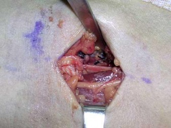
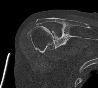
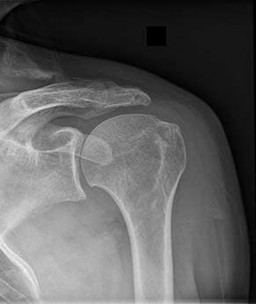
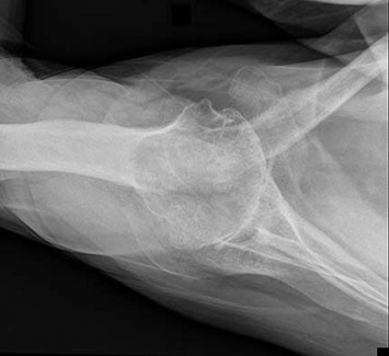
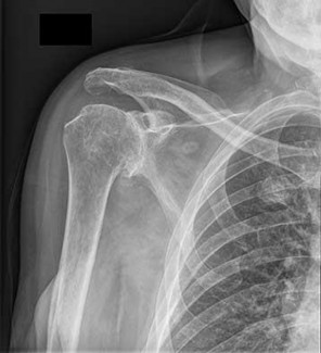
During an anterior approach to the bicipital 53
tuberosity, you encounter a nerve overlying the brachioradialis fascia (Figure 58). It provides innervation to the
1
flexor pollicis longus.
2
skin on the anteromedial forearm.
3
skin on the anterolateral forearm.
4
extensor indicis proprius.
The structure shown is the lateral antebrachial cutaneous nerve (LABC). It is the terminal sensory branch of the musculocutaneous nerve and runs superficial to the brachioradialis. It supplies sensation to the anterolateral surface of the forearm. The flexor pollicis longus is innervated by the anterior interosseous nerve. The extensor indicis proprius is innervated by the radial nerve. The LABC does not innervate the skin of the anteromedial forearm. Careful
identification and protection of this nerve is critical to prevent the most common nerve injury during distal biceps repair.
RECOMMENDED READINGS
1. Agur AM. Grant's Atlas of Human Anatomy. 10th ed. Philadelphia, PA: Lippincott Williams & Wilkins; 1999:460.
2. Hoppenfeld S, deBoer P. Surgical Exposures in Orthopedics. 2nd ed. Philadelphia, PA: Lippincott Williams & Wilkins; 1994:118-125.
RESPONSES FOR QUESTIONS 59 THROUGH 61
Please select the image that represents the most appropriate response to the question or statement below.
54
A B
C D
QUESTION 58
of 100
What degenerative process most often is associated with an intact rotator cuff?
What degenerative process most often is associated with an intact rotator cuff?
1
Figure 59a is the CT image of an 86-year-old woman with acromiohumeral distance of less than 2 mm, night pain, and an inability to actively raise the affected arm above
shoulder level. 55
2
Figure 59b is the radiograph of a 45-year-old man with acromiohumeral distance equal to 7 mm. He is able to actively raise his arm above shoulder level, has lateral arm pain, and abduction and external rotation weakness.
3
Figures 59c and 59d are the radiographs of a 72-year-old man with night pain and reduced range of motion.
- Figures 59c and 59d are the radiographs of a 72-year-old man with night pain and reduced range of motion.
QUESTION 59
of 100
The magnitude of this deformity is directly affected by rotator cuff tear size.
The magnitude of this deformity is directly affected by rotator cuff tear size.
1
Figure 59a is the CT image of an 86-year-old woman with acromiohumeral distance of less than 2 mm, night pain, and an inability to actively raise the affected arm above shoulder level.
2
Figure 59b is the radiograph of a 45-year-old man with acromiohumeral distance equal to 7 mm. He is able to actively raise his arm above shoulder level, has lateral arm pain, and abduction and external rotation weakness.
3
Figures 59c and 59d are the radiographs of a 72-year-old man with night pain and reduced range of motion.
- Figure 59b is the radiograph of a 45-year-old man with acromiohumeral distance equal to 7 mm. He is able to actively raise his arm above shoulder level, has lateral arm pain, and abduction and external rotation weakness.
QUESTION 60
of 100
This image represents the end stage of an uncompensated rotator cuff tear.
This image represents the end stage of an uncompensated rotator cuff tear.
1
Figure 59a is the CT image of an 86-year-old woman with acromiohumeral distance of less than 2 mm, night pain, and an inability to actively raise the affected arm above shoulder level.
2
Figure 59b is the radiograph of a 45-year-old man with acromiohumeral distance equal to 7 mm. He is able to actively raise his arm above shoulder level, has lateral arm pain,
and abduction and external rotation weakness. 56
3
Figures 59c and 59d are the radiographs of a 72-year-old man with night pain and reduced range of motion.
Axillary lateral and anteroposterior (AP) images of the right shoulder (Figures 59c and 59d) reveal osteoarthrosis of the glenohumeral joint, which typically is not associated with significant rotator cuff pathology. An examination often shows limitations in range of motion, crepitance, and pain with motion. An AP radiographic image of the right shoulder (Figure 59b) reveals proximal humeral migration, which normally correlates with rotator cuff tear size. Tears extending into the infraspinatus tendon are associated with more humeral migration than is seen with isolated supraspinatus tears. Presenting complaints are usually of pain and weakness. Examination findings include subacromial crepitance and weakness during rotator cuff testing. Rarely, this may be associated with pseudoparalysis in large uncompensated rotator cuff tears. The CT image of the right shoulder (Figure 59a) shows superior migration of the humerus with respect to the glenoid surface and end-stage
degenerative changes at the glenohumeral joint. These changes are classified as rotator cuff arthropathy. Pain and weakness are common, as is the presence of pseudoparalysis and limited range of motion.
RECOMMENDED READINGS
1. [Kelly JD Jr, Norris TR. Decision making in glenohumeral arthroplasty. J Arthroplasty. 2003 Jan;18(1):75-82. Review. PubMed PMID: 12555187. ](http://www.ncbi.nlm.nih.gov/pubmed/12555187)[View Abstract at PubMed](http://www.ncbi.nlm.nih.gov/pubmed/12555187)
2. Keener JD, Wei AS, Kim HM, Steger-May K, Yamaguchi K. Proximal humeral migration in shoulders with symptomatic and asymptomatic rotator cuff tears. J Bone Joint Surg Am. 2009 Jun;91(6):1405-13. doi: 10.2106/JBJS.H.00854. PubMed PMID:
[19487518/. ](http://www.ncbi.nlm.nih.gov/pubmed/19487518)[View Abstract at PubMed](http://www.ncbi.nlm.nih.gov/pubmed/19487518)
3. [Neer CS 2nd, Craig EV, Fukuda H. Cuff-tear arthropathy. J Bone Joint Surg Am. 1983 Dec;65(9):1232-44. PubMed PMID: 6654936. ](http://www.ncbi.nlm.nih.gov/pubmed/6654936)[View Abstract at PubMed](http://www.ncbi.nlm.nih.gov/pubmed/6654936)
QUESTION 61
of 100
A 35-year-old man who is involved in an improvised explosive device attack is hit by a piece 57 of shrapnel. He has a solitary penetrating wound in his left tibia. Radiographs show no fracture. He has significant pain and his calf is grossly swollen. He has good sensation and palpable dorsalis pedis and tibialis posterior pulses. He has elevated pressure in all 4 compartments. When performing a 4-compartment fasciotomy using the 2-incision technique, which structure is at risk when opening the deep posterior compartment?
A 35-year-old man who is involved in an improvised explosive device attack is hit by a piece 57 of shrapnel. He has a solitary penetrating wound in his left tibia. Radiographs show no fracture. He has significant pain and his calf is grossly swollen. He has good sensation and palpable dorsalis pedis and tibialis posterior pulses. He has elevated pressure in all 4 compartments. When performing a 4-compartment fasciotomy using the 2-incision technique, which structure is at risk when opening the deep posterior compartment?
1
Anterior tibial artery
2
Posterior tibial artery
3
Superficial peroneal nerve
4
Saphenous nerve
Compartment syndrome is commonly seen in lower-extremity trauma, especially in blast injuries or high-energy trauma to the lower extremity. It is important to recall the neurovascular structures present in each of the 4 compartments. The posterior tibial artery is in the deep posterior compartment along with the tibial nerve, the peroneal artery and vein, and the posterior tibial vein. The lateral compartment contains the superficial peroneal nerve.
The anterior compartment contains the deep peroneal nerve and the anterior tibial artery and vein. The superficial posterior compartment does not have any neurovascular structures. The saphenous nerve is superficial.
RECOMMENDED READINGS
1. Thompson JC, ed. Netter’s Concise Atlas of Orthopaedic Anatomy. Teterboro: NJ: Icon Learning Systems; 2002.
2. [Mubarak SJ, Owen CA. Double-incision fasciotomy of the leg for decompression in compartment syndromes. J Bone Joint Surg Am. 1977 Mar;59(2):184-7. PubMed PMID: 15455478.](http://www.ncbi.nlm.nih.gov/pubmed/15455478)[View Abstract at PubMed](http://www.ncbi.nlm.nih.gov/pubmed/15455478)
QUESTION 62
of 100
Figure 63 is the radiograph of a 48-year-old woman with a long history of left forefoot pain and deformity. She says she had surgery as a teenager and that her foot has slowly gotten worse over the years. She recently reached the point at
which she cannot find comfortable shoes. What does the double density at the arrow signify?
58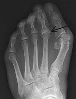
1
An impaction fracture of the metatarsal head
2
A growth disturbance secondary to surgery as a teenager
3
A dislocation of the second metatarsophalangeal joint
4
Chondrocalcinosis or pseudogout
The radiograph shows an overlap of the base of the proximal phalanx over the second metatarsal head and no defined joint space. The base of the proximal phalanx is proximal to the distal aspect of the metatarsal head. The dislocation is chronic secondary to degeneration and disruption of the plantar plate from years of increased stress from the hallux valgus deformity and a long second metatarsal. An impaction fracture or a growth disturbance
would reveal a joint space. Chondrocalcinosis would reveal calcification of the articular cartilage and usually degenerative changes. Ossification of the plantar plate is rare and would look more like a sesamoid.
RECOMMENDED READINGS
1. Coughlin MJ. Lesser toe abnormalities. Instr Course Lect. 2003;52:421-44. Review.
[PubMed PMID: 12690869.](http://www.ncbi.nlm.nih.gov/pubmed/12690869)[View Abstract at PubMed](http://www.ncbi.nlm.nih.gov/pubmed/12690869)
2. Coughlin MJ. Lesser toe deformities. In: Coughlin MJ, Mann RA, Saltzman CL, eds. Surgery of the Foot and Ankle. Vol 1. 8th ed. Philadelphia, PA: Mosby; 2007:407-443.
QUESTION 63
of 100
Figure 64 is the radiograph of a 42-year-old woman who has severe left hip pain and a limp. What procedure may help avoid the postoperative complication of sciatic nerve palsy?
59
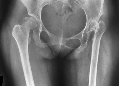
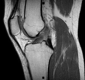
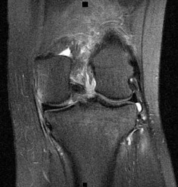

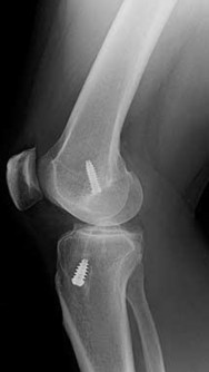
Figure 64 is the radiograph of a 42-year-old woman who has severe left hip pain and a limp. What procedure may help avoid the postoperative complication of sciatic nerve palsy?
59
1
Acetabular osteotomy.
2
Femoral shortening osteotomy.
3
Extended trochanteric osteotomy.
4
Distal femoral rotational osteotomy
This patient has Crowe III acetabular dysplasia with significant shortening (more than 4 cm). With restoration of an anatomic hip center, significant limb lengthening will occur. Femoral shortening osteotomy has been used to decrease risk for sciatic nerve palsy from stretch during total hip arthroplasty with significant limb length restoration. This procedure typically is performed with the use of cementless femoral components, achieving diaphyseal fixation
distal to the osteotomy. The patient has joint incongruity and advanced secondary osteoarthritis, so she is not an optimal candidate for acetabular osteotomy. A high hip center can prevent lengthening and sciatic nerve palsy, but is not among the responses provided. Rotational osteotomy will not protect against sciatic nerve neuropraxia. Extended trochanteric osteotomy is used for revision hip arthroplasty and to manage some femoral deformities, but does not affect tension on the sciatic nerve.
RECOMMENDED READINGS
1. [Jaroszynski G, Woodgate IG, Saleh KJ, Gross AE. Total hip replacement for the dislocated hip. Instr Course Lect. 2001;50:307-16. Review. PubMed PMID: 11372330.](http://www.ncbi.nlm.nih.gov/pubmed/11372330)[View Abstract at PubMed](http://www.ncbi.nlm.nih.gov/pubmed/11372330)
2. [Schmalzried TP, Amstutz HC, Dorey FJ. Nerve palsy associated with total hip replacement. Risk factors and prognosis. J Bone Joint Surg Am. 1991 Aug;73(7):1074-80. PubMed PMID: 1874771. ](http://www.ncbi.nlm.nih.gov/pubmed/1874771)[View Abstract at PubMed](http://www.ncbi.nlm.nih.gov/pubmed/1874771)
3. [Papagelopoulos PJ, Trousdale RT, Lewallen DG. Total hip arthroplasty with femoral osteotomy for proximal femoral deformity. Clin Orthop Relat Res. 1996 Nov;(332):151-62. PubMed PMID: 8913158. ](http://www.ncbi.nlm.nih.gov/pubmed/8913158)[View Abstract at PubMed](http://www.ncbi.nlm.nih.gov/pubmed/8913158)
4. Huo MH, Zatorski LE, Keggi KJ. Oblique femoral osteotomy in cementless total hip arthroplasty. Prospective consecutive series with a 3-year minimum follow-up period.
[J Arthroplasty. 1995 Jun;10(3):319-27. PubMed: 7673911. ](http://www.ncbi.nlm.nih.gov/pubmed/7673911)View Abstract at PubMed 61
CLINICAL SITUATION FOR QUESTIONS 65 THROUGH 70
Figures 65a through 65d are the selected MR images and radiographs of a 29-year-old man who sustained an injury to his left knee.
A
B
C D
QUESTION 64
of 100
Regarding the anteromedial and posterolateral bundles of the injured structure, which relationship is the most accurate?
Regarding the anteromedial and posterolateral bundles of the injured structure, which relationship is the most accurate?
1
Parallel in extension and externally rotated in flexion
61
2
Parallel in extension and internally rotated in flexion
3
Externally rotated in extension and parallel in flexion
4
Internally rotated in extension and parallel in flexion
- Parallel in extension and externally rotated in flexion
QUESTION 65
of 100
What kinematic relationship occurs between the anteromedial and posterolateral bundles of the injured structure identified in these images?
What kinematic relationship occurs between the anteromedial and posterolateral bundles of the injured structure identified in these images?
1
Both are tight in extension and in flexion.
2
Both are tight in extension and loose in flexion.
3
The anteromedial bundles are tight in extension and loose in flexion, and the posterolateral bundle is tight in both flexion and extension.
4
The posterolateral bundle is tight in extension and loose in flexion, and the anteromedial bundle is tight in both flexion and extension.
- The posterolateral bundle is tight in extension and loose in flexion, and the anteromedial bundle is tight in both flexion and extension.
QUESTION 66
of 100
At the time of disruption of the structure identified in Figures 65a and 65b which structure is most commonly injured?
At the time of disruption of the structure identified in Figures 65a and 65b which structure is most commonly injured?
1
Anterior meniscofemoral ligament
2
Intermeniscal ligament
3
Medial meniscus
4
Lateral meniscus
- Lateral meniscus
QUESTION 67
of 100
Which statement best characterizes the injured structure in Figures 65a through 65d?
Which statement best characterizes the injured structure in Figures 65a through 65d?
1
The structure is extrasynovial, contains significant innervation, and is highly
62
vascularized.
2
The structure is extrasynovial, does not contain significant innervation, and is not highly vascularized.
3
The structure is intrasynovial, contains significant innervation, and is not highly vascularized.
4
The structure is intrasynovial, does not contain significant innervation, and is not highly vascularized.
- The structure is intrasynovial, contains significant innervation, and is not highly vascularized.
QUESTION 68
of 100
Reconstruction of the structure injured in Figures 65a and 65b is important to limit which pattern of instability?
Reconstruction of the structure injured in Figures 65a and 65b is important to limit which pattern of instability?
1
Anterior translation only
2
Anterior translation and internal rotation
3
Anterior translation and external rotation
4
Anterior translation and varus-valgus angulation
- Anterior translation and internal rotation
QUESTION 69
of 100
Reconstruction of the injured structure is performed. After surgery, the patient initially notes limitation in motion, and later develops recurrent instability of the knee. Which factor most likely contributed to the development of instability?
Reconstruction of the injured structure is performed. After surgery, the patient initially notes limitation in motion, and later develops recurrent instability of the knee. Which factor most likely contributed to the development of instability?
1
Inadequate graft strength
2
Failure to restore graft length
3
Malposition of the femoral tunnel
4
Malposition of the tibial tunnel
The anteromedial bundle originates on the anterior and proximal aspect of the lateral femoral condyle and inserts on the anteromedial aspect of the anterior cruciate ligament (ACL) footprint on the proximal tibia. The posterolateral bundle originates posterior and distal to 63 the anteromedial bundle and inserts on the posterolateral aspect of the tibial footprint. The fibers are parallel when the knee is in an extended position. As the knee moves into flexion,
the fibers of the anteromedial bundle rotate externally with respect to the posterolateral bundle. The anteromedial bundle is tensioned in both flexion and extension. The posteromedial bundle is tensioned in extension, but relaxes as the knee moves into flexion.
The lateral meniscus is more commonly injured with an acute injury to the ACL. The medial meniscus is injured more commonly when the ACL is chronically unstable.
The ACL is an intra-articular and intrasynovial structure. It is innervated by posterior articular branches from the tibial nerve. Innervation of the ACL involves several types of mechanoreceptors (Ruffini, Pacini, Golgi tendon, and free-nerve endings) that may contribute to proprioceptive function of the knee and modulation of quadriceps function.
Injury to the ACL is predominantly associated with instability to anterior translation of the tibia in extension. The ACL plays a secondary role to limit internal rotation of the tibia, and a loss of ACL stability is confirmed by the reduction of the tibia from a position of anterior translation and internal rotation (pivot shift). The radiographs demonstrate anterior placement of the femoral tunnel. The convex shape of the lateral femoral condyle can make it more difficult to visualize the anatomic femoral origin of the ACL. Failure to identify the
anatomic footprint can result in anterior placement of the femoral tunnel. Anterior ACL graft placement can result in its impingement against the posterior cruciate ligament and early limitation of knee flexion. Over time, impingement on the graft may result in stretching of the graft and recurrent knee instability symptoms.
RECOMMENDED READINGS
1. [Duthon VB, Barea C, Abrassart S, Fasel JH, Fritschy D, Ménétrey J. Anatomy of the anterior cruciate ligament. Knee Surg Sports Traumatol Arthrosc. 2006 Mar;14(3):204-13. Epub 2005 Oct 19. Review. PubMed PMID: 16235056. ](http://www.ncbi.nlm.nih.gov/pubmed/16235056)[View](http://www.ncbi.nlm.nih.gov/pubmed/16235056)[ ](http://www.ncbi.nlm.nih.gov/pubmed/16235056)[Abstract at PubMed](http://www.ncbi.nlm.nih.gov/pubmed/16235056)
2. [Zantop T, Petersen W, Sekiya JK, Musahl V, Fu FH. Anterior cruciate ligament anatomy and function relating to anatomical reconstruction. Knee Surg Sports Traumatol Arthrosc. 2006 Oct;14(10):982-92. Epub 2006 Aug 5. Review. PubMed PMID: 16897068. ](http://www.ncbi.nlm.nih.gov/pubmed/16897068)[View Abstract at PubMed](http://www.ncbi.nlm.nih.gov/pubmed/16897068)
QUESTION 70
of 100
64
What is the optimal biomechanical screw placement position to treat a waist-level scaphoid fracture?
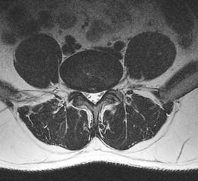
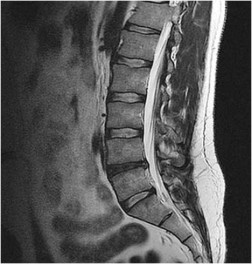
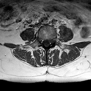
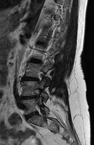
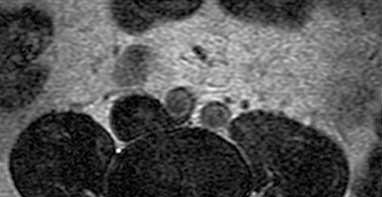

64
What is the optimal biomechanical screw placement position to treat a waist-level scaphoid fracture?
1
Dorsal one-third of the distal and proximal fragments
2
Dorsal half of the distal and proximal fragments
3
Central axis of the distal and proximal fragments
4
Volar half of the distal and proximal fragments
Dodds and associates looked at short (compared to long) scaphoid screws placed down the central axis in cadavers. A long screw placed along the central axis was significantly more stable when tested in all planes of motion about the wrist. McCallister and associates examined central placement and eccentric placement in a cadaver model. Central positioning produced 43% more stiffness and 113% more load (P < .01).
RECOMMENDED READINGS
1. [Bedi A, Jebson PJ, Hayden RJ, Jacobson JA, Martus JE. Internal fixation of acute, nondisplaced scaphoid waist fractures via a limited dorsal approach: an assessment of radiographic and functional outcomes. J Hand Surg Am. 2007 Mar;32(3):326-33. PubMed PMID: 17336838.](http://www.ncbi.nlm.nih.gov/pubmed/17336838)[View Abstract at PubMed](http://www.ncbi.nlm.nih.gov/pubmed/17336838)
2. [Dodds SD, Panjabi MM, Slade JF 3rd. Screw fixation of scaphoid fractures: a biomechanical assessment of screw length and screw augmentation. J Hand Surg Am. 2006 Mar;31(3):405-13. PubMed PMID: 16516734. ](http://www.ncbi.nlm.nih.gov/pubmed/16516734)[View Abstract at PubMed](http://www.ncbi.nlm.nih.gov/pubmed/16516734)
3. [McCallister WV, Knight J, Kaliappan R, Trumble TE. Central placement of the screw in simulated fractures of the scaphoid waist: a biomechanical study. J Bone Joint Surg Am.2003 Jan;85-A(1):72-7.PubMed: 12533575.](http://www.ncbi.nlm.nih.gov/pubmed/12533575)[View Abstract at PubMed](http://www.ncbi.nlm.nih.gov/pubmed/12533575)
RESPONSES FOR QUESTIONS 72 THROUGH 75
The figures listed above depict the scenarios described below. Please match the appropriate scenarios and figures.
65
A B
C
D
E F
66
G H
QUESTION 71
of 100
A 45-year-old man has motor weakness of the quadriceps and tibialis anterior, sensory loss of the medial calf, and loss of the knee jerk reflex on his left.
A 45-year-old man has motor weakness of the quadriceps and tibialis anterior, sensory loss of the medial calf, and loss of the knee jerk reflex on his left.
1
Figure 72a Figure 72b
2
Figure 72c Figure 72d
3
Figure 72e Figure 72f
4
Figure 72g Figure 72h
- Figure 72c Figure 72d
QUESTION 72
of 100
This finding has been shown to be variably present in asymptomatic patients, and its incidence increases with age.
This finding has been shown to be variably present in asymptomatic patients, and its incidence increases with age.
1
Figure 72a Figure 72b
2
Figure 72c Figure 72d
3
Figure 72e Figure 72f
4
Figure 72g Figure 72h
- Figure 72a Figure 72b
QUESTION 73
of 100
This pathology most likely causes mechanical low-back pain.
This pathology most likely causes mechanical low-back pain.
1
Figure 72a Figure 72b
2
Figure 72c Figure 72d
3
Figure 72e Figure 72f
4
Figure 72g Figure 72h 67
- Figure 72e Figure 72f
QUESTION 74
of 100
A 47-year-old man has left-sided motor weakness in the extensor digitorum longus and extensor hallucis longus, sensory loss in the lateral calf and dorsal foot, and no discernible reflex loss.
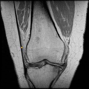
A 47-year-old man has left-sided motor weakness in the extensor digitorum longus and extensor hallucis longus, sensory loss in the lateral calf and dorsal foot, and no discernible reflex loss.
1
Figure 72a Figure 72b
2
Figure 72c Figure 72d
3
Figure 72e Figure 72f
4
Figure 72g Figure 72h
Figures 72a and 72b are the T1-weighted MR images of a disk bulge. Disk bulges are common in asymptomatic people and are not predictive of pathology. Figures 72c and 72d show T1 MR images of a far lateral disk herniation at the L4-L5 level. This would affect the exiting or L4 nerve root. Radicular symptoms would occur at the L4 level. Figures 72e and 72f show an annular tear at the L4-L5 level. This pathology is associated with discogenic low-back pain. Figures 72g and 72h show the T1 MR images of a central lateral disk herniation at the L4-L5 level. This would affect the traversing or L5 nerve root. Radicular symptoms would occur at the L5 level.
RECOMMENDED READINGS
1. [View Abstract at PubMed](http://www.ncbi.nlm.nih.gov/pubmed/15990670)
2. Hoppenfeld S. Physical Exam of the Spine and Extremities. Stamford, CT: Appleton and Lange; 1995.
3. [Madigan L, Vaccaro AR, Spector LR, Milam RA. Management of symptomatic lumbar degenerative disk disease. J Am Acad Orthop Surg. 2009 Feb;17(2):102-11. Review. PubMed PMID: 19202123.](http://www.ncbi.nlm.nih.gov/pubmed/19202123)[View Abstract at PubMed](http://www.ncbi.nlm.nih.gov/pubmed/19202123)
68
RESPONSES FOR QUESTIONS 76 THROUGH 82
For each MR image above, match the correct structure. The structure is labeled with a yellow dot.
QUESTION 75
of 100
Figure 76
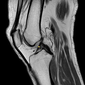
Figure 76
1
Anterior cruciate ligament
2
Biceps femoris tendon
3
Iliotibial band
4
Lateral collateral ligament
5
Ligament of Wrisberg
- Iliotibial band
QUESTION 76
of 100
Figure 77
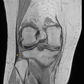
Figure 77
1
Anterior cruciate ligament
2
Biceps femoris tendon
3
Iliotibial band
4
Lateral collateral ligament
5
Ligament of Wrisberg
- Anterior cruciate ligament
QUESTION 77
of 100
Figure 78
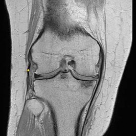
Figure 78
1
Anterior cruciate ligament
2
Biceps femoris tendon
3
Iliotibial band
4
Lateral collateral ligament 69
5
Ligament of Wrisberg
- Biceps femoris tendon
QUESTION 78
of 100
Figure 79
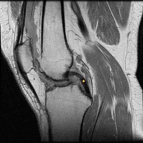
Figure 79
1
Anterior cruciate ligament
2
Biceps femoris tendon
3
Iliotibial band
4
Lateral collateral ligament
5
Ligament of Wrisberg
- Lateral collateral ligament
QUESTION 79
of 100
Figure 80
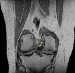
Figure 80
1
Anterior cruciate ligament
2
Biceps femoris tendon
3
Iliotibial band
4
Lateral collateral ligament
5
Ligament of Wrisberg
- Posterior cruciate ligament
QUESTION 80
of 100
Figure 81
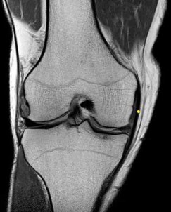
Figure 81
1
Anterior cruciate ligament
2
Biceps femoris tendon
3
Iliotibial band
4
Lateral collateral ligament 71
5
Ligament of Wrisberg
- Ligament of Wrisberg
QUESTION 81
of 100
Figure 82
Figure 82
1
Anterior cruciate ligament
2
Biceps femoris tendon
3
Iliotibial band
4
Lateral collateral ligament
5
Ligament of Wrisberg
- Medial collateral ligament
The figures show coronal and sagittal T1 MRI scan images of a knee. Figure 76 shows the Iliotibial band on the lateral side of the knee and its insertion on Gerdy tubercle. Figure 77 shows the anterior cruciate ligament. Figure 78 is the biceps femoris tendon insertion into the fibular head. Figure 79 shows the lateral collateral ligament. Figure 80 is the posterior cruciate ligament. Figure 81 is the ligament of Wrisberg, which runs from the posterior horn of the lateral meniscus to the medial femoral condyle. Figure 82 shows the medial collateral ligament.
RECOMMENDED READINGS
1. [Erbagci H, Yildirim H, Kizilkan N, Gümüsburun E. An MRI study of the meniscofemoral and transverse ligaments of the knee. Surg Radiol Anat. 2002 May;24(2):120-4. PubMed PMID: 12197021. ](http://www.ncbi.nlm.nih.gov/pubmed/12197021)[View Abstract at PubMed](http://www.ncbi.nlm.nih.gov/pubmed/12197021)
2. Watanabe AT, Carter BC, Teitelbaum GP, Bradley WG Jr. Common pitfalls in magnetic resonance imaging of the knee. J Bone Joint Surg Am. 1989 Jul;71(6):857-
[62/. PubMed PMID: 2745483. ](http://www.ncbi.nlm.nih.gov/pubmed/2745483)[View Abstract at PubMed](http://www.ncbi.nlm.nih.gov/pubmed/2745483)
3. Hoppenfeld S, deBoer P. Surgical Exposures in Orthopedics. 3rd ed. Philadelphia. PA: Lippincott Williams & Wilkins; 2003:493-568.
71
QUESTION 82
of 100
As a surgeon approaches the medial midfoot to harvest the flexor digitorum longus tendon for a transfer, two muscles are reflected plantarly off of the first metatarsal: the medial cuneiform and the navicular, as shown in Figure 83 as A and B. Muscle A is short and broad and muscle B is long and tendinous.
Which nerve is at risk during this approach?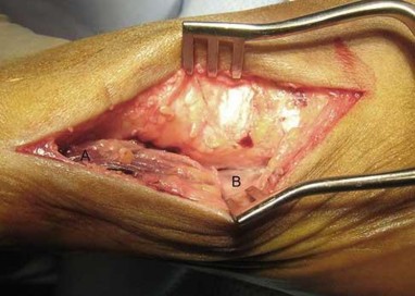
1
Deep peroneal nerve
2
Lateral plantar nerve
3
Saphenous nerve
4
Medial plantar nerve
- Medial plantar nerve
The medial plantar nerve lies between the medial head of the flexor hallucis brevis (A) and the abductor hallucis muscles (B). The medial approach to the midfoot requires that the medial plantar nerve be retracted plantarly to reach the flexor hallucis longus or flexor digitorum longus for a transfer. The deep peroneal nerve is a dorsal structure. The lateral plantar nerve should be safe. The saphenous nerve is proximal and dorsal.
RECOMMENDED READINGS
1. Hoppenfeld S, deBoer P. Surgical Exposures in Orthopaedics: The Anatomic Approach. Philadelphia, PA: JB Lippincott; 1984:508-520.
2. Bauer R, Kerschbaumer F, Poisel S. Operative Approaches in Orthopedic Surgery and Traumatology. New York, NY: Thieme; 1987: 193-198.
RESPONSES FOR QUESTIONS 84 THROUGH 86
For each surgical approach to the hip, please identify the nerve most likely to be injured. 72
QUESTION 83
of 100
Posterior approach
Posterior approach
1
Lateral femoral cutaneous nerve
2
Inferior gluteal nerve
3
Superior gluteal nerve
4
Sciatic nerve
- Sciatic nerve
QUESTION 84
of 100
Direct lateral approach
Direct lateral approach
1
Lateral femoral cutaneous nerve
2
Inferior gluteal nerve
3
Superior gluteal nerve
4
Sciatic nerve
- Superior gluteal nerve
QUESTION 85
of 100
Anterior approach

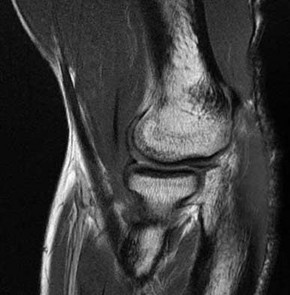
Anterior approach
1
Lateral femoral cutaneous nerve
2
Inferior gluteal nerve
3
Superior gluteal nerve
4
Sciatic nerve
The nerve most commonly injured in the posterior approach to the hip is the sciatic nerve. Overall injury prevalence is 1% to 2%. This nerve is more commonly injured in cases of hip dysplasia with excessive leg lengthening. The superior gluteal nerve is at highest risk with the direct lateral approach to the hip. This nerve courses in the gluteus medius muscle and is
at risk when splitting the muscle 5 cm proximal to the greater trochanter. The lateral femoral 73
cutaneous nerve is commonly damaged with anterior total hip replacement surgery. Neuropraxia has been reported in 81% of patients. The inferior gluteal nerve travels from the greater sciatic notch and enters the gluteus maximus muscle. It is at risk when the posterior approach to the hip is used.
RECOMMENDED READINGS
1. Hoppenfeld S, deBoer P. Surgical Exposures in Orthopedics. 3rd ed. Philadelphia.
PA: Lippincott Williams & Wilkins; 2003:365-453.
2. [DeHart MM, Riley LH Jr. Nerve injuries in total hip arthroplasty. J Am Acad Orthop Surg. 1999 Mar-Apr;7(2):101-11. Review. PubMed PMID: 10217818. ](http://www.ncbi.nlm.nih.gov/pubmed/10217818)[View Abstract](http://www.ncbi.nlm.nih.gov/pubmed/10217818)[ at PubMed](http://www.ncbi.nlm.nih.gov/pubmed/10217818)
3. Goulding K, Beaulé PE, Kim PR, Fazekas A. Incidence of lateral femoral cutaneous nerve neuropraxia after anterior approach hip arthroplasty. Clin Orthop Relat Res. 2010 Sep;468(9):2397-404. doi: 10.1007/s11999-010-1406-5. PubMed PMID:
[20532717.](http://www.ncbi.nlm.nih.gov/pubmed/20532717)[View Abstract at PubMed](http://www.ncbi.nlm.nih.gov/pubmed/20532717)
Figures 87a and 87b are sagittal and coronal MR images of the affected elbow of a 36-year-old man who has a history of painful mechanical symptoms in his dominant arm when extending his elbow in full supination. What is the most likely cause of his painful snapping?
A
B
1. Lacertus fibrosis contracture
2. Intra-articular loose bodies 74
3. Olecranon fossa impingement
4. Radiocapitellar plica
PREFERRED RESPONSE: 4- Radiocapitellar plica
DISCUSSION
The MRI studies show a radiocapitellar plica. This anomalous structure has been associated with symptomatic snapping. Lacertus fibrosis contracture will not cause painful snapping. An intra-articular pathology such as loose bodies is not present on these imaging studies. Olecranon fossa impingement causes posterior pain in extension and is not shown in the images.
RECOMMENDED READINGS
1. [Antuna SA, O'Driscoll SW. Snapping plicae associated with radiocapitellar chondromalacia. Arthroscopy. 2001 May;17(5):491-5. PubMed 11337715. ](http://www.ncbi.nlm.nih.gov/pubmed/11337715)[View Abstract](http://www.ncbi.nlm.nih.gov/pubmed/11337715)[ ](http://www.ncbi.nlm.nih.gov/pubmed/11337715)[at PubMed](http://www.ncbi.nlm.nih.gov/pubmed/11337715)
2. [Ruch DS, Papadonikolakis A, Campolattaro RM. The posterolateral plica: a cause of refractory lateral elbow pain. J Shoulder Elbow Surg. 2006 May-Jun;15(3):367-70. PubMed PMID: 16679240. ](http://www.ncbi.nlm.nih.gov/pubmed/16679240)[View Abstract at PubMed](http://www.ncbi.nlm.nih.gov/pubmed/16679240)
QUESTION 86
of 100
Figure 88a is an anteroposterior pelvis radiograph of a 50-year-old obese man who has right groin pain. He is a former college athlete. Examination reproduces pain with right hip flexion and internal rotation and adduction, and his Stinchfield test result is positive. Figure 88b is a lateral view of his right hip. What is the most likely cause of his hip pain?
A B
75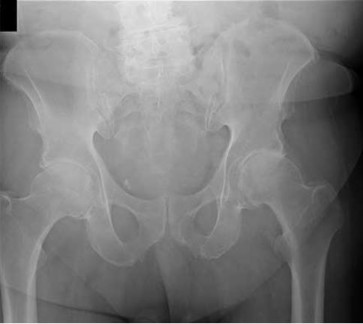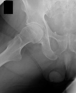
1
Osteonecrosis
2
Obesity
3
Femoroacetabular impingement, Cam type
4
Femoroacetabular impingement, pincer type
The patient’s history and examination is consistent with an intra-articular hip disorder. The radiographs show a large Cam deformity on the anterosuperior femoral neck. Pincer deformities are seen when there is overcoverage of the acetabulum on the femoral head or a deep socket. Femoroacetabular impingement can also occur in cases of acetabular retroversion. The radiographs do not demonstrate these findings. Signs of femoral head collapse and sclerotic lesions are seen in avascular necrosis but are not present here. Obesity plays a role in development of hip arthritis, but its contribution is not fully known and not the major cause of pain in this patient.
RECOMMENDED READINGS
1. [Leunig M, Parvizi J, Ganz R. Nonarthroplasty surgical treatment of hip osteoarthritis. Instr Course Lect. 2006;55:159-66. Review. PubMed PMID: 16958448. ](http://www.ncbi.nlm.nih.gov/pubmed/16958448)[View](http://www.ncbi.nlm.nih.gov/pubmed/16958448)[ ](http://www.ncbi.nlm.nih.gov/pubmed/16958448)[Abstract at PubMed](http://www.ncbi.nlm.nih.gov/pubmed/16958448)
2. [Ganz R, Parvizi J, Beck M, Leunig M, Nötzli H, Siebenrock KA. Femoroacetabular impingement: a cause for osteoarthritis of the hip. Clin Orthop Relat Res. 2003 Dec;(417):112-20. Review. PubMed PMID: 14646708. ](http://www.ncbi.nlm.nih.gov/pubmed/14646708)[View Abstract at PubMed](http://www.ncbi.nlm.nih.gov/pubmed/14646708)
QUESTION 87
of 100
Among the ankle arthroscopy portals described below, which portal is at highest risk for serious complications?
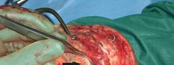
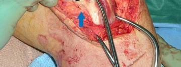
Among the ankle arthroscopy portals described below, which portal is at highest risk for serious complications?
1
Posterolateral: lateral to the Achilles tendon
2
Anterolateral: lateral to the peroneus tertius tendon
3
Anteromedial: medial to the tibialis anterior tendon
4
Posteromedial: medial to the Achilles tendon
All of the portals listed pose risk for some structures. Because they have been shown to be the safest, the most common anterior portals are the anteromedial and the anterolateral. The safest posterior portal is the posterolateral portal. Because of the location of the posterior medial tendons and the neurovascular bundle, the posteromedial portal is at highest risk for serious complications.
RECOMMENDED READINGS
1. [Golanó P, Vega J, Pérez-Carro L, Götzens V. Ankle anatomy for the arthroscopist. Part I: The portals. Foot Ankle Clin. 2006 Jun;11(2):253-73, v. Review. PubMed PMID: 16798511.](http://www.ncbi.nlm.nih.gov/pubmed/16798511)[View Abstract at PubMed](http://www.ncbi.nlm.nih.gov/pubmed/16798511)
2. Ferkel RD, Hommen JP. Arthroscopy of the ankle and foot. In: Coughlin MJ, Mann RA, Saltzman CL, eds. Surgery of the Foot and Ankle. Vol 2. 8th ed. Philadelphia, PA: Mosby; 2007:1641-1726.
Figure 90 is an intraoperative image showing the medial approach to the elbow. The arm is proximal and to the right, and the forearm is distal and to the left. The blue arrow points to the medial epicondyle. A black arrow points to a piece of glass in a nervous structure. This injury most likely would affect which distal muscular structure?
1. Extensor digiti minimi
2. Flexor digitorum superficialis (FDS) to the index finger 77
3. Abductor pollicis brevis
4. First dorsal interossei
PREFERRED RESPONSE: 4- First dorsal interossei
DISCUSSION
The intraoperative image shows a piece of glass splitting the ulnar nerve in the cubital tunnel. The extensor digiti minimi is innervated by the radial nerve. The FDS to the index finger and the abductor pollicis brevis are innervated by the median nerve. The first dorsal interossei is the last muscle innervated by the ulnar nerve.
RECOMMENDED READINGS
1. Miller MD. Review of Orthopaedics. 3rd ed. New York, NY: Saunders; 2000.
2. Anderson JE. Grant’s Atlas of Anatomy. 8th ed. Baltimore, MD: Williams & Wilkins; 1983.
RECOMMENDED READINGS
1. [Golanó P, Vega J, Pérez-Carro L, Götzens V. Ankle anatomy for the arthroscopist. Part I: The portals. Foot Ankle Clin. 2006 Jun;11(2):253-73, v. Review. PubMed PMID: 16798511.](http://www.ncbi.nlm.nih.gov/pubmed/16798511)[View Abstract at PubMed](http://www.ncbi.nlm.nih.gov/pubmed/16798511)
2. Ferkel RD, Hommen JP. Arthroscopy of the ankle and foot. In: Coughlin MJ, Mann RA, Saltzman CL, eds. Surgery of the Foot and Ankle. Vol 2. 8th ed. Philadelphia, PA: Mosby; 2007:1641-1726.
Figure 90 is an intraoperative image showing the medial approach to the elbow. The arm is proximal and to the right, and the forearm is distal and to the left. The blue arrow points to the medial epicondyle. A black arrow points to a piece of glass in a nervous structure. This injury most likely would affect which distal muscular structure?
1. Extensor digiti minimi
2. Flexor digitorum superficialis (FDS) to the index finger 77
3. Abductor pollicis brevis
4. First dorsal interossei
PREFERRED RESPONSE: 4- First dorsal interossei
DISCUSSION
The intraoperative image shows a piece of glass splitting the ulnar nerve in the cubital tunnel. The extensor digiti minimi is innervated by the radial nerve. The FDS to the index finger and the abductor pollicis brevis are innervated by the median nerve. The first dorsal interossei is the last muscle innervated by the ulnar nerve.
RECOMMENDED READINGS
1. Miller MD. Review of Orthopaedics. 3rd ed. New York, NY: Saunders; 2000.
2. Anderson JE. Grant’s Atlas of Anatomy. 8th ed. Baltimore, MD: Williams & Wilkins; 1983.
QUESTION 88
of 100
From which artery does the princeps pollicis artery branch?
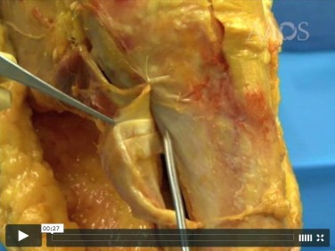
From which artery does the princeps pollicis artery branch?
1
Dorsal radial carpal artery
2
Dorsal ulnar carpal artery
3
Ulnar artery
4
Radial artery
The princeps pollicis artery can be located on the palmar aspect of the adductor pollicis and emerges into the subcutaneous tissue at the thumb metacarpophalangeal flexion crease. It branches from the radial artery just distal to the location of the deep palmar arch.
RECOMMENDED READINGS
1. Coleman SS, Anson B: Arterial patterns in the hand based on a study of 650 specimens. Surg Gynec Obstet 1961;4:409-424.
2. Ames EL, Bissonnette M, Acland R, Lister G, Firrell J. Arterial anatomy of the thumb. 78 [J Hand Surg Br. 1993 Aug;18(4):427-36. PubMed PMID: 8409651. ](http://www.ncbi.nlm.nih.gov/pubmed/8409651)[View Abstract at](http://www.ncbi.nlm.nih.gov/pubmed/8409651)[ ](http://www.ncbi.nlm.nih.gov/pubmed/8409651)[PubMed](http://www.ncbi.nlm.nih.gov/pubmed/8409651)
QUESTION 89
of 100
Which 2 tendons are identified in the dissection shown in Video 92?
Which 2 tendons are identified in the dissection shown in Video 92?
1
Semitendinosus and gracilis
2
Semitendinosus and semimembranosus
3
Semimembranosus and gracilis
4
Semimembranosus and sartorius
- Semitendinosus and gracilis
The demonstration in Video 92 shows the tendons of the semitendinosus and gracilis muscles. They insert on the tibia deep to the sartorial fascia. The semimembranosus inserts more proximal and posterior on the tibia.
RECOMMENDED READINGS
1. Babb JR, Detterline AJ, Noyes FR. AAOS Orthopaedic Video Theater. The Key to the Knee: A Layer-by-Layer Video Demonstration of Medial and Anterior Anatomy. Rosemont, IL: American Academy of Orthopaedic Surgeons; 2009.
2. Hoppenfeld S, deBoer P. Surgical Exposures in Orthopedics. 3rd ed. Philadelphia, PA: Lippincott Williams & Wilkins; 2003:493-568.
QUESTION 90
of 100
A 17-year-old high school athlete comes in with a 6-month history of right midfoot pain. She has been treated with cast immobilization, crutches, and physical therapy. She still has significant pain with activities and cannot participate in sports. Her radiograph is shown in
Figure 93a, and MR images are shown in Figures 93b and 93c. What is the most appropriate 79
next step?
A B
C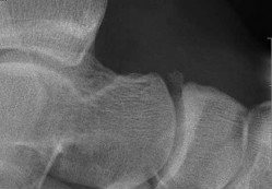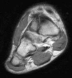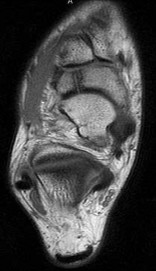
1
Repeat treatment with a nonweight-bearing cast
2
Percutaneous lag screw fixation
3
Addition of a bone stimulator
4
Injection of bone morphogenic protein
- Percutaneous lag screw fixation
This patient’s MR images are indicative of a nondisplaced navicular stress fracture, which is best treated with percutaneous lag screw fixation. She has persistent symptoms despite appropriate nonsurgical treatment. Although all of the above choices may allow successful healing of her navicular, surgery has been shown to result in a shorter recovery and a more predictable outcome, which is especially important to serious athletes. Use of bone morphogenic protein has not been established as a treatment for this injury.
RECOMMENDED READINGS
1. [Lee S, Anderson RB. Stress fractures of the tarsal navicular. Foot Ankle Clin. 2004 Mar;9(1):85-104. Review. PubMed PMID: 15062216. ](http://www.ncbi.nlm.nih.gov/pubmed/15062216)[View Abstract at PubMed](http://www.ncbi.nlm.nih.gov/pubmed/15062216)
2. Anderson RB, Cohen BE. Stress fractures of the foot and ankle. In: Coughlin MJ, Mann RA, Saltzman CL, eds. Surgery of the Foot and Ankle. Vol 2. 8th ed. Philadelphia, PA: Mosby; 2007:1590-1597.
QUESTION 91
of 100
81
Figures 94a and 94b show T1 sagittal and coronal MR images of the right shoulder of a 45-year-old woman. She has insidious onset of dull, aching right shoulder pain localized at the superior aspect of her shoulder. The nerve that supplies the atrophied muscle arises from the upper trunk from contributions of which nerve roots?
A
B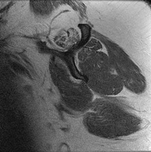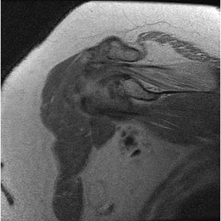
1
C4 and C5 with occasional contribution from C3
2
C4 and C5 with occasional contribution from C6
3
C5 and C6 with occasional contribution from C4
4
C5 and C6 with occasional contribution from C7
The suprascapular nerve innervates the supraspinatus muscle. Patients with suprascapular neuropathy usually have insidious onset of dull, aching shoulder pain at the superior or posterior aspect of the shoulder. There can be several causes of nerve compression, and the nerve is susceptible to compression at the suprascapular and spinoglenoid notches. Extrinsic compression can be secondary to joint-related fluid filled cysts of soft-tissue masses. Traction neuropathy may occur as the result of excessive nerve excursion during athletic activity (usually overhead sports) or after a massive, retracted rotator cuff tear. The suprascapular nerve originates from the upper trunk from predominantly the C5 and C6 nerve roots, with an occasional contribution from the C4 nerve root.
RECOMMENDED READINGS
1. Boykin RE, Friedman DJ, Higgins LD, Warner JJ. Suprascapular neuropathy. J Bone Joint Surg Am. 2010 Oct 6;92(13):2348-64. doi: 10.2106/JBJS.I.01743. Review. 81
[PubMed PMID: 20926731. ](http://www.ncbi.nlm.nih.gov/pubmed/20926731)[View Abstract at PubMed](http://www.ncbi.nlm.nih.gov/pubmed/20926731)
2. [Piasecki DP, Romeo AA, Bach BR Jr, Nicholson GP. Suprascapular neuropathy. J Am Acad Orthop Surg. 2009 Nov;17(11):665-76. Review. PubMed PMID: 19880677 ](http://www.ncbi.nlm.nih.gov/pubmed/19880677)[View Abstract at PubMed](http://www.ncbi.nlm.nih.gov/pubmed/19880677)
QUESTION 92
of 100
Which osseous landmark indicates the proximal border of the femoral insertion of the posterior cruciate ligament?
Which osseous landmark indicates the proximal border of the femoral insertion of the posterior cruciate ligament?
1
Medial intercondylar ridge
2
Medial interfemoral ridge
3
Medial interfascicular ridge
4
Medial bifurcate ridge
The posterior cruciate ligament is composed of the anterolateral and posteromedial bundles. The medial intercondylar ridge marks the proximal border of the femoral insertion. Occasionally, a medial bifurcate ridge will separate the anterolateral and posteromedial bundles, and a clear change in slope occurs between the bundles. Knowledge of these osseous landmarks may aid in anatomic reconstruction of the posterior cruciate ligament. The medial interfemoral and interfascicular ridges are not accepted nomenclature.
RECOMMENDED READINGS
1. Lopes OV Jr, Ferretti M, Shen W, Ekdahl M, Smolinski P, Fu FH. Topography of the femoral attachment of the posterior cruciate ligament. J Bone Joint Surg Am. 2008 Feb;90(2):249-55. doi: 10.2106/JBJS.G.00448. PubMed PMID: 18245582.
[View Abstract at PubMed](http://www.ncbi.nlm.nih.gov/pubmed/18245582)
2. Forsythe B, Harner C, Martins CA, Shen W, Lopes OV Jr, Fu FH. Topography of the femoral attachment of the posterior cruciate ligament. Surgical technique. J Bone Joint Surg Am. 2009 Mar 1;91 Suppl 2 Pt 1:89-100. doi: 10.2106/JBJS.H.01514.
[PubMed PMID: 19255202. ](http://www.ncbi.nlm.nih.gov/pubmed/19255202)[View Abstract at PubMed](http://www.ncbi.nlm.nih.gov/pubmed/19255202)
82
QUESTION 93
of 100
A 15-year-old girl is thrown from a snowmobile and has severe left foot and ankle pain. Her CT image is shown in Figure 96a, and a lateral radiograph is shown in Figure 96b. The arrow in Figure 96a points to which structure?
A
B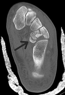
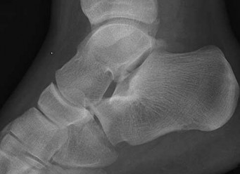
A 15-year-old girl is thrown from a snowmobile and has severe left foot and ankle pain. Her CT image is shown in Figure 96a, and a lateral radiograph is shown in Figure 96b. The arrow in Figure 96a points to which structure?
A
B
1
Base of the fourth metatarsal
2
Base of the fifth metatarsal
3
Cuboid
4
Anterior process of the calcaneus
The image shown is a transverse cut of the foot, which shows the inferior calcaneus, the cuboid, and the three cuneiform bones. The arrow points to a fractured cuboid. The lateral radiograph also shows fractures of the anterior process of the calcaneus and the lateral process of the talus.
RECOMMENDED READINGS
1. Sarrafian SK. Anatomy of the Foot and Ankle: Descriptive, Topographic, Functional.
2nd ed. Philadelphia, PA: Lippincott Williams & Wilkins; 1993:393-406.
2. Hoppenfeld S, deBoer P. Surgical Exposures in Orthopaedics: The Anatomic Approach.
Philadelphia, PA: JB Lippincott; 1984:495-520.
83
QUESTION 94
of 100
Figure 97 is the clinical photograph of a 74-year-old man 6 months after undergoing an acute anterior inferior shoulder dislocation. The patient notes weakness in shoulder flexion, abduction, and extension. This clinical picture represents damage to a nerve that courses
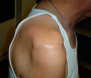
Figure 97 is the clinical photograph of a 74-year-old man 6 months after undergoing an acute anterior inferior shoulder dislocation. The patient notes weakness in shoulder flexion, abduction, and extension. This clinical picture represents damage to a nerve that courses
1
through the suprascapular notch underneath the transverse scapular ligament to enter the supraspinatus fossa, exiting the fossa through the spinoglenoid notch to terminate in the infraspinatus fossa.
2
through the sternocleidomastoid muscle and continuing inferiorly to terminate within the trapezius.
3
Anterior to the scalenus posterior muscle, coursing distal and laterally
deep to the clavicle and superficial to the first and second rib, then running inferiorly on the chest wall in the mid axillary line to terminate within the serratus anterior.
4
on the anterior surface of the subscapularis and then traveling inferior to the glenohumeral joint capsule and coursing from posterior to anterior on the deep surface of the deltoid muscle.
The axillary nerve supplies innervation to the deltoid muscle; this image shows atrophy of this muscle. It arises from the posterior cord of the brachial plexus, coursing on the anterior surface of the subscapularis and then traveling inferior to the glenohumeral joint capsule and coursing from posterior to anterior on the deep surface of the deltoid muscle. Deltoid atrophy
can mimic infraspinatus atrophy as shown in the image, but the latter would not cause the
84
physical symptoms or lateral and anterior atrophy.
Response 1 describes the course of the suprascapular nerve. Damage to this nerve causes loss of abduction and external rotation strength as well as dull, aching superior shoulder pain. Response 2 describes the course of the spinal accessory nerve. Damage to this nerve causes trapezius atrophy, depressed shoulder girdle, lateral scapular winging, scapular dyskinesis, trapezius weakness, and limited active shoulder abduction. Response 3 describes the course of the long thoracic nerve. The superficial location and the length of this nerve are thought to predispose it to injury. Damage to this nerve causes weakness of the serratus anterior which results in a depressed shoulder girdle, medial scapular winging, scapular dyskinesis, and weakened forward flexion.
RECOMMENDED READINGS
1. [Uz A, Apaydin N, Bozkurt M, Elhan A. The anatomic branch pattern of the axillary nerve. J Shoulder Elbow Surg. 2007 Mar-Apr;16(2):240-4. Epub 2006 Nov 9. PubMed PMID: 17097311.](http://www.ncbi.nlm.nih.gov/pubmed/17097311)[View Abstract at PubMed](http://www.ncbi.nlm.nih.gov/pubmed/17097311)
2. [Piasecki DP, Romeo AA, Bach BR Jr, Nicholson GP. Suprascapular neuropathy. J Am Acad Orthop Surg. 2009 Nov;17(11):665-76. Review. PubMed PMID: 19880677. ](http://www.ncbi.nlm.nih.gov/pubmed/19880677)[View Abstract at PubMed](http://www.ncbi.nlm.nih.gov/pubmed/19880677)
QUESTION 95
of 100
The asterisks on Figures 98a through 98c represent which anatomic structure?
A
B
C
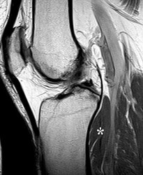
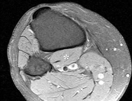
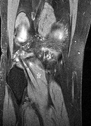
The asterisks on Figures 98a through 98c represent which anatomic structure?
A
B
C
1
Medial head of the gastrocnemius
2
Lateral head of the gastrocnemius
3
Semimembranosus
4
Popliteus
The popliteus muscle arises from the posteromedial part of the tibia, and the tendon continues to attach to the lateral femoral condyle. The tendon is an intra-articular, extra synovial structure coursing through the popliteus hiatus, then deep to the fibular collateral ligament before inserting in the anterior portion of the popliteal sulcus.
RECOMMENDED READINGS
1. Clarke HD, Scott WN, Insall JN, et al. Anatomy. In: Insall JN, Scott WN, eds. Surgery of the Knee. Vol 1. 4th ed. Philadelphia, PA: Churchill Livingstone; 2006:3-66.
2. Miller TT: Magnetic resonance imaging of the knee. In: Insall JN, Scott WN, eds. Surgery of the Knee. Vol 1. 4th ed. Philadelphia, PA: Churchill Livingstone; 2006:201-224.
QUESTION 96
of 100
Figure 99 shows a dorsal approach for a midfoot arthrodesis following a Lisfranc injury in a 43-year-old woman. The base of the second metatarsal is labeled with the letter B. The interval used to create this exposure is
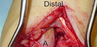
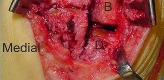
Figure 99 shows a dorsal approach for a midfoot arthrodesis following a Lisfranc injury in a 43-year-old woman. The base of the second metatarsal is labeled with the letter B. The interval used to create this exposure is
1
an internervous plane between muscles innervated by the deep and superficial peroneal nerves.
2
an internervous plane between muscles innervated by the deep peroneal and the medial plantar nerves.
3
an interval between the extensor digitorum longus and the extensor digitorum brevis.
4
not an internervous plane.
The interval shown is between the extensor hallucis longus (left) and the extensor hallucis brevis (right), which is not an internervous plane because both are innervated by the deep peroneal nerve. The neurovascular bundle is under the extensor hallucis brevis muscle. Both
muscles are innervated by branches of the deep peroneal nerve. The superficial peroneal
86
nerve supplies muscles in the lateral compartment, and the medial plantar nerve supplies
muscles in the plantar medial aspect of the foot. The deep peroneal nerve innervates the anterior compartment muscles, the extensor hallucis brevis, and the extensor digitorum brevis muscle and supplies sensation to the dorsal first web space.
RECOMMENDED READINGS
1. Hoppenfeld S, deBoer P. Surgical Exposures in Orthopaedics: The Anatomic Approach. Philadelphia, PA: JB Lippincott; 1984:511-513.
2. Bellabarba C, Barei DP, Sanders RW. Dislocations of the foot. In: Coughlin MJ, Mann RA, Saltzman CL, eds. Surgery of the Foot and Ankle. Vol 2. 8th ed. Philadelphia, PA: Mosby; 2007:2171-2180.
QUESTION 97
of 100
Rupture of the structure shown in the axial cross and the sagittal sections in Figures 100a and 100b causes weakness in
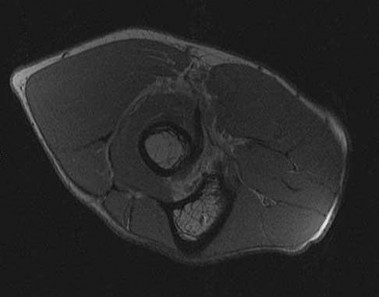
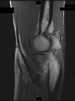
Rupture of the structure shown in the axial cross and the sagittal sections in Figures 100a and 100b causes weakness in
1
extension and supination.
2
pronation.
3
flexion and pronation.
4
flexion and supination.
87
The structure identified is the distal biceps tendon. Rupture of this tendon causes weakness in both flexion and supination. The biceps tendon does not affect extension or pronation.
RECOMMENDED READINGS
1. [Baker BE, Bierwagen D. Rupture of the distal tendon of the biceps brachii. Operative versus non-operative treatment. J Bone Joint Surg Am. 1985 Mar;67(3):414-7. PubMed PMID: 3972865. ](http://www.ncbi.nlm.nih.gov/pubmed/3972865)[View Abstract at PubMed](http://www.ncbi.nlm.nih.gov/pubmed/3972865)
2. [Mazzocca AD, Spang JT, Arciero RA. Distal biceps rupture. Orthop Clin North Am. 2008 Apr;39(2):237-49, vii. doi: 10.1016/j.ocl.2008.01.001. Review. PubMed PMID: 18374814. ](http://www.ncbi.nlm.nih.gov/pubmed/18374814)[View Abstract at PubMed](http://www.ncbi.nlm.nih.gov/pubmed/18374814)
