Part of the Master Guide

Orthopedic Surgery MCQs: Arthroplasty, Fracture & Hip Exam Prep | Part 18

Orthopedic MCQ Exam: Foot, Hip & Knee Practice Questions Part 88

23 Apr 2026 68 min read 56 Views
OITE & ABOS Orthopedic Board Prep: Practice Exam Part 88

Key Takeaway

This page offers Part 88 of an interactive orthopedic surgery board review quiz, specifically designed for orthopedic residents and surgeons preparing for OITE/AAOS/ABOS certification. It includes 50 high-yield MCQs, formatted like real exams, covering Foot, Hip, and Knee, with detailed explanations for comprehensive exam preparation.

Orthopedic MCQ Exam: Foot, Hip & Knee Practice Questions Part 88

Comprehensive 100-Question Exam


00:00

Start Quiz

Question 1

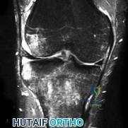
A 55-year-old female presents with acute knee pain after performing a deep squat. MRI demonstrates a complete radial tear of the medial meniscus posterior root.

Biomechanically, this injury pattern most closely mimics which of the following conditions?


Explanation

A complete tear of the medial meniscus posterior root disrupts the hoop stresses of the meniscus. Biomechanical studies have demonstrated that this leads to extrusion of the meniscus under load, rendering the knee biomechanically equivalent to a total meniscectomy. This results in significantly increased peak contact pressures in the medial compartment, rapidly predisposing the patient to osteoarthritis if left untreated.

Question 2

A 65-year-old female undergoes primary total hip arthroplasty (THA) via a posterior approach. Post-operatively, she experiences recurrent anterior dislocations.

Which of the following component malpositions is the most likely cause of her anterior instability?


Explanation

Anterior dislocation of a THA is most commonly caused by excessive combined anteversion (acetabular anteversion + femoral anteversion). When the combined anteversion is too high, the femoral head is driven anteriorly out of the acetabulum, especially during hip extension and external rotation. Conversely, excessive retroversion typically leads to posterior instability.

Question 3

A 60-year-old male with long-standing, poorly controlled diabetes presents with a red, hot, and swollen left foot of 2 weeks duration. He denies any systemic symptoms or open wounds. Radiographs reveal fragmentation, periarticular debris, and subluxation at the tarsometatarsal joint.

What is the most appropriate initial management?


Explanation

The patient is presenting with acute Stage 1 (developmental/fragmentation) Charcot arthropathy (Eichenholtz classification). The clinical presentation of a red, hot, swollen foot in a diabetic patient without an open ulcer is highly suggestive of acute Charcot. The gold standard for initial management is immobilization with a total contact cast to offload the foot, prevent further deformity, and allow the acute inflammatory process to consolidate. Surgery is generally contraindicated in the acute phase due to high rates of hardware failure and complication.

Question 4

An 18-year-old female with recurrent patellar dislocations is scheduled for medial patellofemoral ligament (MPFL) reconstruction. Correct placement of the femoral tunnel is critical to ensure anisometry is minimized. Fluoroscopically, the correct femoral attachment (Schöttle point) is best identified by which of the following landmarks on a true lateral radiograph?


Explanation

The Schöttle point is a highly reliable radiographic landmark for the femoral origin of the MPFL. On a true lateral radiograph, it is located 1 mm anterior to a line extending the posterior femoral cortex, 2.5 mm distal to the posterior origin of the medial femoral condyle, and just proximal to the posterior point of Blumensaat line. Proper placement prevents the graft from becoming inappropriately tight or loose during knee range of motion.

Question 5

A 55-year-old female presents with medial ankle pain and a progressive flatfoot deformity. She cannot perform a single-leg heel raise. Passive correction of the hindfoot valgus is possible. Radiographs demonstrate a talonavicular uncoverage of 20% without arthritic changes.

Which of the following surgical procedures is most appropriate?


Explanation

This patient has a flexible Stage IIA adult-acquired flatfoot deformity (posterior tibial tendon dysfunction). Because the deformity is flexible and there is mild forefoot abduction (<30% talonavicular uncoverage), joint-sparing procedures are indicated. The standard of care is a flexor digitorum longus (FDL) transfer to the navicular combined with a medial displacement calcaneal osteotomy (MDCO) to correct the mechanical axis. If severe forefoot abduction was present (>30% uncoverage, Stage IIB), a lateral column lengthening would also be indicated. Arthrodesis is reserved for rigid deformities or arthritis (Stage III).

Question 6

A 28-year-old male hockey player presents with chronic groin pain that worsens with deep hip flexion and internal rotation. A standing AP pelvis radiograph demonstrates a 'crossover sign'. A frog-leg lateral radiograph shows an alpha angle of 65 degrees.

Which of the following morphologies is predominantly present?


Explanation

The patient exhibits findings of both Cam and Pincer morphology, which is the most common presentation of femoroacetabular impingement (FAI). The 'crossover sign' indicates acetabular retroversion or focal overcoverage (Pincer morphology). An alpha angle greater than 50-55 degrees on a lateral radiograph indicates an aspherical femoral head-neck junction (Cam morphology). Thus, it is a combined FAI.

Question 7

A 30-year-old male sustains an isolated posterior cruciate ligament (PCL) injury during a motor vehicle collision. On physical examination, the posterior drawer test is utilized to assess posterior tibial translation. At what degree of knee flexion is the PCL subjected to the highest in situ forces, making it the most reliable position for this test?


Explanation

The primary restraint to posterior tibial translation is the posterior cruciate ligament (PCL). Biomechanical studies have shown that the PCL experiences the highest in situ forces at 90 degrees of knee flexion. Consequently, the posterior drawer test is performed at 90 degrees of flexion to most accurately assess the integrity of the PCL. The anterolateral bundle is the larger, tighter bundle in flexion.

Question 8

A 45-year-old active male presents with chronic dorsal foot pain localized to the first metatarsophalangeal (MTP) joint. Radiographs show dorsal osteophytes with mild to moderate joint space narrowing and preservation of the plantar joint space (Coughlin and Shurnas Grade 2).

Non-operative management has failed. Which surgical intervention is the most appropriate first-line treatment?


Explanation

This patient has Grade 2 hallux rigidus. For active patients with mild to moderate disease (Grades 1 and 2), a cheilectomy (removal of the dorsal one-third of the metatarsal head and osteophytes) is the preferred initial surgical procedure, as it preserves motion and provides reliable pain relief. First MTP arthrodesis is the gold standard for severe, end-stage disease (Grades 3 and 4).

Question 9

A 12-year-old obese male presents with left knee pain and a limp for 3 weeks. Examination reveals an antalgic gait. When the hip is passively flexed, it falls into obligatory external rotation.

What is the most appropriate definitive management for the left hip?


Explanation

The clinical presentation (obese adolescent, knee/thigh pain, obligatory external rotation with hip flexion) is classic for a Slipped Capital Femoral Epiphysis (SCFE). The standard of care for a stable or unstable SCFE is in situ percutaneous screw fixation (usually a single cannulated screw placed centrally in the epiphysis) to prevent further slippage and promote physeal closure.

Question 10

A 22-year-old female undergoes an anterior cruciate ligament (ACL) reconstruction. Post-operatively, she complains of a persistent lack of full knee extension. Imaging reveals that the graft is impinging against the intercondylar roof. What is the most likely technical error leading to this complication?


Explanation

A tibial tunnel that is placed too far anteriorly will result in the graft impinging on the intercondylar roof (Blumensaat line) during knee extension, leading to a loss of full extension and potential graft abrasion/failure. Conversely, a femoral tunnel placed too anteriorly results in a graft that becomes unphysiologically tight in flexion, limiting knee flexion.

Question 11

A 25-year-old professional soccer player sustains a fracture at the metaphyseal-diaphyseal junction of the fifth metatarsal.

Given his athletic status, what is the recommended treatment to minimize nonunion risk and expedite return to play?


Explanation

A fracture at the metaphyseal-diaphyseal junction of the fifth metatarsal is a true Jones fracture. This area represents a vascular watershed zone, predisposing the fracture to a high rate of delayed union or nonunion. In high-demand athletes, early intramedullary screw fixation is recommended to achieve the highest union rates and the fastest return to sport, compared to non-operative casting.

Question 12

A 40-year-old female with systemic lupus erythematosus on chronic corticosteroids presents with progressive groin pain. MRI reveals a crescent sign in the anterosuperior aspect of the femoral head.

According to the Ficat and Arlet classification of osteonecrosis, what stage does this radiographic finding represent?


Explanation

The Ficat and Arlet classification for osteonecrosis of the femoral head is as follows: Stage I has normal radiographs but abnormal MRI/bone scan; Stage II shows cystic/sclerotic changes on radiographs but a spherical head; Stage III is characterized by subchondral collapse, which is radiographically visible as the 'crescent sign'; Stage IV involves complete collapse of the femoral head with secondary degenerative changes in the acetabulum (joint space narrowing).

Question 13

A 70-year-old male presents with global knee stiffness 6 months after a primary total knee arthroplasty (TKA). His range of motion is 15 to 70 degrees. Radiographs demonstrate appropriate component sizing, but the joint line has been elevated by 8 mm compared to the contralateral knee.

What is the most likely biomechanical consequence of elevating the joint line in TKA?


Explanation

Elevating the joint line during TKA (often due to excessive distal femoral resection compensated by a thicker polyethylene insert) leads to a condition known as pseudo-patella baja. The patella sits lower relative to the elevated joint line, which alters patellofemoral kinematics, causes patellar impingement against the tibial insert, and significantly decreases postoperative knee flexion.

Question 14

A 38-year-old recreational athlete sustains an acute Achilles tendon rupture. He opts for non-operative management utilizing an early functional rehabilitation protocol. Compared to traditional open surgical repair, what does high-level literature demonstrate regarding rerupture rates when early functional rehabilitation is strictly followed?


Explanation

Historically, non-operative management of Achilles tendon ruptures (via prolonged cast immobilization) was associated with higher rerupture rates than surgical repair. However, modern level I evidence (such as the Willits et al. trial) has shown that when non-operative treatment is combined with a strict early functional rehabilitation protocol (early weight-bearing in a functional brace), the rerupture rates are statistically similar to operative repair, while avoiding surgical complications like infection and nerve injury.

Question 15

A 65-year-old female presents with intractable lateral hip pain and weakness in hip abduction. MRI confirms a severe abductor tendon tear. Anatomically, the gluteus minimus tendon primarily inserts onto which specific facet of the greater trochanter?


Explanation

The greater trochanter has distinct anatomical facets for tendon insertions. The gluteus minimus tendon inserts primarily onto the anterior facet. The gluteus medius tendon has a broader footprint, inserting onto the lateral and superoposterior facets. Knowledge of these footprints is critical for accurate MRI interpretation and surgical repair of abductor tears.

Question 16

A 14-year-old male presents with knee pain and catching. Radiographs reveal a classic osteochondritis dissecans (OCD) lesion.

What is the most common anatomic location for an OCD lesion in the knee?


Explanation

The most common location for osteochondritis dissecans (OCD) of the knee is the lateral aspect of the medial femoral condyle, accounting for roughly 70-80% of cases. A helpful mnemonic is 'LAME' (Lateral Aspect of Medial Epicondyle/condyle). The underlying etiology is thought to be repetitive microtrauma to a susceptible area of subchondral bone.

Question 17

A 32-year-old male sustains a severe midfoot injury. Imaging shows a complete lateral displacement of the second through fifth metatarsals, while the first metatarsal remains in anatomic alignment with the medial cuneiform.

According to the Hardcastle and Myerson classification of Lisfranc injuries, this pattern is best described as:


Explanation

The Hardcastle and Myerson classification organizes Lisfranc injuries into three types: Type A (total incongruity/displacement of all 5 metatarsals in one direction), Type B (partial incongruity), and Type C (divergent). This specific case, where the 1st metatarsal is uninjured but the lesser metatarsals (2-5) are laterally displaced, is a Type B2 injury.

Question 18

A 78-year-old active female sustains a displaced femoral neck fracture (Garden IV). When counseling her on surgical options, comparing total hip arthroplasty (THA) to hemiarthroplasty, THA is associated with which of the following outcomes?


Explanation

For active, independent elderly patients with a displaced femoral neck fracture, Total Hip Arthroplasty (THA) provides superior long-term functional outcomes and lower reoperation rates compared to hemiarthroplasty. Hemiarthroplasty has a higher reoperation rate primarily due to subsequent acetabular wear and erosion. However, THA does carry a higher risk of dislocation, longer operative time, and greater blood loss than hemiarthroplasty.

Question 19

A 25-year-old male suffers a varus blow to his anteromedial knee while his foot is planted. Examination demonstrates increased external rotation of the tibia at 30 degrees of knee flexion compared to the uninjured side, but symmetrical tibial rotation at 90 degrees of flexion. Which structure is predominantly injured?


Explanation

The physical exam described is the Dial test. Increased external rotation (>10 degrees compared to the contralateral side) at 30 degrees of flexion, but not at 90 degrees, indicates an isolated injury to the posterolateral corner (PLC). If the test were positive at both 30 and 90 degrees, it would suggest a combined PLC and posterior cruciate ligament (PCL) injury, as the PCL becomes the primary restraint to external rotation at 90 degrees.

Question 20

A 60-year-old male with end-stage post-traumatic ankle osteoarthritis is undergoing a tibiotalar arthrodesis.

What is the optimal position for the ankle fusion to maximize functional outcomes and gait?


Explanation

The optimal position for an ankle arthrodesis is neutral dorsiflexion (0 degrees), 0 to 5 degrees of hindfoot valgus, and 5 to 10 degrees of external rotation (matching the contralateral side). Plantarflexion leads to a back-knee (genu recurvatum) thrust during gait, while varus positioning locks the transverse tarsal joints, accelerating adjacent joint arthritis in the midfoot.

Question 21

A 65-year-old male is 3 months post-operative from a primary total hip arthroplasty (THA) via a posterior approach. He presents with a history of three posterior dislocations. Radiographs demonstrate a well-fixed acetabular component with 45 degrees of abduction and 20 degrees of anteversion. The femoral stem is stable but appears retroverted by 10 degrees. What is the most appropriate surgical intervention to definitively address this instability?


Explanation

Combined anteversion is the critical factor in preventing THA instability. The ideal combined anteversion is typically 25 to 35 degrees for males. The acetabular component is already well-positioned (45 deg abduction, 20 deg anteversion). The femoral stem, however, is in 10 degrees of retroversion, resulting in a low combined anteversion (10 degrees total) that predisposes the patient to posterior dislocation. Revising the femoral component to appropriate anteversion (10-15 degrees) restores normal biomechanics. Over-anteverting the cup (Option B) can lead to anterior instability. A constrained liner (Option D) is a salvage option but does not address the underlying significant malposition.

Question 22

A 28-year-old male presents with right knee pain following a dashboard mechanism injury. On examination, the posterior drawer test is negative. The dial test demonstrates 15 degrees of increased external rotation at 30 degrees of knee flexion compared to the uninjured side. At 90 degrees of knee flexion, external rotation is symmetric bilaterally. Which of the following structures is most likely injured?


Explanation

The dial test is used to evaluate the integrity of the posterolateral corner (PLC) and the posterior cruciate ligament (PCL). Increased external rotation (>10-15 degrees difference from the contralateral side) at 30 degrees of flexion with symmetric rotation at 90 degrees indicates an isolated PLC injury. If the dial test is positive at both 30 degrees and 90 degrees of flexion, it suggests a combined injury to both the PLC and the PCL. The negative posterior drawer test further confirms the PCL is intact.

Question 23

A 55-year-old female with poorly controlled type 2 diabetes mellitus presents with a warm, swollen, and erythematous left foot and ankle. She denies any systemic symptoms, fevers, or open wounds. White blood cell count and inflammatory markers are mildly elevated. Radiographs reveal fragmentation, periarticular debris, and subluxation of the tarsometatarsal joints.

Which of the following is the most appropriate initial management?


Explanation

This patient is presenting with acute Eichenholtz stage 1 (developmental/fragmentation) Charcot arthropathy, characterized by a red, hot, swollen foot with radiographic evidence of fragmentation, osteopenia, and subluxation. The mainstay of initial treatment is strict offloading and immobilization using a total contact cast (TCC). Surgery during the acute inflammatory phase carries a very high risk of hardware failure, infection, and worsening of the Charcot process. Arthrodesis or exostectomy is reserved for the chronic/consolidation phase (Stage 3) if there is an unbraceable deformity or recurrent ulceration.

Question 24

A 24-year-old male professional soccer player complains of chronic groin pain exacerbated by kicking. Physical examination reveals a positive impingement test (pain with flexion, adduction, and internal rotation). Radiographs display a "pistol-grip" deformity of the proximal femur and an alpha angle of 68 degrees. What is the primary pathoanatomy responsible for this condition?


Explanation

The clinical picture describes femoroacetabular impingement (FAI). The radiographic findings of a pistol-grip deformity and an elevated alpha angle (>50-55 degrees) are pathognomonic for Cam-type impingement. This is caused by a loss of the normal concave junction between the femoral head and neck (reduced head-neck offset), which creates an aspherical head that abrades the acetabular cartilage during flexion and internal rotation. Pincer impingement is caused by acetabular overcoverage (e.g., coxa profunda, acetabular retroversion, positive crossover sign).

Question 25

A 70-year-old female is 5 years status post total knee arthroplasty (TKA). She complains of anterior knee pain and a "clunking" sensation when actively extending the knee from a flexed position. Physical examination reveals a palpable and audible clunk at roughly 35 degrees of knee flexion. Radiographs demonstrate a well-fixed posterior stabilized (PS) TKA with no evidence of loosening. What is the most likely diagnosis?


Explanation

Patellar clunk syndrome is a complication seen primarily in posterior stabilized (PS) TKA designs. It is caused by the formation of a fibrotic tissue nodule at the superior pole of the patella or within the suprapatellar pouch. As the knee extends from a flexed position (usually around 30-45 degrees of flexion), this nodule catches in the intercondylar box of the femoral component and then abruptly pops out, creating a painful clunk. Treatment is typically arthroscopic debridement of the fibrotic nodule.

Question 26

A 32-year-old male sustains a midfoot injury while playing football. Weight-bearing radiographs demonstrate a 3 mm diastasis between the medial and middle cuneiforms and a "fleck sign" in the first intermetatarsal space. Which ligament complex is primarily disrupted in this injury?


Explanation

The Lisfranc ligament is an oblique interosseous ligament that connects the lateral aspect of the medial cuneiform to the medial aspect of the base of the second metatarsal. It is the strongest and most critical ligament stabilizing the tarsometatarsal joint complex. The "fleck sign" is pathognomonic for a Lisfranc injury and represents a bony avulsion of this ligament, usually from the base of the second metatarsal.

Question 27

A 13-year-old obese male presents with insidious onset of left groin and knee pain. He walks with an externally rotated gait. When his left hip is passively flexed, it obligatory goes into external rotation. Radiographs confirm a stable slipped capital femoral epiphysis (SCFE). He undergoes in situ single-screw fixation. Which of the following is the most common long-term complication following successful fixation of this condition?


Explanation

The obligatory external rotation with hip flexion (Drehmann sign) is classic for SCFE. Following in situ fixation of a SCFE, the residual prominent anterior-superior femoral metaphysis frequently leads to Cam-type femoroacetabular impingement (FAI). While avascular necrosis (AVN) is the most devastating complication, it is far more common in unstable SCFE. Chondrolysis is less common today, historically associated with unrecognized intra-articular screw penetration. Thus, FAI is the most common long-term complication even after successful stable in situ pinning.

Question 28

A 35-year-old female sustains a high-energy knee dislocation. On examination in the emergency department, the foot is cool with diminished pulses. An ankle-brachial index (ABI) is 0.7. A subsequent CT angiogram confirms a complete popliteal artery occlusion. What is the most appropriate sequence of surgical intervention?


Explanation

In a knee dislocation with hard signs of vascular compromise (ABI < 0.9, absent pulses, confirmed occlusion), emergent restoration of blood flow is critical. The standard sequence is temporary skeletal stabilization (typically a spanning external fixator) to protect the vascular repair, followed immediately by vascular repair (or temporary shunting if ischemia time is critical), and finally prophylactic four-compartment fasciotomies to prevent compartment syndrome secondary to reperfusion injury. Performing ligament reconstruction in the acute ischemic/vascular repair setting is contraindicated.

Question 29

A 42-year-old weekend athlete sustains an acute Achilles tendon rupture. He is managed nonoperatively with a functional rehabilitation protocol. Based on recent Level I evidence, which of the following statements is true regarding nonoperative functional rehabilitation compared to operative management?


Explanation

Historically, nonoperative treatment with cast immobilization had a higher re-rupture rate than surgery. However, modern Level I evidence (such as the Willits trial) demonstrates that nonoperative treatment utilizing early functional rehabilitation (early weight-bearing in a functional brace) yields re-rupture rates that are not significantly different from operative management, while entirely avoiding the surgical risks of wound breakdown, infection, and sural nerve injury.

Question 30

A 45-year-old male on chronic corticosteroids for systemic lupus erythematosus presents with severe groin pain. Plain radiographs show a crescent sign, and MRI confirms Ficat Stage III avascular necrosis (AVN) of the femoral head involving 45% of the weight-bearing area. What is the most reliable definitive surgical treatment for this patient?


Explanation

Ficat Stage III AVN is characterized by subchondral collapse (the 'crescent sign') with preserved joint space. Once the subchondral bone has mechanically collapsed, joint-preserving procedures such as core decompression or vascularized fibular grafting have a high failure rate and are generally no longer indicated, particularly for large lesions (>30% weight-bearing area). Total hip arthroplasty (THA) provides the most reliable pain relief and functional improvement for post-collapse AVN.

Question 31

A 21-year-old male sustains a bucket-handle tear of the medial meniscus. The tear is located in the peripheral red-white zone. The surgeon elects to perform an arthroscopic repair using an inside-out technique for the posterior horn. During the passage of the sutures through the posterior medial joint capsule, which anatomic structure is at greatest risk of iatrogenic injury?


Explanation

During an inside-out repair of the medial meniscus, the needles are passed from inside the joint to a posterior medial incision. The structure at greatest risk during a posteromedial approach/needle passage is the sartorial branch of the saphenous nerve, which runs posteromedial to the sartorius. The popliteal artery and tibial nerve are at risk if needles are passed too centrally/posteriorly. The common peroneal nerve is at risk during a posterolateral inside-out repair for the lateral meniscus.

Question 32

A 22-year-old collegiate football running back hyper-extends his great toe during a tackle. Examination reveals exquisite tenderness at the plantar aspect of the first metatarsophalangeal (MTP) joint, swelling, and ecchymosis. MRI demonstrates a complete disruption of the plantar plate and capsuloligamentous complex with proximal retraction of the sesamoids. What is the most appropriate management for this athlete?


Explanation

This describes a Grade 3 "turf toe" injury, defined as a complete tear of the plantar plate and MTP capsuloligamentous complex, evidenced by proximal migration of the sesamoids. In an elite competitive athlete, nonoperative management of a Grade 3 injury often results in chronic pain, loss of push-off strength, and progressive hallux valgus or rigidus. Surgical repair of the plantar plate is indicated to restore anatomy and function. Arthrodesis is a salvage procedure, and complete sesamoidectomy disrupts the intrinsic flexor mechanics.

Question 33

A 68-year-old female presents with a grossly loose total hip arthroplasty and severe pelvic osteolysis. Preoperative evaluation suspects a pelvic discontinuity. Which of the following radiographic findings is the most reliable indicator of an ipsilateral pelvic discontinuity?


Explanation

Pelvic discontinuity is a complete separation of the superior half of the pelvis (ilium) from the inferior half (ischium and pubis) through the acetabulum. Radiographic signs include a visible transverse fracture line crossing the acetabulum and a medial translation of the inferior hemipelvis relative to the superior hemipelvis. Breakage of screws or superior migration indicates component loosening but not necessarily complete pelvic discontinuity.

Question 34

A 40-year-old male fell from a ladder 6 weeks ago. He presents now with an inability to actively extend his knee. Radiographs reveal patella alta and no fractures. MRI confirms a complete, chronically retracted patellar tendon rupture. If primary surgical repair is attempted, what adjunctive soft-tissue procedure is most likely required due to the chronicity of the injury?


Explanation

In chronic or delayed (>2-6 weeks) patellar tendon ruptures, the quadriceps muscle contracts significantly, pulling the patella proximally (patella alta). During surgery, it is often impossible to mobilize the patella distally enough to achieve a tension-free primary repair. Therefore, an extensive release or a V-Y quadriceps lengthening (quadricepsplasty) is often required. Additionally, due to poor tendon tissue quality, augmentation with allograft or autograft (e.g., hamstrings) is usually necessary to protect the repair.

Question 35

A 50-year-old female complains of a painful bunion. Weight-bearing radiographs show a hallux valgus angle (HVA) of 38 degrees and an intermetatarsal angle (IMA) of 16 degrees. Clinical examination reveals no hypermobility of the first tarsometatarsal (TMT) joint and no evidence of degenerative joint disease. What is the most appropriate surgical intervention?


Explanation

The patient has a moderate-to-severe hallux valgus deformity (IMA >13-15 degrees, HVA >30-40 degrees). A distal chevron osteotomy is generally reserved for mild deformities (IMA <13 deg) because it provides limited correction. For an IMA of 16 degrees, a proximal osteotomy (e.g., crescentic, Ludloff, or SCARF) combined with a distal soft tissue release is indicated to achieve adequate correction. A Lapidus procedure is preferred if there is first TMT hypermobility or arthritis. First MTP arthrodesis is used for severe deformity with concomitant arthritis.

Question 36

A 60-year-old male with a metal-on-metal (MoM) total hip arthroplasty presents with new-onset groin pain and a palpable anterior thigh mass. Serum cobalt and chromium levels are elevated. MRI demonstrates a large fluid collection and cystic pseudotumor around the hip.

What type of hypersensitivity reaction is primarily responsible for this adverse local tissue reaction (ALTR)?


Explanation

Adverse local tissue reactions (ALTR) or aseptic lymphocyte-dominated vasculitis-associated lesions (ALVAL) associated with metal-on-metal bearing surfaces are primarily characterized as a Type IV delayed hypersensitivity reaction. This is a T-cell mediated immune response against metal ions (cobalt and chromium) that act as haptens, leading to massive perivascular lymphocytic infiltration, tissue necrosis, and pseudotumor formation.

Question 37

A 25-year-old football player sustains a valgus blow to his right knee. Examination reveals pain along the medial joint line, 5 mm of medial opening at 30 degrees of knee flexion with a firm endpoint, and no opening at 0 degrees of flexion. MRI confirms an isolated tear of the superficial medial collateral ligament (MCL). What is the recommended treatment?


Explanation

The patient has a Grade II injury to the MCL (laxity at 30 degrees with a firm endpoint, stable at 0 degrees). Isolated Grade I, II, and even most Grade III MCL injuries have excellent healing potential and are best treated nonoperatively. The standard of care is a hinged knee brace to protect against valgus stress while allowing early range of motion and weight-bearing as tolerated, combined with a structured physical therapy program.

Question 38

A 60-year-old female presents with a progressive, painful flatfoot deformity. She is unable to perform a single-leg heel raise on the affected side. Examination reveals a flexible pes planovalgus deformity. Radiographs demonstrate 40% talonavicular uncoverage but no significant degenerative joint disease. After failing 6 months of orthotics and bracing, which surgical procedure is most appropriate?


Explanation

The patient has Stage II posterior tibial tendon dysfunction (PTTD), defined by a painful, flexible flatfoot with an inability to perform a single-leg heel raise. Because the deformity is flexible and there is no arthritis, joint-sparing surgery is indicated. The gold standard is replacing the dysfunctional posterior tibial tendon with an FDL transfer, combined with a medial displacement calcaneal osteotomy (MDCO) to restore the biomechanical axis of the hindfoot and protect the transfer. Stage III (rigid flatfoot or arthritis) requires arthrodesis.

Question 39

In a patient with a typical slipped capital femoral epiphysis (SCFE), what is the true anatomic displacement of the femoral metaphysis (femoral neck) relative to the epiphysis?


Explanation

In a SCFE, the clinical and radiographic appearance is often described as the epiphysis slipping "posterior and inferior." However, biomechanically and anatomically, the epiphysis remains relatively fixed in the acetabulum (tethered by the ligamentum teres). It is the femoral neck (metaphysis) that physically translates anteriorly and superiorly (and externally rotates). This anterior metaphyseal prominence is the classic source of Cam impingement post-SCFE.

Question 40

A 14-year-old male presents with vague knee pain and occasional catching. Radiographs reveal a 1.5 cm osteochondritis dissecans (OCD) lesion on the lateral aspect of the medial femoral condyle. MRI confirms the lesion is completely intact with no high T2 fluid signal behind the fragment. His distal femoral physes remain wide open. What is the initial treatment of choice?


Explanation

This is a case of juvenile osteochondritis dissecans (JOCD) characterized by open physes and a stable lesion on MRI (no fluid behind the fragment, intact overlying cartilage). Stable JOCD lesions have a very high rate of spontaneous healing (up to 70-80%) with conservative management. The initial treatment is strict activity modification and weight-bearing restriction. Surgical intervention (drilling, fixation, or cartilage restoration) is indicated only if the lesion is unstable (fluid behind fragment), if the fragment is detached, or if there is failure of 6 months of nonoperative management.

Question 41

A 68-year-old woman presents with recurrent posterior dislocations of her total hip arthroplasty. Radiographic evaluation demonstrates an acetabular component positioned at 45 degrees of inclination and 0 degrees of anteversion.

Which of the following is the most appropriate surgical intervention to prevent further posterior dislocation?


Explanation

Posterior dislocation is most commonly associated with inadequate anteversion of the acetabular component. The target 'safe zone' (Lewinnek) is 40+/-10 degrees of inclination and 15+/-10 degrees of anteversion. Correcting the 0 degrees of anteversion to a more anteverted position will help prevent posterior dislocation.

Question 42

In ceramic-on-ceramic total hip arthroplasty, which of the following component malpositions is most strongly associated with the complication of squeaking?


Explanation

Squeaking in ceramic-on-ceramic THA is often attributed to edge loading or microseparation. Excessive acetabular inclination (a steep cup, typically >50 degrees) or extreme versions lead to edge loading, disrupting the fluid film lubrication, causing stripe wear, and resulting in squeaking.

Question 43

During a posterior-stabilized (PS) total knee arthroplasty, trial reduction reveals that the knee is well-balanced in full extension but excessively tight in 90 degrees of flexion. Which of the following adjustments is the most appropriate next step?


Explanation

A knee that is tight in flexion but balanced in extension has an isolated tight flexion gap. Options to increase the flexion gap without affecting the extension gap include downsizing the femoral component using anterior referencing (which removes more posterior condylar bone), dropping the posterior slope of the tibia (increases flexion gap more than extension), or translating the femoral component anteriorly. Increasing distal femoral resection would affect the extension gap.

Question 44

A 24-year-old athlete sustains a midfoot injury. Weight-bearing radiographs demonstrate subtle widening between the medial and middle cuneiforms and a 'fleck sign' at the base of the second metatarsal. The torn ligament responsible for this pathognomonic sign connects which two structures?


Explanation

The Lisfranc ligament is an interosseous ligament that spans from the lateral aspect of the medial cuneiform to the medial aspect of the base of the second metatarsal. The 'fleck sign' represents a bony avulsion of this ligament and is highly indicative of a Lisfranc injury.

Question 45

A 55-year-old man presents with dorsal foot pain and limited dorsiflexion of his great toe. Radiographs show a dorsal osteophyte at the first metatarsophalangeal (MTP) joint with preservation of the plantar joint space. He has failed conservative management. What is the most appropriate surgical treatment?


Explanation

The patient has Grade 2 hallux rigidus (dorsal osteophyte, preserved plantar cartilage, pain primarily at the end range of dorsiflexion). Cheilectomy (removal of the dorsal osteophyte and the dorsal third of the metatarsal head) is the treatment of choice for early to mid-stage hallux rigidus with preserved plantar articular cartilage.

Question 46

Regarding the native anterior cruciate ligament (ACL), which of the following statements correctly describes the biomechanical function of its two distinct bundles?


Explanation

The native ACL has two main bundles: the anteromedial (AM) and posterolateral (PL) bundles. The AM bundle is tight in flexion and is the primary restraint to anterior tibial translation. The PL bundle is tight in extension and is the primary restraint to rotational instability.

Question 47

A 32-year-old man sustains a completely displaced Pauwels type III femoral neck fracture in a motor vehicle accident.

Which of the following fixation constructs provides the most biomechanically stable fixation against the dominant vertical shear forces present in this fracture pattern?


Explanation

Pauwels III fractures (>50 degrees to horizontal) are characterized by high vertical shear forces. Fixed-angle constructs, such as a sliding hip screw (dynamic hip screw), provide superior biomechanical stability against vertical shear and varus collapse compared to multiple cancellous screws. A derotation screw is often added to control rotation during insertion and postoperatively.

Question 48

During an ankle fracture-dislocation with suspected syndesmotic injury, it is critical to understand the stabilizing structures. Which of the following ligaments provides the greatest resistance to lateral displacement of the fibula?


Explanation

The posterior inferior tibiofibular ligament (PITFL) is the strongest component of the syndesmosis, providing approximately 42% of the resistance to lateral displacement of the fibula. The AITFL provides about 35%, and the interosseous ligament provides about 22%.

Question 49

A 60-year-old woman presents with isolated medial compartment knee osteoarthritis. Which of the following clinical or radiographic findings is a classic contraindication to performing a medial unicompartmental knee arthroplasty (UKA)?


Explanation

Classic indications/contraindications for UKA (Kozinn and Scott criteria) state that a deficient ACL is a contraindication to medial UKA because it leads to eccentric wear and early failure. While modern indications have expanded somewhat, ACL deficiency remains a strong classic contraindication for standard fixed-bearing UKA. Age and weight limits have largely been abandoned as strict contraindications.

Question 50

A 28-year-old male hockey player presents with groin pain exacerbated by hip flexion and internal rotation. An AP pelvis radiograph demonstrates a 'crossover sign' and a 'prominent ischial spine sign.' These radiographic findings are most indicative of which of the following pathologies?


Explanation

The crossover sign (anterior wall of the acetabulum crossing the posterior wall), prominent ischial spine sign, and posterior wall sign are radiographic markers of acetabular retroversion. This focal overcoverage leads to pincer-type femoroacetabular impingement (FAI).

Question 51

According to the Young-Burgess classification, an anteroposterior compression type II (APC-II) pelvic ring injury is characterized by the disruption of the symphysis pubis and which of the following posterior structures?


Explanation

In an APC-II injury, there is widening of the symphysis pubis > 2.5 cm, with tearing of the anterior sacroiliac (SI) ligaments, sacrotuberous, and sacrospinous ligaments. The posterior SI ligaments remain intact, providing vertical stability but allowing rotational instability ('open book'). An APC-III injury involves disruption of both anterior and posterior SI ligaments.

Question 52

When comparing the open tibial inlay technique to the arthroscopic transtibial tunnel technique for posterior cruciate ligament (PCL) reconstruction, biomechanical and clinical outcome studies have demonstrated which of the following?


Explanation

While the tibial inlay technique was designed to avoid the 'killer turn' (the acute angle of the graft at the posterior tibial aperture) seen in the transtibial technique, systematic reviews and meta-analyses have shown no significant differences in clinical outcomes, functional scores, or anteroposterior stability between the two techniques.

Question 53

A 56-year-old man with long-standing, poorly controlled diabetes presents with a unilaterally warm, erythematous, and swollen foot for 3 weeks. Radiographs display marked osteopenia and periarticular fragmentation without signs of consolidation.

According to the Eichenholtz classification, what is the most appropriate initial management for this condition?


Explanation

The clinical presentation and radiographic findings (osteopenia, fragmentation, debris) represent Eichenholtz Stage 1 (Developmental/Fragmentation phase) of Charcot neuroarthropathy. The gold standard for initial management is immobilization with total contact casting (TCC) and strict non-weight-bearing to arrest the inflammatory process and prevent further deformity.

Question 54

A 16-year-old female presents with recurrent lateral patellar dislocations. MRI evaluation of the knee demonstrates a tibial tubercle-trochlear groove (TT-TG) distance of 24 mm. In addition to a medial patellofemoral ligament (MPFL) reconstruction, which of the following procedures is most indicated to correct her underlying pathoanatomy?


Explanation

A TT-TG distance > 20 mm is considered pathologic and a significant risk factor for patellar instability. An MPFL reconstruction alone in the setting of a highly elevated TT-TG has a high risk of failure. A tibial tubercle osteotomy (medialization) is indicated to correct the lateralized extensor mechanism pull and normalize patellofemoral tracking.

Question 55

Based on the 2018 International Consensus Meeting (ICM) on Periprosthetic Joint Infection criteria, which of the following synovial fluid profiles strongly supports the diagnosis of a chronic PJI following total hip arthroplasty?


Explanation

According to the 2018 ICM criteria, minor criteria for chronic PJI include elevated synovial fluid WBC (>3,000 cells/uL) and elevated PMN percentage (>80%). The values in option C (3,500 cells/uL, 85%) exceed these thresholds, strongly supporting the diagnosis of a chronic PJI.

Question 56

A 40-year-old construction worker falls from a ladder and sustains an intra-articular calcaneus fracture.

Based on the Sanders classification, a coronal CT image showing two articular fragments (one primary fracture line) through the posterior facet is classified as:


Explanation

The Sanders classification is based on coronal CT images of the posterior facet. Type I: non-displaced. Type II: two articular fragments (one fracture line). Type III: three articular fragments (two lines). Type IV: four or more articular fragments (highly comminuted).

Question 57

A 22-year-old male sustains an acute knee dislocation resulting in disruption of the ACL, PCL, and the posterolateral corner (Schenck KD III-L). Which of the following physical exam findings must be carefully evaluated due to its high incidence in this specific injury pattern?


Explanation

A KD III-L injury (ACL, PCL, and lateral/PLC disruption) has a high association with common peroneal nerve injury (up to 40% in posterolateral corner injuries). The common peroneal nerve innervates the anterior compartment (deep peroneal), which is responsible for ankle dorsiflexion and great toe extension via the extensor hallucis longus (EHL).

Question 58

A 45-year-old female presents with severe end-stage osteoarthritis secondary to developmental dysplasia of the hip (DDH). Radiographs demonstrate complete dislocation of the femoral head with proximal migration greater than 100% of the femoral head height (Crowe IV).

During total hip arthroplasty, the acetabular component is placed at the level of the true acetabulum. Which of the following is the most appropriate technique to safely reduce the hip and minimize the risk of sciatic nerve palsy?


Explanation

In Crowe IV DDH, restoring the anatomic center of rotation (true acetabulum) often requires distalizing the femur several centimeters. To accomplish this without causing catastrophic stretching of the sciatic nerve (lengthening > 4cm is high risk), a subtrochanteric shortening osteotomy is frequently required.

Question 59

A 48-year-old male runner complains of chronic posterior heel pain that worsens with activity. MRI confirms insertional Achilles tendinopathy with a large retrocalcaneal exostosis (Haglund's deformity) and calcification within the tendon insertion. During surgical debridement, 60% of the Achilles tendon insertion is detached to remove the diseased tissue and bone. What is the most appropriate next step in surgical management?


Explanation

In insertional Achilles tendinopathy, if more than 50% of the tendon insertion is detached during debridement of the diseased tendon and Haglund's exostosis, augmentation is indicated to prevent avulsion and restore plantarflexion strength. Flexor hallucis longus (FHL) transfer is the gold standard for this augmentation.

Question 60

A 70-year-old man with severe osteoarthritis of the right hip walks with a cane. To maximally decrease the joint reaction force across his right hip, in which hand should he hold the cane and what is the primary biomechanical reason?


Explanation

Using a cane in the contralateral (left) hand reduces the joint reaction force across the affected (right) hip. The cane pushes down on the ground, creating an upward ground reaction force on the left side. This counter-torque supports the pelvis, drastically decreasing the force that the right hip abductor muscles must generate. Since abductor muscle force is the primary contributor to the hip joint reaction force, decreasing it leads to a significant decrease in the overall joint reaction force.

Question 61

A 12-year-old boy presents with left hip pain and an acutely worsening limp. Radiographs confirm a severe, unstable slipped capital femoral epiphysis (SCFE) of the left hip. The right hip is radiographically normal and asymptomatic. Which of the following is the strongest universally accepted indication for prophylactic in situ pinning of the contralateral asymptomatic hip?


Explanation

The primary indications for prophylactic pinning of a contralateral asymptomatic hip in a patient with SCFE include an underlying endocrine or metabolic disorder (e.g., hypothyroidism, renal osteodystrophy), patient age less than 10 years, and open triradiate cartilage. These factors represent a high risk for subsequent contralateral slip.

Question 62

A 24-year-old male sustains an isolated posterior cruciate ligament (PCL) tibial avulsion fracture. Surgical fixation is planned via an open posteromedial approach (Burks and Schaffer). Which internervous or intermuscular interval is utilized in this specific surgical approach?


Explanation

The classic posteromedial approach to the knee, as described by Burks and Schaffer, utilizes the interval between the medial head of the gastrocnemius and the semimembranosus. This provides excellent exposure to the posteromedial corner and the tibial attachment of the PCL while protecting the neurovascular bundle laterally.

Question 63

A 22-year-old professional football player sustains a severe hyperextension injury to his first metatarsophalangeal (MTP) joint. Clinical examination reveals significant ecchymosis and gross instability. MRI confirms a complete rupture of the plantar plate with proximal migration of the sesamoids. What is the most appropriate management for this injury?


Explanation

This is a Grade III turf toe injury. In high-level athletes, Grade III injuries (complete plantar plate tear with capsular disruption and proximal sesamoid migration) generally require primary surgical repair to restore the push-off strength and stability of the first MTP joint.

Question 64

A 55-year-old female with a metal-on-metal total hip arthroplasty presents with new-onset groin pain and a palpable anterior mass 5 years postoperatively. Radiographs show a well-fixed implant. A MARS MRI reveals a large cystic lesion.

What is the classic histologic finding associated with this specific complication?


Explanation

The clinical scenario and imaging describe an ALVAL (aseptic lymphocyte-dominated vasculitis-associated lesion) or pseudotumor, which is an adverse local tissue reaction to metal debris in metal-on-metal hips. Histologically, it represents a Type IV delayed hypersensitivity reaction characterized by perivascular lymphocytic infiltration and necrosis.

Question 65

A 45-year-old active female presents with isolated lateral compartment knee osteoarthritis and an anatomic valgus alignment of 12 degrees. A lateral opening-wedge distal femoral osteotomy is planned. Compared to a medial closing-wedge osteotomy, what is a primary biomechanical or surgical advantage of the lateral opening-wedge technique?


Explanation

A lateral opening-wedge distal femoral osteotomy preserves or slightly increases limb length, whereas a medial closing-wedge osteotomy causes limb shortening. However, an opening-wedge technique generally has a higher risk of nonunion, requires bone grafting, and entails a longer period of protected weight-bearing.

Question 66

A 35-year-old construction worker falls from a roof, sustaining an intra-articular calcaneal fracture.

The Sanders classification is utilized to grade this injury. This classification system is based on the number and location of fracture lines through which anatomic structure on the coronal CT scan?


Explanation

The Sanders classification is based on coronal CT images through the widest portion of the posterior facet of the calcaneus. It dictates surgical decision-making by evaluating the number of primary fracture lines through this articular surface (Types I through IV).

Question 67

A 28-year-old male hockey player presents with anterior groin pain worsened by hip flexion, adduction, and internal rotation. Radiographs reveal a "pistol grip" deformity of the proximal femur. An alpha angle is measured on the lateral radiograph to quantify the cam lesion. In the context of Femoroacetabular Impingement (FAI), an alpha angle greater than what value is traditionally considered the threshold for abnormal?


Explanation

An alpha angle greater than 50 to 55 degrees on a lateral radiograph or axial MRI is traditionally considered indicative of a cam deformity. This angle measures the loss of sphericity of the anterior femoral head-neck junction.

Question 68

During a medial patellofemoral ligament (MPFL) reconstruction for recurrent patellar instability, accurate placement of the femoral tunnel is critical to ensure proper graft isometry. According to Schottle's point, where should the optimal femoral attachment be positioned on a true lateral radiograph?


Explanation

Schottle described a radiographic landmark for the femoral origin of the MPFL on a true lateral radiograph: 1 mm anterior to the posterior cortex extension line, 2.5 mm distal to the posterior origin of the medial femoral condyle, and proximal to the level of the posterior aspect of Blumensaat's line.

Question 69

A 22-year-old collegiate basketball player sustains a Zone 2 fracture of the proximal fifth metatarsal (true Jones fracture). He is treated with intramedullary screw fixation. To optimize biomechanical stability and reduce the risk of nonunion, which of the following screw characteristics is highly recommended?


Explanation

For intramedullary screw fixation of Jones fractures, the literature supports using the largest diameter solid screw that fits the intramedullary canal (often 4.5 mm, 5.5 mm, or larger). All threads must pass completely distal to the fracture site to achieve adequate interfragmentary lag compression.

Question 70

A 65-year-old male is undergoing a total hip arthroplasty (THA). He has a history of severe heterotopic ossification (Brooker Class IV) following a previous contralateral THA. Which of the following prophylactic regimens is most appropriate and supported by the highest level of evidence?


Explanation

Prophylaxis against heterotopic ossification in high-risk patients is best achieved with either a single fraction of localized radiation therapy (700-800 cGy) given within 24 hours pre-op or 72 hours post-op, or a prolonged course of NSAIDs (e.g., Indomethacin 75 mg daily for 2 to 6 weeks). Three days of indomethacin is insufficient.

Question 71

A 60-year-old male with isolated medial compartment knee osteoarthritis is evaluated for a unicompartmental knee arthroplasty (UKA).

According to classical indications, which of the following is considered a primary contraindication to performing a medial UKA?


Explanation

Historically, absolute contraindications for UKA included inflammatory arthropathy, ACL deficiency, fixed varus >10-15 degrees, and flexion contracture >15 degrees. ACL deficiency leads to excessive AP translation, accelerating wear and early failure of the relatively unconstrained medial UKA components. Note: Age and weight limits have been largely relaxed in modern practice.

Question 72

A 56-year-old male with uncontrolled type II diabetes presents with an acute, warm, swollen right foot. Radiographs reveal fragmentation, osteopenia, and subluxation exclusively involving the talonavicular and calcaneocuboid joints. The tarsometatarsal joints are entirely spared. According to the Brodsky anatomic classification of Charcot neuroarthropathy, what type of injury is this?


Explanation

In the Brodsky classification for Charcot arthropathy: Type 1 involves the tarsometatarsal (Lisfranc) joints (most common). Type 2 involves the hindfoot (Chopart) joints: talonavicular, calcaneocuboid, and subtalar joints. Type 3a involves the tibiotalar joint. Type 3b is a pathologic fracture of the calcaneal tuberosity.

Question 73

Ceramic-on-ceramic (CoC) bearing surfaces in total hip arthroplasty offer extremely low volumetric wear rates. However, they are associated with unique complications not seen in other bearing couples. Which of the following is a recognized and unique complication of CoC bearings?


Explanation

Squeaking is a unique, well-documented complication of ceramic-on-ceramic bearings, occurring in approximately 1% to 10% of patients. It is thought to be multifactorial, related to edge loading, microseparation, component malposition, and loss of fluid film lubrication.

Question 74

A 52-year-old male undergoes MRI of the knee after a deep squatting injury, which reveals a complete posterior medial meniscal root tear. Biomechanical studies have demonstrated that a complete tear of the medial meniscus posterior root is mechanically equivalent to which of the following conditions regarding tibiofemoral contact pressures?


Explanation

A complete tear of the posterior root of the medial meniscus results in an inability of the meniscus to convert axial loads into hoop stresses. The meniscus essentially extrudes, leading to a profound increase in contact pressures equivalent to a total medial meniscectomy.

Question 75

The Lisfranc ligament complex is critical for maintaining the stability of the midfoot. Which of the following accurately describes the anatomic attachments of the primary Lisfranc ligament?


Explanation

The Lisfranc ligament is an interosseous ligament that attaches from the lateral surface of the medial cuneiform to the medial surface of the base of the second metatarsal. Notably, there is no direct transverse ligamentous connection between the bases of the first and second metatarsals.

Question 76

A 32-year-old female in her third trimester of pregnancy presents with the insidious onset of severe left groin pain. She has no history of trauma. Radiographs show focal, severe osteopenia of the left femoral head and neck with a preserved joint space. MRI reveals diffuse bone marrow edema in the femoral head and neck without subchondral collapse.

What is the most likely diagnosis and appropriate initial management?


Explanation

Transient osteoporosis of the hip classically affects women in the third trimester of pregnancy and middle-aged men. It presents with severe pain and diffuse marrow edema on MRI without focal necrosis or collapse. It is a self-limiting condition managed with protected weight-bearing to prevent a completion fracture.

Question 77

The posterior cruciate ligament (PCL) provides primary restraint against posterior tibial translation. It is composed of two main functional bundles. During knee range of motion, how do the tension patterns of these bundles behave?


Explanation

The PCL consists of the larger anterolateral (AL) bundle and the smaller posteromedial (PM) bundle. Biomechanically, the AL bundle is tightest in knee flexion, whereas the PM bundle is tightest in knee extension.

Question 78

A 26-year-old male sustains a pronation-external rotation ankle fracture with syndesmotic disruption. He undergoes open reduction internal fixation, including the placement of two solid 3.5 mm trans-syndesmotic screws. According to current prospective literature, what is the recommendation regarding the routine removal of these syndesmotic screws prior to initiating weight-bearing?


Explanation

Recent level I and II evidence has shown no significant clinical or functional outcome differences between patients who have their syndesmotic screws routinely removed versus those who retain them, even in instances where the retained screws ultimately loosen or break. Routine removal is generally no longer mandated unless symptomatic.

Question 79

A 40-year-old male sustained a traumatic posterior hip dislocation 2 years ago. He now presents with worsening groin pain.

Radiographs demonstrate a sclerotic femoral head with a clear subchondral lucent line (crescent sign), but the articular surface has not collapsed. According to the Ficat and Arlet classification for avascular necrosis, what stage is this disease?


Explanation

The Ficat and Arlet classification evaluates plain radiographs for osteonecrosis. Stage 0: Normal. Stage I: Normal x-ray, abnormal MRI/bone scan. Stage II: Cystic/sclerotic changes, normal contour. Stage III: Subchondral radiolucency (crescent sign) representing subchondral fracture, possibly with mild collapse but preserved joint space. Stage IV: Joint space narrowing and secondary osteoarthritis.

Question 80

A 42-year-old male undergoes a medial opening-wedge high tibial osteotomy (HTO) for medial compartment osteoarthritis with a varus deformity. During the procedure, the osteotomy gap is opened equally at the anterior and posterior cortex with a rectangular distractor. What is the most likely consequence of this maneuver on the sagittal profile of the proximal tibia?


Explanation

Because the proximal tibia is triangular in cross-section (narrower anteriorly than posteriorly), opening an osteotomy gap equally from anterior to posterior results in a relatively greater angular opening anteriorly. This geometrically leads to an unintended increase in the posterior tibial slope. To preserve the slope, the gap must typically be opened approximately half as much anteriorly as posteriorly.

Question 81

A 28-year-old male sustains a knee injury during a soccer match. On physical examination, the dial test reveals 15 degrees of increased external rotation on the injured side compared to the normal side when tested at 30 degrees of knee flexion. When tested at 90 degrees of knee flexion, the external rotation is symmetric between both knees. Which of the following is the most likely diagnosis?


Explanation

A positive dial test at 30 degrees of flexion with symmetry at 90 degrees of flexion indicates an isolated posterolateral corner (PLC) injury. If asymmetry is present at both 30 and 90 degrees, it indicates a combined PCL and PLC injury.

Question 82

A 65-year-old male who underwent a primary total hip arthroplasty 5 years ago presents with persistent groin pain. Workup reveals elevated serum cobalt levels and a pseudotumor on MRI, consistent with trunnionosis. Which of the following factors is biomechanically most associated with an increased risk of mechanically assisted crevice corrosion at the head-neck junction?


Explanation

Larger femoral head diameters increase the frictional torque at the bearing surface, translating to greater stress and micromotion at the head-neck junction (trunnion). This increased toggle predisposes the construct to mechanically assisted crevice corrosion.

Question 83

A 15-year-old boy with Charcot-Marie-Tooth disease presents with a progressive cavovarus foot deformity. On examination, his hindfoot varus corrects to neutral when standing on a Coleman block, indicating a flexible hindfoot driven by forefoot pathology. Overactivity of which of the following tendons is the primary deforming force driving his plantarflexed first ray?


Explanation

In Charcot-Marie-Tooth disease, there is early weakness of the tibialis anterior and peroneus brevis. The relative overpull of the intact peroneus longus forcefully plantarflexes the first ray, driving the forefoot-driven cavovarus deformity.

Question 84

A 22-year-old female undergoes a medial patellofemoral ligament (MPFL) reconstruction for recurrent patellar instability. Postoperatively, she complains of a severe loss of knee flexion, and examination reveals a tight graft in deeper degrees of flexion. Which of the following femoral tunnel malpositions is the most likely cause of this complication?


Explanation

The isometric point for the MPFL femoral origin (Schöttle's point) is strictly defined. Placement of the femoral tunnel too proximal results in a graft that becomes excessively tight in knee flexion, leading to a flexion deficit and increased medial patellofemoral cartilage pressures.

Question 85

A 40-year-old male with a history of high-dose corticosteroid use presents with insidious onset groin pain. MRI of the hip reveals a subchondral crescentic lesion with a "double-line sign" on T2-weighted imaging. The inner hyperintense line of this classic sign represents which of the following?


Explanation

The double-line sign on T2-weighted MRI is pathognomonic for avascular necrosis (AVN) of the femoral head. The outer low-signal band represents sclerotic reactive bone, while the inner high-signal band represents vascularized granulation tissue trying to repair the necrotic zone.

Question 86

During a primary total knee arthroplasty using a measured resection technique, the trial components are placed. The surgeon notes that the joint is well-balanced and symmetric in full extension, but the flexion gap is unacceptably tight. Which of the following is the most appropriate intraoperative step to balance the knee?


Explanation

A tight flexion gap with a balanced extension gap implies the posterior femoral condyles are effectively "too thick." Decreasing the femoral component size (often with an anterior referencing system) translates the posterior condyles anteriorly, safely loosening the flexion gap without affecting extension.

Question 87

A 26-year-old male athlete presents with deep anterior groin pain exacerbated by hip flexion and internal rotation. Radiographs demonstrate a pistol-grip deformity and an alpha angle of 65 degrees. During hip arthroscopy for this condition, where is the most common anatomic location of articular cartilage damage?


Explanation

The patient has Cam-type femoroacetabular impingement (FAI), characterized by an aspherical femoral head lacking normal offset. The classic pattern of injury is sheer force causing chondral delamination at the anterosuperior aspect of the acetabulum.

Question 88

A 38-year-old male undergoes percutaneous repair of an acute Achilles tendon rupture. During the passage of sutures in the proximal stump, the surgeon must be particularly careful to avoid injury to a nerve. Which nerve is most at risk, and what is its typical anatomical relationship to the Achilles tendon in this region?


Explanation

The sural nerve crosses from midline to the lateral border of the Achilles tendon approximately 10 cm proximal to the calcaneal insertion. It is the structure most at risk during percutaneous or minimally invasive Achilles tendon repairs.

Question 89

A 30-year-old male presents to the trauma bay with a visibly deformed knee after a motorcycle collision. Radiographs confirm a multi-ligamentous knee dislocation (KD-III). After prompt closed reduction, the patient's Ankle-Brachial Index (ABI) is calculated to be 0.85, though distal pulses are palpable. What is the most appropriate next step in management?


Explanation

In the setting of a knee dislocation, an ABI < 0.9 is highly suspicious for a vascular injury (such as a popliteal intimal flap) and mandates advanced imaging, typically CT angiography, to precisely locate and define the injury before definitive management.

Question 90

A 78-year-old osteoporotic female falls and sustains a reverse obliquity intertrochanteric femur fracture (AO/OTA 31-A3). Which of the following fixation constructs is biomechanically optimal and associated with the lowest failure rate for this specific fracture pattern?


Explanation

Reverse obliquity intertrochanteric fractures are highly unstable because the main fracture line runs from proximal-medial to distal-lateral. A cephalomedullary nail is biomechanically superior as it prevents medial displacement of the femoral shaft, a common failure mode seen when sliding hip screws are improperly used here.

Question 91

A 62-year-old male complains of severe first metatarsophalangeal (MTP) joint pain present throughout the entire arc of motion. Radiographs show complete obliteration of the joint space, extensive dorsal and lateral osteophytes, and subchondral cystic changes. He has failed rigid-soled shoe modifications. What is the gold-standard surgical treatment?


Explanation

The patient has Coughlin and Shurnas Grade 4 hallux rigidus (pain throughout motion, end-stage radiographic changes). First MTP arthrodesis is the most reliable, gold-standard procedure for pain relief and functional restoration in Grade 4 disease.

Question 92

A 12-year-old gymnast complains of poorly localized knee pain. Radiographs demonstrate a well-circumscribed osteochondral defect with a stable subchondral bone fragment. In juvenile osteochondritis dissecans (JOCD) of the knee, what is the most common anatomical location of the lesion?


Explanation

The classic and most common location for osteochondritis dissecans (OCD) in the knee is the lateral aspect of the medial femoral condyle. Initial management for a stable lesion in a patient with open physes is typically non-operative.

Question 93

A 12-year-old boy presents with right-sided groin pain and an externally rotated leg. Radiographs confirm a unilateral slipped capital femoral epiphysis (SCFE). Which of the following conditions constitutes the strongest absolute indication for prophylactic in situ pinning of the asymptomatic contralateral hip?


Explanation

Patients with an underlying endocrinopathy (such as hypothyroidism) or renal osteodystrophy have an exceptionally high risk of developing contralateral SCFE. Prophylactic pinning of the contralateral hip is strongly recommended in these systemic conditions.

Question 94

A 55-year-old female presents with Stage IIb adult acquired flatfoot deformity, demonstrating a flexible hindfoot valgus and greater than 40% talonavicular uncoverage on AP weight-bearing radiographs. In addition to a flexor digitorum longus (FDL) transfer and medial displacement calcaneal osteotomy, which procedure is specifically indicated to address her profound forefoot abduction?


Explanation

Stage IIb posterior tibial tendon dysfunction (PTTD) is characterized by severe forefoot abduction (>40% TN uncoverage). A lateral column lengthening (such as an Evans calcaneal osteotomy) is required to restore the lateral column length and swing the forefoot out of abduction.

Question 95

A 6-year-old girl is evaluated for a painless "snapping" sensation in her lateral knee with extension. MRI confirms a Wrisberg-variant discoid lateral meniscus. By definition, this specific meniscal variant lacks which of the following normal posterior stabilizing attachments?


Explanation

The Wrisberg-variant of a discoid lateral meniscus lacks the normal posterior coronary (meniscotibial) ligament attachments. Its only posterior tether is the meniscofemoral ligament of Wrisberg, leading to hypermobility and the classic 'snapping knee' phenomenon.

Question 96

A 45-year-old male sustains a severe pelvic ring injury after a crush accident. Radiographs reveal an anteroposterior compression (APC) injury. According to the Young-Burgess classification, which finding differentiates an APC III injury from an APC II injury?


Explanation

An APC II injury involves disruption of the anterior sacroiliac ligaments with an intact posterior SI hinge. An APC III injury implies complete dissociation of the hemipelvis due to concurrent disruption of the robust posterior sacroiliac ligaments, severely increasing pelvic volume and instability.

Question 97

A 32-year-old female sustains a Hawkins Type II fracture of the talar neck after a fall from a height. The surgeon counsels her on the significant risk of avascular necrosis (AVN). Which artery provides the primary, most abundant blood supply to the talar body that is typically disrupted in displaced talar neck fractures?


Explanation

The artery of the tarsal canal, a branch of the posterior tibial artery, provides the dominant blood supply to the talar body. It enters the talar neck inferiorly and runs retrograde; thus, fractures through the neck frequently disrupt this critical vascular supply.

Question 98

During a primary total knee arthroplasty, a surgeon accidentally sets the tibial component with 15 degrees of excessive internal rotation relative to the tibial tubercle. Which of the following complications is most directly caused by this rotational malalignment?


Explanation

Internal rotation of the tibial component effectively externally rotates the tibial tubercle relative to the trochlear groove. This increases the Q-angle dynamically, leading to lateral patellar maltracking and subluxation.

None

Dr. Mohammed Hutaif
Medically Verified Content by
Prof. Dr. Mohammed Hutaif
Consultant Orthopedic & Spine Surgeon
Chapter Index