Mastering Multiligament Knee Reconstruction: An Intraoperative Guide to PCL Inlay and Collateral Repair
Key Takeaway
Step into the operating room for an unparalleled masterclass on multiligament knee reconstruction. This guide provides a granular, real-time walkthrough of the procedure, emphasizing critical surgical anatomy, precise intraoperative techniques, and essential pearls for managing complex knee injuries. Fellows will learn advanced PCL inlay reconstruction, graft selection, and strategies for avoiding common pitfalls and ensuring optimal patient outcomes.
Welcome, colleagues, to the operating theater. Today, we are undertaking a comprehensive exploration of one of the most intellectually and technically demanding procedures in orthopaedic traumatology and sports medicine: the reconstruction of the multiligament-injured knee. These devastating injuries, often the sequelae of frank tibiofemoral dislocations, demand a profound understanding of three-dimensional knee anatomy, meticulous surgical technique, and sound, evidence-based clinical judgment. Our overarching objective is the restoration of native kinematics, stability, and functional longevity. Achieving this requires exactitude in preoperative planning, vigilance in neurovascular assessment, and precision during every intraoperative maneuver.
Comprehensive Introduction and Patho-Epidemiology
The multiligament-injured knee (MLKI) represents a catastrophic failure of the knee's static and dynamic stabilizing structures. By definition, an MLKI involves the rupture of at least two of the four primary ligamentous restraints: the anterior cruciate ligament (ACL), the posterior cruciate ligament (PCL), the medial collateral ligament (MCL) complex, and the posterolateral corner (PLC) or fibular collateral ligament (FCL). These injuries are traditionally classified using the Schenck classification system, ranging from KD I (single cruciate with collateral involvement) to KD V (multiligament injury with an associated periarticular fracture). Furthermore, the addition of "C" or "N" to the classification denotes the presence of concomitant arterial or nerve injuries, respectively.
The patho-epidemiology of these injuries spans a bimodal distribution of energy mechanisms. High-energy trauma, such as motor vehicle collisions or severe industrial accidents, frequently results in overt knee dislocations, profound soft tissue envelope compromise, and a high incidence of neurovascular traction injuries. Conversely, ultra-low-velocity mechanisms, such as morbidly obese patients sustaining a simple mechanical fall or athletes involved in high-velocity contact sports, can also result in multiligamentous disruption. Crucially, the tibiofemoral joint often spontaneously reduces prior to presentation in the emergency department. This spontaneous reduction can mask the true magnitude of the initial displacement, leading to a dangerous underestimation of the potential for occult vascular injury.
The initial evaluation must therefore proceed with a high index of suspicion. The presence of a "loose knee" in multiple planes, extensive ecchymosis, or a hemarthrosis that has escaped the capsule into the calf or thigh compartments are hallmark signs of a spontaneously reduced dislocation. The treating orthopaedic surgeon must approach every multiligament knee injury as a limb-threatening emergency until definitive vascular assessment proves otherwise. The failure to recognize and promptly address a popliteal artery intimal tear or occlusion remains one of the most perilous pitfalls in orthopaedic surgery, carrying a devastatingly high rate of above-knee amputation if ischemia time exceeds six to eight hours.
Detailed Surgical Anatomy and Biomechanics
Before a scalpel is ever passed, the orthopaedic surgeon must possess a masterful, three-dimensional command of the knee's intricate anatomy. The surgical management of the MLKI requires navigating some of the most unforgiving anatomic corridors in the human body. The posterior aspect of the knee, particularly the popliteal fossa, harbors critical neurovascular structures that dictate our surgical approaches and limit our margins of error.
The Popliteal Fossa and Neurovascular Proximity
The popliteal fossa is a diamond-shaped surgical minefield. Understanding its boundaries and contents is paramount to safe posteromedial and posterolateral dissection. The distal borders are defined by the medial and lateral heads of the gastrocnemius muscle. The medial border is formed by the pes anserinus tendons (sartorius, gracilis, semitendinosus) and the semimembranosus, while the lateral border is defined by the biceps femoris tendon. The floor of this fossa is formed by the popliteus muscle, the posterior joint capsule, the oblique popliteal ligament, and the posterior femoral cortex.
The contents of the popliteal fossa are arranged in a specific, predictable orientation that must be respected. The popliteal artery enters superiorly through the adductor hiatus (Hunter’s canal), courses deep within the fossa directly against the posterior joint capsule, and exits distally beneath the soleal arch. The popliteal vein enters superolateral to the artery and courses superficial to it, but remains deep to the neural structures. The tibial nerve and common peroneal nerve are the most superficial and lateral neurovascular structures in the popliteal fossa.
Surgically, the popliteal artery is tethered proximally at the adductor hiatus and distally at the soleus arch. This tethering makes it highly susceptible to traction injuries, intimal flaps, and complete transections during hyperextension or anterior tibial dislocations. Furthermore, the vascular structures lie directly behind the posterior horns of the medial and lateral menisci and the PCL tibial footprint. When utilizing a posteromedial approach for a PCL inlay, the surgeon must remain strictly anterior to the medial head of the gastrocnemius muscle. The gastrocnemius acts as a protective shield; retracting it posteriorly safely displaces the neurovascular bundle away from the posterior capsule and the surgical working zone.
Layered Anatomy of the Medial and Lateral Compartments
The soft tissue envelope of the knee is best conceptualized through the classic layered anatomical models described by Warren and Marshall (medial) and Seebacher (lateral). Medially, Layer I consists of the deep crural fascia and the sartorius fascia. Layer II contains the superficial medial collateral ligament (sMCL) and the medial patellofemoral ligament (MPFL). Layer III is the deep capsule, which thickens posteromedially to form the posterior oblique ligament (POL). The sMCL is the primary restraint to valgus stress, while the POL is a critical secondary restraint to valgus and internal rotation, particularly near extension.
Laterally, Layer I comprises the iliotibial band (ITB) and the biceps femoris fascia. Layer II includes the fibular collateral ligament (FCL) and the patellar retinaculum. Layer III encompasses the lateral joint capsule, the popliteus tendon, the popliteofibular ligament (PFL), and the fabellofibular ligament. The FCL, popliteus tendon, and PFL collectively form the posterolateral corner (PLC), which is the primary restraint to varus stress and external tibial rotation. A Segond fracture, an avulsion of the anterolateral ligament (ALL) or the thickened middle third of the lateral capsule, is a pathognomonic radiographic sign of a severe rotational injury, often accompanying ACL and multiligament ruptures.
Biomechanics of the Posterior Cruciate Ligament
The PCL is the primary restraint to posterior tibial translation, providing 95% of the resisting force to posterior displacement. It is composed of two functional bundles: the larger, stiffer anterolateral (AL) bundle and the smaller posteromedial (PM) bundle. The AL bundle is taut in flexion and lax in extension, whereas the PM bundle is taut in extension and lax in flexion. This reciprocal tensioning pattern is critical for maintaining stability throughout the entire arc of motion.
The anatomic footprint of the PCL on the posterior tibia is vast, extending over the "champagne glass" drop-off of the posterior tibial plateau. Traditional transtibial PCL reconstruction techniques force the graft to negotiate an acute, 90-degree angle at the posterior tibial aperture—a phenomenon known as the "killer curve." This sharp angle creates high shear stresses on the graft, leading to attenuation, elongation, and ultimate failure over time. The tibial inlay technique, which we are focusing on today, elegantly bypasses this killer curve by securing a bone block directly into the anatomic tibial footprint, thereby replicating native biomechanics and reducing graft attrition.
Exhaustive Indications and Contraindications
The decision-making process for surgical intervention in multiligament knee injuries is highly nuanced. It requires a delicate balance between stabilizing the joint to prevent early-onset osteoarthritis and avoiding catastrophic complications such as arthrofibrosis or neurovascular compromise.
| Parameter | Indications for Surgical Reconstruction | Absolute / Relative Contraindications |
|---|---|---|
| Patient Profile | Young, active individuals; high-demand laborers; athletes; patients with chronic functional instability. | Advanced age with low physical demands; severe medical comorbidities precluding prolonged anesthesia; non-compliant patients unable to adhere to strict rehabilitation. |
| Injury Pattern | KD II, III, IV, and V injuries; combined cruciate and collateral injuries; irreducible dislocations; open dislocations. | Isolated KD I injuries in low-demand patients (often treated non-operatively with bracing); active local or systemic infection. |
| Vascular Status | Intact vascularity or successfully revascularized limbs (post-vascular bypass/repair). | Untreated popliteal artery occlusion; ischemic limb requiring emergent vascular intervention (orthopaedic surgery must be deferred until perfusion is restored). |
| Soft Tissue | Resolved acute edema; healed traumatic incisions/abrasions; restored passive range of motion (often 2-4 weeks post-injury). | Massive soft tissue compromise, active blistering, or compromised skin bridges that cannot support surgical incisions (relative contraindication requiring delay or external fixation). |
The timing of surgical intervention remains a topic of significant academic debate, but a consensus has emerged favoring a delayed approach. While emergent reduction of a dislocated knee is mandatory, immediate ligamentous reconstruction within the first few days is generally contraindicated unless it is an open dislocation or associated with a vascular repair requiring immediate skeletal stabilization. Early reconstruction in the setting of an acute, massive inflammatory response significantly increases the risk of severe arthrofibrosis. Our protocol dictates a "prehabilitation" phase, allowing the knee to "calm down," regain passive range of motion, and permit the soft tissue envelope to recover, typically operating at the 2- to 4-week mark.
Pre-Operative Planning, Templating, and Patient Positioning
Thorough preoperative planning is the cornerstone of a successful multiligament reconstruction. The complexity of the procedure demands a pre-formulated strategy for graft selection, tunnel trajectory, and sequence of fixation.
Advanced Imaging and Vascular Assessment
The initial clinical examination must prioritize neurovascular integrity. An ankle-brachial index (ABI) should be performed on all suspected MLKIs. An ABI of less than 0.9 is highly sensitive for occult arterial injury and mandates immediate further investigation with a CT angiogram (CTA). Even in the presence of a normal ABI, serial examinations are required, as intimal flaps can propagate and cause delayed occlusion. Neurologically, the common peroneal nerve is injured in up to 25% of knee dislocations, particularly those involving posterolateral corner disruption.
Radiographic evaluation begins with orthogonal weight-bearing views (if possible) or high-quality AP and lateral radiographs in a splint. These are scrutinized for joint congruity, avulsion fractures (e.g., Segond, arcuate sign, tibial spine), and osteochondral defects. Magnetic Resonance Imaging (MRI) is the gold standard for delineating the specific ligamentous, meniscal, and capsular pathology. High-resolution, non-contrast MRI allows the surgeon to map the exact pattern of injury, guiding the decision between repair versus reconstruction for collateral structures. However, MRI findings must always be correlated with a meticulous Examination Under Anesthesia (EUA) immediately prior to prepping the patient, as EUA provides the definitive assessment of dynamic joint laxity.
Graft Selection Strategy
For multiligament reconstructions, the sheer volume of required tissue often necessitates the use of allografts to minimize donor-site morbidity. Harvesting multiple autografts (e.g., ipsilateral bone-patellar tendon-bone, quadriceps tendon, and bilateral hamstrings) in a single setting significantly increases surgical trauma, exacerbates postoperative pain, and elevates the risk of profound arthrofibrosis and extensor mechanism dysfunction.
For the PCL tibial inlay, a fresh-frozen Achilles tendon allograft is our preferred choice. The calcaneal bone block provides rigid, bone-to-bone healing at the tibial inlay site, while the robust tendinous portion provides ample collagen to recreate both the anterolateral and posteromedial bundles of the PCL. For collateral ligament reconstructions, we frequently utilize tibialis anterior or posterior allografts due to their length and biomechanical strength, preserving the native hamstrings which act as crucial dynamic stabilizers of the knee.
Patient Positioning and Operating Room Setup
For a combined PCL inlay and collateral ligament reconstruction, the patient is positioned supine on a radiolucent operating table. A standard, well-padded lateral leg post is utilized, positioned at the level of the mid-thigh tourniquet. This setup allows the knee to be freely manipulated from full extension to past 90 degrees of flexion, providing stable counter-resistance during tibial and femoral tunnel drilling.
A sterile pneumatic tourniquet is applied high on the proximal thigh. While we attempt to minimize tourniquet time to reduce ischemic muscle damage and postoperative pain, its inflation during the initial deep posteromedial dissection is invaluable for maintaining a bloodless field and clearly identifying tissue planes. The C-arm fluoroscopy unit is positioned on the contralateral side of the table, draped sterilely, and must be able to freely roll in and out to provide precise AP and lateral views. Standard sterile draping is applied, ensuring exposure from the proximal thigh down to the toes to allow for continuous assessment of distal perfusion and unrestricted manipulation of the limb.
Step-by-Step Surgical Approach and Fixation Technique
The execution of a multiligament reconstruction requires a highly choreographed surgical sequence. We generally adhere to the following order: diagnostic arthroscopy, preparation of femoral tunnels (ACL/PCL), posteromedial approach for PCL tibial inlay, collateral ligament tunnel preparation, graft passage, and finally, sequential graft tensioning and fixation.
Achilles Allograft Preparation on the Back Table
While the surgeon performs the initial diagnostic arthroscopy and tunnel localization, the surgical assistant meticulously prepares the Achilles tendon allograft on the back table. The calcaneal bone plug is fashioned into a trapezoidal shape using a microsaw. The ideal dimensions are approximately 20 to 25 mm in length, 10 to 12 mm in width at its base, and 8 to 10 mm in depth. The trapezoidal shape ensures a wedge-like, press-fit into the tibial trough.
The tendinous portion of the Achilles allograft is then longitudinally split to recreate the native two-bundle anatomy of the PCL. The larger portion, measuring approximately 8 to 9 mm in diameter, will serve as the anterolateral (AL) bundle. The smaller portion, measuring 6 to 7 mm in diameter, will serve as the posteromedial (PM) bundle. The free ends of both bundles are tubularized and whipstitched using high-strength, non-absorbable sutures (e.g., #2 FiberWire or equivalent) to facilitate passage through the femoral tunnels.
The Posteromedial Approach and Deep Dissection
With the knee flexed to approximately 70 degrees, a 7 to 8 cm longitudinal incision is made over the posteromedial aspect of the proximal tibia, beginning just distal to the joint line and extending distally along the medial border of the tibia.
The subcutaneous tissue is sharply incised. Great care is taken to identify and protect the saphenous nerve and the greater saphenous vein, which are mobilized and retracted anteriorly. The deep crural fascia is incised, exposing the pes anserinus tendons and the medial head of the gastrocnemius muscle. The interval for deep dissection lies between the medial border of the medial gastrocnemius (posteriorly) and the semimembranosus tendon and posterior joint capsule (anteriorly).
Using blunt finger dissection and careful sharp release of fascial adhesions, the medial gastrocnemius is mobilized laterally. A broad, blunt retractor (such as a Fukuda or a specialized PCL retractor) is carefully placed deep to the medial gastrocnemius, retracting it laterally and posteriorly. This maneuver is the crux of the procedure: by retracting the gastrocnemius, the surgeon simultaneously retracts and protects the popliteal artery, popliteal vein, and tibial nerve, which lie immediately deep to the muscle belly.
The posterior joint capsule and the oblique popliteal ligament are now clearly visible. These structures are incised longitudinally over the posterior aspect of the proximal tibia, directly exposing the anatomic footprint of the PCL. A fiberoptic retractor or a well-placed surgical headlight is essential at this stage to illuminate the deep, cavernous surgical field.
Tibial Trough Preparation and Bone Plug Fixation
Once the PCL footprint is exposed, the remnants of the native PCL are debrided using a combination of electrocautery and a pituitary rongeur. The exact location of the PCL insertion—distal to the articular cartilage of the tibial plateau, residing on the posterior tibial declivity (the PCL facet)—is identified.
Using a high-speed burr or a sharp, narrow osteotome, a rectangular trough is created within the native PCL footprint. The dimensions of this trough must precisely match the prepared calcaneal bone block (approximately 20x10 mm). The trough is deepened to the cancellous bone bed, ensuring adequate decortication to promote rapid, robust bone-to-bone healing. The depth should allow the bone block to sit flush with, or slightly recessed beneath, the surrounding posterior tibial cortex to prevent any mechanical impingement during terminal extension.
The prepared Achilles allograft is brought into the surgical field. The tendinous tails are passed intra-articularly from posterior to anterior, traversing the intercondylar notch, and retrieved through the anterior portal. The calcaneal bone block is then firmly seated into the prepared tibial trough.
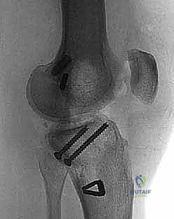
Fixation is achieved using two cannulated, fully threaded 4.0-mm or 4.5-mm cancellous screws. Guidewires are placed through the bone block and into the anterior tibia, aiming slightly distally to avoid penetrating the articular surface.
At this critical juncture, the C-arm fluoroscopy unit is brought into the field. True AP and lateral radiographic views are obtained to confirm the precise trajectory of the guidewires, ensuring they do not breach the anterior tibial cortex or violate the joint space. Once confirmed, the screws are advanced over the wires, utilizing washers if the bone block quality is marginal. The screws are tightened to achieve a rigid, bicortical-equivalent compression of the bone block into the cancellous trough.
Collateral Ligament Reconstruction and Final Tensioning
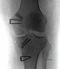
Following the secure fixation of the PCL tibial inlay, attention is turned to the collateral ligaments. If an MCL/POL reconstruction is indicated, we utilize an anatomic, double-bundle technique utilizing a tibialis anterior allograft. Femoral tunnels are drilled at the anatomic origins of the sMCL and POL on the medial epicondyle. Tibial fixation is achieved distally for the sMCL (beneath the pes anserinus) and proximally for the POL (near the semimembranosus insertion).
If a posterolateral corner (PLC) reconstruction is required, an anatomic technique recreating the FCL, popliteus tendon, and popliteofibular ligament is performed. This involves precise tunnel placement at the fibular head, the proximal posterolateral tibia, and the lateral femoral epicondyle.
The sequence of graft tensioning is of paramount importance to restore native knee kinematics. We adhere to the following sequence:
1. PCL Tensioning: The anterolateral bundle is tensioned and fixed at 90 degrees of flexion, restoring the normal anterior tibial step-off. The posteromedial bundle is tensioned and fixed near full extension (0 to 10 degrees).
2. ACL Tensioning: The ACL graft (if reconstructed concurrently) is tensioned and fixed in full extension.
3. Collateral Tensioning: The PLC/FCL is tensioned in full extension with neutral rotation and slight valgus force. The MCL/POL complex is tensioned at 20 degrees of flexion with a slight varus force to ensure appropriate medial compartment gapping.
Complications, Incidence Rates, and Salvage Management
The multiligament knee reconstruction is an inherently high-risk endeavor. The magnitude of the initial trauma, combined with the extensive surgical dissection required, predisposes the patient to a myriad of potential complications. A profound understanding of these risks and their salvage pathways is mandatory.
| Complication | Estimated Incidence | Etiology and Pathophysiology | Prevention and Salvage Management |
|---|---|---|---|
| Arthrofibrosis | 15% - 30% | Exaggerated fibroblastic response to trauma and surgery; operating too early in the acute inflammatory phase; prolonged postoperative immobilization. | Prevention: Delay surgery 2-4 weeks; early, controlled passive ROM. Salvage: Aggressive physical therapy; arthroscopic lysis of adhesions and manipulation under anesthesia (MUA) if ROM plateaus at 12 weeks. |
| Popliteal Artery Injury | < 1% (Surgical) | Direct laceration, traction, or compression by retractors during posteromedial dissection or transtibial drilling. | Prevention: Maintain strict plane anterior to medial gastrocnemius; avoid plunging drills. Salvage: Immediate intraoperative vascular surgery consultation; temporary shunting; saphenous vein interposition grafting; fasciotomies. |
| Common Peroneal Nerve Palsy | 10% - 25% (Traumatic); 2% (Surgical) | Traction injury during initial varus/hyperextension trauma; iatrogenic injury during PLC reconstruction (fibular head drilling). | Prevention: Direct visualization and neurolysis of the nerve during lateral approaches; meticulous retractor placement. Salvage: Baseline EMG at 6 weeks; ankle-foot orthosis (AFO); tendon transfers (e.g., Bridle procedure) if no recovery by 12-18 months. |
| Graft Failure / Laxity | 10% - 15% | Non-anatomic tunnel placement; premature weight-bearing; failure to recognize and reconstruct all injured structures (e.g., missed PLC leading to cruciate failure). | Prevention: Anatomic reconstruction techniques; secure cortical fixation; strict adherence to rehab protocols. Salvage: Revision reconstruction utilizing alternative allografts; potential staging with osteotomies (e.g., High Tibial Osteotomy) to correct underlying malalignment prior to revision. |
| Deep Joint Infection | 1% - 3% | Prolonged surgical time; massive surgical exposure; use of multiple allografts; compromised soft tissue envelope. | Prevention: Meticulous soft tissue handling; judicious use of tourniquet; prophylactic antibiotics. Salvage: Emergent arthroscopic or open irrigation and debridement (I&D); graft retention if caught early (within days); graft explantation and antibiotic spacers if delayed or intractable. |
Phased Post-Operative Rehabilitation Protocols
The success of a multiligament reconstruction is inextricably linked to the postoperative rehabilitation protocol. The rehabilitation must walk a tightrope between protecting the healing allografts and preventing debilitating arthrofibrosis. The protocol is highly individualized, dictated by the specific ligaments reconstructed and the security of fixation.
Phase I: Maximum Protection (Weeks 0 - 6)
The patient is placed in a hinged knee brace locked in full extension. Weight-bearing is strictly restricted to toe-touch or non-weight-bearing to protect the PCL and collateral repairs. Passive range of motion (PROM) is initiated early, typically within the first week, but is strictly limited. For PCL inlay reconstructions, flexion is limited to 90 degrees to prevent excessive tension on the healing bone block. To prevent posterior tibial subluxation during passive flexion, all ROM exercises must be performed in the prone position, utilizing gravity to assist in maintaining anterior tibial translation.
Phase II: Controlled Mobilization (Weeks 6 - 12)
The brace is gradually unlocked to allow progressive range of motion. Weight-bearing is advanced linearly, typically reaching full weight-bearing by week 8 to 10. Active quadriceps strengthening is initiated, focusing on closed-kinetic-chain exercises (e.g., mini-squats, leg presses) to minimize shear forces across the joint. Active hamstring exercises are strictly prohibited during this phase, as hamstring contraction actively translates the tibia posteriorly, placing direct stress on the PCL graft.
Phase III: Strengthening and Proprioception (Months 3 - 6)
The patient is transitioned out of the hinged brace. The focus shifts to restoring normal gait mechanics, progressive resistance training, and intensive proprioceptive drills. Open-kinetic-chain quadriceps exercises can be cautiously introduced. Hamstring strengthening is gradually incorporated, starting with isometric contractions and progressing to isotonic exercises.
Phase IV: Return to Function/Sport (Months 6 - 12+)
Return to high-impact activities and sports is delayed until a minimum of 9 to 12 months postoperatively. Progression is contingent upon achieving full, painless range of motion, resolution of all effusions, and achieving at least 85% to 90% limb symmetry on isokinetic strength testing and functional hop tests. A custom functional knee brace may be utilized during the first year of return to play, particularly for contact sports.
Summary of Landmark Literature and Clinical Guidelines
The evolution of multiligament knee reconstruction has been guided by several landmark biomechanical and clinical studies. The transition from transtibial PCL reconstruction to the tibial inlay technique was heavily influenced by the work of Markolf et al., who demonstrated the deleterious biomechanical effects of the "killer curve" on graft attrition. Their cadaveric studies proved that the inlay technique significantly reduced graft thinning and failure under cyclic loading.
Clinically, the work of Fanelli and colleagues has established the efficacy of the posteromedial approach for the tibial inlay, demonstrating excellent long-term restoration of posterior stability with a low complication rate. Their comprehensive outcomes data underscored the superiority of anatomic, double-bundle PCL reconstruction in restoring native knee kinematics compared to single-bundle techniques.
Regarding the timing of surgery, Stannard and the team at the University of Missouri have published extensively on the benefits of delayed reconstruction. Their prospective cohorts demonstrated a statistically significant reduction in the incidence of severe arthrofibrosis when surgery was delayed for 2 to 3 weeks to allow for soft tissue recovery, compared to acute interventions. Furthermore, Engebretsen's extensive registry data from Scandinavia has highlighted the critical importance of recognizing and concurrently reconstructing the posterolateral corner; failure to address PLC instability is the leading cause of early cruciate graft failure in the multiligament knee.
In conclusion, mastering the multiligament knee reconstruction requires a lifelong commitment to anatomical study, technical refinement, and evidence-based practice. The PCL tibial inlay, combined with anatomic collateral ligament reconstruction, offers a robust, biomechanically sound solution for these devastating injuries, providing our patients with the best possible chance to restore function and reclaim their quality of life.
