Mastering Meniscus Basic Science & Biomechanics: Orthopedic Board Prep MCQs

Key Takeaway
The menisci primarily consist of longitudinal (circumferential) type I collagen fibers, essential for converting vertical compressive knee loads into 'hoop stresses.' These stresses are then efficiently resisted by the circumferential fibers, allowing the meniscus to transmit load, absorb shock, and protect articular cartilage. Radial fibers act as tie-ropes, enhancing structural integrity.
You are presented with this patient who has suffered a twisting injury to the knee. Please examine this radiograph and discuss the relevant surgical anatomy and the biomechanical implications of injury to this structure.
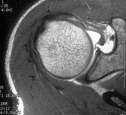
Candidate: The radiograph shows an avulsion of the posterior horn of the medial meniscus root. The meniscus is primarily composed of type I collagen. Its function is to convert axial loads into hoop stresses. A root tear effectively acts like a total meniscectomy, leading to joint extrusion and rapid osteoarthritis.
Failing to mention the specific anatomical classification of the meniscus (fibrocartilage vs. hyaline). Simply stating "it's a tear" without quantifying the functional loss (e.g., "loss of hoop stress capability") or failing to mention that this is a biomechanical equivalent to a total meniscectomy.
Structured as: 1. Pathology: Identify the posterior medial meniscus root avulsion. 2. Anatomy/Histology: Note it is fibrocartilage (Type I collagen) with circumferential fibers. 3. Biomechanics: Explain the conversion of axial compressive loads into circumferential hoop stresses. 4. Clinical Consequence: State that root avulsion causes loss of circumferential integrity, leading to meniscal extrusion, increased contact pressure (up to 200-300%), and rapid progression to symptomatic osteoarthritis.
What are the key anatomical differences between the medial and lateral meniscus that influence their surgical management?
Candidate: The medial is C-shaped and firmly attached to the capsule and MCL, so it's less mobile. The lateral is O-shaped, more mobile, and doesn't attach to the LCL.
Only describing shape and mobility. Failing to mention the clinical implication: the medial meniscus's lack of mobility makes it more prone to injury in valgus stress/MCL-involved injuries, while the lateral meniscus's hyper-mobility (and the popliteus hiatus) influences its susceptibility to specific tear patterns.
Structural differences: - Morphology: C-shaped (medial) vs O-shaped (lateral). - Mobility: Medial is anchored tightly to the deep MCL; lateral is highly mobile, with gaps at the popliteus tendon. - Clinical Implications: The medial meniscus is more susceptible to injury in combined MCL/ACL scenarios. The lateral meniscus has a greater excursion, which protects it in some instances but makes it more susceptible to rotational shear in ACL deficiency.
