Internal Fixation of Fifth Metatarsal Fractures: A Masterclass Surgical Guide
Key Takeaway
Internal fixation of fifth metatarsal fractures, particularly Jones and diaphyseal stress fractures, requires precise intramedullary screw placement. This technique provides robust biomechanical stability, facilitating early rehabilitation in high-demand athletes. Critical steps include protecting the sural nerve, achieving an optimal starting trajectory parallel to the hindfoot, and addressing medullary sclerosis with inlay bone grafting in cases of nonunion to ensure reliable osseous consolidation.
Comprehensive Introduction and Patho-Epidemiology
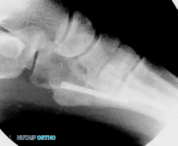
Fractures of the fifth metatarsal represent a highly complex, biomechanically demanding spectrum of injuries that necessitate a profound understanding of foot kinematics, regional vascular anatomy, and patient-specific functional requirements. Historically categorized by Lawrence and Botte into three distinct anatomic zones, these fractures present unique challenges to the orthopedic surgeon. Zone 1 encompasses the tuberosity, typically resulting from an avulsion injury involving the lateral band of the plantar fascia and the peroneus brevis tendon. Zone 2, the classic Jones fracture, occurs at the metaphyseal-diaphyseal junction and involves the fourth-fifth intermetatarsal articulation. Zone 3 fractures occur in the proximal diaphysis distal to the intermetatarsal articulation and are predominantly stress fractures secondary to chronic repetitive microtrauma. Fractures occurring in Zones 2 and 3 are notoriously prone to delayed union, nonunion, and refracture, making their management a subject of intense orthopedic scrutiny.
The vulnerability of the proximal fifth metatarsal to nonunion is primarily attributed to a well-documented vascular "watershed" area combined with the immense, repetitive tensile forces exerted during the terminal stance and heel-off phases of the human gait cycle. During these phases, the foot transitions from a flexible shock absorber to a rigid lever arm. The peroneus brevis and the lateral band of the plantar fascia exert massive distractive forces across the proximal fifth metatarsal, effectively pulling the fracture fragments apart. When this mechanical distraction is superimposed upon a region with inherently tenuous blood supply, the biologic potential for endosteal and periosteal callus formation is severely compromised. Consequently, what may appear as a simple, undisplaced transverse fracture on initial radiographs can rapidly evolve into a recalcitrant nonunion characterized by profound medullary sclerosis and cortical hypertrophy.
Epidemiologically, these injuries are ubiquitous across both the general population and elite athletic cohorts, though the patho-etiology differs significantly between the two. In the general population, acute Jones fractures typically result from an acute adduction force applied to a plantarflexed foot, such as stepping off a curb or twisting the ankle on an uneven surface. Conversely, in the athletic population—particularly among professional football, basketball, and soccer players—Zone 3 stress fractures develop insidiously. These athletes subject their feet to extraordinary cyclical loading, leading to microtrabecular failure that outpaces the osteoblastic remodeling capacity. The resulting stress fracture is often heralded by weeks of prodromal lateral foot pain before a catastrophic completion of the fracture line occurs during a sudden change of direction or explosive jump.
For high-demand patients, professional athletes, and cases of established nonunion, conservative management with cast immobilization often yields unacceptable rates of clinical failure, prolonged absence from sport, and high rates of refracture upon return to play. Consequently, internal fixation utilizing a solid or cannulated intramedullary malleolar screw has firmly established itself as the gold standard of care. This operative intervention fundamentally alters the biomechanical environment, neutralizing the tensile forces and converting them into interfragmentary compression. This masterclass chapter comprehensively details the evidence-based surgical techniques, critical anatomical considerations, and meticulously phased postoperative protocols required to achieve reliable, durable osseous consolidation and facilitate a safe return to pre-injury activity levels.
Detailed Surgical Anatomy and Biomechanics
Osteology and Three-Dimensional Morphology
The osteology of the fifth metatarsal is uniquely adapted to its role as the lateral column's primary weight-bearing and stabilizing strut. Unlike the relatively straight central metatarsals, the fifth metatarsal exhibits a complex three-dimensional morphology characterized by a distinct lateral and plantar bow. The proximal aspect consists of the base, which articulates medially with the fourth metatarsal and proximally with the cuboid. Extending laterally and proximally from the base is the prominent styloid process (tuberosity), which serves as a critical lever arm for eversion and plantarflexion. Understanding this inherent curvature is paramount during intramedullary fixation; a straight trajectory initiated from the anatomic center of the tuberosity will inevitably abut the medial or plantar cortex of the diaphysis, leading to iatrogenic cortical blowout or malreduction.
The Vascular Watershed
The blood supply to the proximal fifth metatarsal dictates its healing potential and is the primary anatomic factor driving the high rate of nonunion in Zone 2 and Zone 3 fractures. The bone receives its arterial supply from three distinct sources: the principal nutrient artery, metaphyseal perforating vessels, and a delicate periosteal network. The nutrient artery enters the medial cortex in the middle third of the diaphysis and arborizes proximally to supply the endosteum. Conversely, the metaphyseal vessels enter the tuberosity and supply the proximal base. Between these two robust vascular networks lies a relative avascular zone at the metaphyseal-diaphyseal junction. Disruption of the intraosseous microcirculation at this specific watershed zone during a traumatic event or chronic stress severely impairs the biologic healing response, necessitating rigid mechanical stability to allow for revascularization.
Musculotendinous and Ligamentous Forces
The biomechanical environment of the proximal fifth metatarsal is dominated by powerful musculotendinous and ligamentous attachments that exert continuous, multiaxial forces across the bone. The peroneus brevis tendon inserts broadly onto the dorsolateral aspect of the tuberosity, acting as the primary evertor of the foot. The peroneus tertius inserts onto the dorsal aspect of the proximal metaphysis, contributing to dorsiflexion and eversion. Plantarly, the robust lateral band of the plantar fascia attaches to the plantar aspect of the tuberosity. During the propulsive phase of gait, as the heel rises and the foot supinates, these structures undergo maximal tension. This creates a powerful bending moment and tensile stress across the lateral cortex of the proximal fifth metatarsal. Intramedullary fixation must be robust enough to act as an internal tension band, resisting these distractive forces until solid cortical bridging occurs.
Neurologic Considerations and the Sural Nerve
The surgical approach to the base of the fifth metatarsal places the sural nerve at significant and well-documented risk. As demonstrated in landmark cadaveric studies by Donley et al., the sural nerve courses distally along the lateral aspect of the foot and bifurcates into a dorsal branch and a straight lateral branch. The dorsolateral branch, in particular, courses dangerously close to the optimal percutaneous insertion point for an intramedullary screw. Iatrogenic injury, transection, or entrapment of this nerve via aggressive retraction or sharp dissection can lead to debilitating postoperative neuromas. These neuropathic pain syndromes are often refractory to conservative treatment and can completely overshadow the successful osseous union of the fracture, leading to a disastrous clinical outcome.
Exhaustive Indications and Contraindications
The decision to proceed with operative intervention for a fifth metatarsal fracture requires a meticulous assessment of the fracture pattern, chronicity, and the physiological and functional demands of the patient. Acute Zone 1 tuberosity avulsion fractures are almost universally managed nonoperatively with a hard-soled shoe or walking boot, as they possess an excellent blood supply and a vast cancellous bed that ensures rapid healing. However, the paradigm shifts dramatically when evaluating Zone 2 (Jones) and Zone 3 (diaphyseal stress) fractures. In these zones, the inherent biologic disadvantage necessitates a more aggressive posture, particularly in patients who cannot tolerate prolonged immobilization or the high risk of delayed union.
For acute Zone 2 fractures in the general population, a trial of non-weight-bearing cast immobilization for 6 to 8 weeks remains a viable option, provided the patient is thoroughly counseled on the approximately 15% to 25% risk of nonunion. However, in high-demand patients, elite athletes, manual laborers, and military personnel, acute intramedullary screw fixation is strongly indicated. Operative intervention in this cohort significantly reduces the time to clinical union, minimizes the risk of muscle atrophy secondary to prolonged immobilization, and drastically lowers the incidence of nonunion. Furthermore, any Zone 2 or Zone 3 fracture that presents with evidence of delayed union (lack of radiographic progression at 8 weeks) or established nonunion (sclerotic fracture margins, canal obliteration) is an absolute indication for surgical intervention, often requiring biologic augmentation in addition to mechanical stabilization.
Torg et al. provided a highly useful radiographic classification for proximal fifth metatarsal fractures that directly guides operative indications. Torg Type I represents acute fractures with sharp margins and no intramedullary sclerosis; these can be treated conservatively in low-demand patients but are often fixed in athletes. Torg Type II indicates delayed union, characterized by a widened fracture line and early intramedullary sclerosis; these are strong candidates for operative fixation. Torg Type III indicates established nonunion, defined by complete obliteration of the medullary canal by dense sclerotic bone; these require mandatory operative intervention, typically involving canal reaming, intramedullary fixation, and often an inlay bone graft to breach the sclerotic barrier.
Contraindications to intramedullary fixation must be rigorously respected to prevent catastrophic complications. Active local or systemic infection is an absolute contraindication. Severe peripheral vascular disease, profound neuropathy (such as advanced Charcot neuroarthropathy), and inadequate soft tissue coverage over the lateral border of the foot represent significant relative contraindications that require careful multidisciplinary optimization before any surgical intervention is considered. Additionally, patients who are non-ambulatory or possess extremely low functional demands may not benefit from the risks associated with surgical intervention, making benign neglect or chronic bracing a more appropriate strategy.
| Clinical Scenario | Operative Indication Status | Rationale / Recommended Approach |
|---|---|---|
| Acute Zone 1 (Tuberosity Avulsion) | Rarely Indicated | Excellent blood supply; heals reliably with symptomatic weight-bearing in a stiff-soled shoe or boot. |
| Acute Zone 2 (Jones) in Athlete | Strongly Indicated | Minimizes time to return to play; prevents high rate of nonunion associated with conservative care. Intramedullary screw. |
| Acute Zone 2 (Jones) in Low-Demand | Relative Indication | Trial of NWB casting is acceptable. Surgery offered for faster rehab or if patient refuses prolonged NWB status. |
| Zone 3 Stress Fracture (Torg Type II/III) | Absolutely Indicated | High risk of complete failure with conservative care. Requires intramedullary screw ± canal reaming ± bone grafting. |
| Distal Spiral (Dancer's Fracture) | Rarely Indicated | Robust periosteal blood supply. Heals excellently with conservative care despite displacement. Surgery only for severe open/crush injuries. |
| Active Soft Tissue Infection | Absolute Contraindication | High risk of deep hardware infection and osteomyelitis. Treat infection first. |
Pre-Operative Planning, Templating, and Patient Positioning
Advanced Imaging and Radiographic Assessment
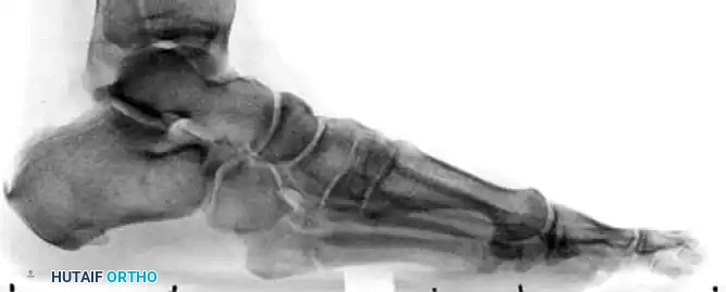
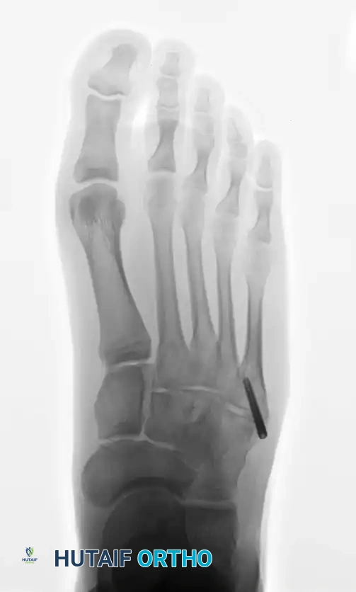
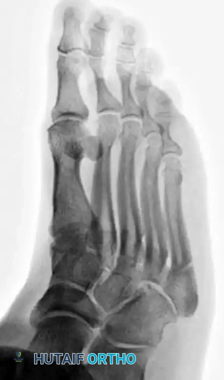
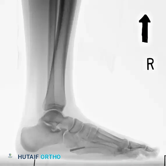
Meticulous preoperative planning begins with high-quality, weight-bearing orthogonal radiographs of the foot, including anteroposterior (AP), lateral, and oblique views. The oblique view is particularly critical, as it perfectly profiles the fracture line at the metaphyseal-diaphyseal junction and allows for accurate assessment of fracture displacement and comminution. In cases of chronic pain, delayed union, or established nonunion, standard radiographs often underestimate the degree of medullary sclerosis and cortical hypertrophy. In these complex scenarios, a fine-cut Computed Tomography (CT) scan is highly recommended. CT imaging provides unparalleled detail regarding the precise diameter of the medullary canal, the extent of the sclerotic plug, and the three-dimensional morphology of the bone, which is essential for accurate preoperative templating.
Digital Templating and Hardware Selection
Preoperative digital templating is a non-negotiable step to prevent intraoperative disasters such as lateral cortical blowout or inadequate distal purchase. The surgeon must measure the narrowest portion of the medullary canal (the isthmus) on the AP and lateral radiographs to determine the appropriate screw diameter. Historically, 4.0mm and 4.5mm screws were utilized; however, modern biomechanical literature strongly advocates for the use of the largest diameter screw that the canal can safely accommodate—typically 4.5mm to 5.5mm, and occasionally up to 6.5mm in large athletes. A screw that is too small will fail to achieve adequate cortical purchase and is at high risk for fatigue failure. The length of the screw must also be templated; it must be long enough to bypass the fracture site and engage the diaphyseal isthmus, ensuring that all threads are positioned distal to the fracture line to facilitate true interfragmentary compression.
Patient Positioning and Operating Room Setup
Optimal patient positioning is critical for obtaining unobstructed fluoroscopic views and facilitating a seamless surgical workflow. The patient may be placed in the lateral decubitus position, which naturally presents the lateral border of the foot to the surgeon. Alternatively, and often preferred for ease of anesthesia management, the patient can be positioned supine with a large, dense bump placed beneath the ipsilateral hip. This internally rotates the lower extremity, bringing the lateral border of the foot directly toward the ceiling. A completely radiolucent operating table is mandatory to allow the C-arm to swing freely between AP, lateral, and oblique planes without interference from table posts or radiopaque seams.
Anesthesia and Hemostasis
The procedure is typically performed under general anesthesia or a regional anesthetic block. A popliteal sciatic nerve block is highly advantageous, providing excellent intraoperative anesthesia and profound postoperative analgesia, thereby minimizing the need for systemic opioids. To ensure a bloodless surgical field and permit meticulous identification of the sural nerve branches, a pneumatic tourniquet is applied to either the proximal thigh or the calf. The limb is exsanguinated with an Esmarch bandage prior to tourniquet inflation. Prophylactic intravenous antibiotics (typically a first-generation cephalosporin) must be administered within one hour prior to the surgical incision.
Step-by-Step Surgical Approach and Fixation Technique
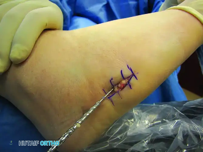
1. Incision and Sural Nerve Protection
The surgical approach demands precision to avoid devastating iatrogenic neurologic injury. A 1.5 to 2.0 cm longitudinal incision is made starting precisely at the palpable tip of the fifth metatarsal tuberosity and extending proximally in line with the metatarsal shaft. The initial incision must traverse the skin only. The surgeon must strictly avoid using a scalpel for deep dissection. Instead, meticulous blunt dissection using a small hemostat is employed to gently spread the subcutaneous adipose tissue down to the periosteum. During this blunt dissection, the dorsal and straight lateral branches of the sural nerve must be actively sought, directly visualized, and gently retracted away from the planned drill portal.
2. Establishing the "High and Inside" Starting Point
Achieving the correct starting point is arguably the most critical and technically demanding step of the entire operation. Because of the inherent lateral and plantar bow of the fifth metatarsal, initiating the guidewire directly on the anatomic tip of the tuberosity will invariably result in a trajectory that breaches the medial or plantar cortex. To counteract this, the surgeon must utilize the "high and inside" rule. The optimal starting portal is located slightly dorsal and medial to the anatomic center of the tuberosity. The tendon of the peroneus brevis, which inserts broadly over this area, may obscure the portal. The surgeon should longitudinally split the tendon in line with its fibers or elevate a small dorsal cuff of the tendon off the bone to gain unobstructed access to the starting point.
3. Guidewire Insertion and Trajectory Optimization
Once the starting point is established, a stout guidewire (typically 1.4mm or 1.6mm, depending on the cannulated system) is introduced. The trajectory of the wire must be almost perfectly parallel to the plantar aspect of the foot in the sagittal plane and parallel to the medial border of the foot in the axial plane. The wire is advanced under continuous or frequent multi-planar fluoroscopy. The goal is to pass the wire precisely down the center of the medullary canal, crossing the fracture site, and resting centrally within the distal diaphyseal isthmus. If the guidewire meets hard, unyielding resistance before crossing the fracture, the surgeon must immediately stop; the wire is likely abutting the dense medial or plantar cortex. The wire should be withdrawn, the starting point adjusted slightly more dorsal and medial, and the trajectory corrected.
4. Drilling, Tapping, and Canal Preparation
With the guidewire perfectly positioned and confirmed via orthogonal fluoroscopy, a cannulated drill bit is advanced over the wire into the medullary canal. The drill must cross the fracture site and engage the diaphyseal cortex to prepare the canal for the screw threads. In young, athletic patients with dense, hypertrophic cortical bone, the canal must be meticulously tapped over the wire trajectory. Attempting to insert a large-diameter screw into dense cortical bone without prior tapping is a frequent cause of intraoperative screw breakage or catastrophic lateral cortical blowout. If a solid screw system is preferred for its superior fatigue strength, the guidewire is removed after drilling, and the solid screw is inserted along the prepared track.
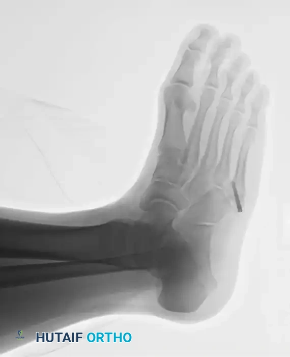
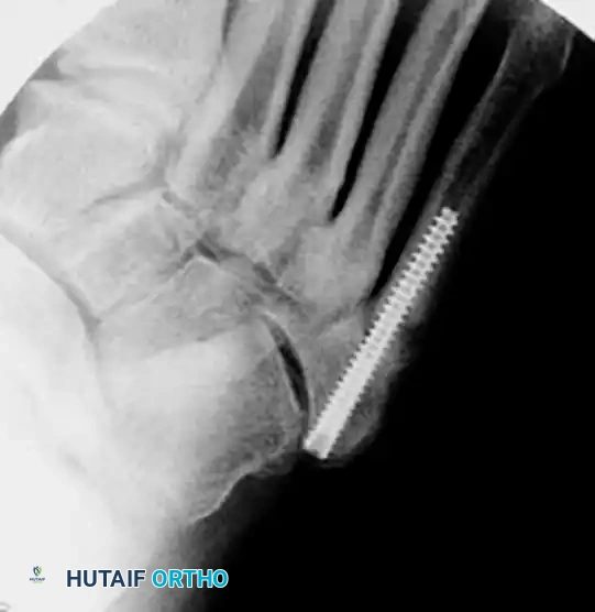
5. Screw Insertion and Head Burial
The selected partially threaded screw (typically 4.5mm to 5.5mm in diameter) is advanced over the guidewire. The surgeon must ensure that all threads of the screw pass completely distal to the fracture line; if threads bridge the fracture, interfragmentary compression cannot be achieved, and the fracture will be held in distraction. As the screw head approaches the tuberosity, it is absolutely imperative to countersink or bury the head deep into the bone. A prominent screw head left proud on the lateral border of the foot will cause severe, unrelenting irritation to the overlying peroneus brevis tendon and the thin subcutaneous tissues, often necessitating a secondary operation for hardware removal once the fracture has united.
6. Management of Nonunions: The Torg Inlay Graft Technique
While acute fractures respond excellently to isolated intramedullary screw fixation, chronic Torg Type III nonunions present a profound biologic deficit that mechanical stabilization alone cannot overcome. The complete obliteration of the medullary canal by dense, avascular sclerotic bone acts as an impenetrable barrier to osteogenesis. In these cases, the Torg inlay bone graft technique is highly effective. A longitudinal incision is made directly over the nonunion. A rectangular window (approximately 0.5 cm x 1.5 cm) is meticulously outlined across the fracture site using a microsaw. The sclerotic bone plug is then aggressively resected using a high-speed burr or sharp curettes until healthy, punctate bleeding cancellous bone is encountered both proximally and distally, effectively re-establishing the patency of the medullary canal. A precisely contoured corticocancellous autograft, harvested from the anteromedial proximal tibia, is tamped into the defect. This biologic augmentation is then stabilized with an intramedullary screw, providing the ultimate biomechanical and biologic synergy.
Complications, Incidence Rates, and Salvage Management
Despite meticulous surgical technique, the operative management of fifth metatarsal fractures carries a distinct complication profile. The most frequent complication is symptomatic hardware prominence, occurring in up to 20% of patients if the screw head is not adequately countersunk. This typically presents as focal pain and swelling over the tuberosity, exacerbated by tight-fitting footwear. Management is straightforward: once robust clinical and radiographic union is confirmed (usually after 6 months), the screw can be removed in a brief outpatient procedure. However, premature hardware removal before complete medullary consolidation invites a massive risk of refracture.
Iatrogenic fractures during screw insertion represent a severe intraoperative complication. Lateral cortical blowout occurs when the starting point is too lateral or the screw diameter selected is too large for the native canal. Medial cortical breach occurs when the starting point is too plantar or lateral, driving the trajectory medially. If a blowout occurs intraoperatively, the surgeon must immediately reassess. If the fracture is still stable, a smaller diameter screw or a longer screw that bypasses the blowout may be utilized. If stability is compromised, the surgeon must be prepared to salvage the fixation using a low-profile lateral locking plate to span the fracture and the iatrogenic defect.
Sural nerve injury is a devastating complication that can result in complex regional pain syndrome (CRPS) or a painful localized neuroma. Incidence is reported between 2% and 5%. Prevention through meticulous blunt dissection is paramount. If a neuroma develops and is refractory to conservative measures (gabapentinoids, targeted corticosteroid injections, desensitization therapy), surgical excision of the neuroma with proximal burying of the nerve stump into the belly of the peroneus brevis muscle may be required.
Delayed union and nonunion, despite operative fixation, still occur in approximately 5% of cases, most commonly due to the use of an undersized screw (e.g., a 4.0mm screw in a canal that could accommodate a 5.5mm screw) or premature return to high-impact activities. Refracture with hardware failure (a broken screw inside the canal) is the ultimate nightmare scenario. Salvage of a broken intramedullary screw requires specialized extraction instruments (trephines, hollow reamers) to remove the distal threaded segment. Once removed, the canal must be over-reamed, biologically augmented with autograft or bone morphogenetic protein (BMP), and stabilized with a significantly larger diameter solid screw or a robust lateral plating construct.
| Complication | Estimated Incidence | Prevention Strategy | Salvage Management |
|---|---|---|---|
| Symptomatic Hardware | 10% - 20% | Aggressive countersinking of screw head into the tuberosity. | Hardware removal only after complete radiographic union (bridging trabeculae). |
| Sural Nerve Neuroma | 2% - 5% | Blunt dissection only; avoid sharp scalpels deep to the dermis. | Neuroma excision and proximal nerve stump burial into muscle. |
| Lateral Cortical Blowout | 1% - 3% | "High and inside" starting point; accurate preoperative CT templating. | Intraoperative conversion to lateral locking plate fixation. |
| Nonunion / Delayed Union | 3% - 5% | Use largest possible screw diameter; ensure threads cross fracture line. | Revision with larger screw, canal reaming, and Torg inlay autograft. |
| Hardware Failure (Breakage) | < 2% | Avoid undersized screws; strictly enforce NWB and RTP protocols. | Trephine extraction of broken distal tip; over-reaming; massive screw or plate. |
Phased Post-Operative Rehabilitation Protocols
The postoperative rehabilitation protocol following intramedullary screw fixation of a proximal fifth metatarsal fracture is a delicate balancing act. The surgeon must provide rigid immobilization to protect the tenuous mechanical construct during the early phases of biologic healing, while simultaneously encouraging early functional rehabilitation to prevent profound disuse atrophy, joint stiffness, and deep vein thrombosis. A meticulously phased approach is essential for optimal outcomes.
Phase I: Immediate Postoperative (Weeks 0-2)
Immediately following the procedure, while still in the operating room, the patient is placed in a well-padded, short-leg, non-walking cast or a rigid fracture boot locked in neutral dorsiflexion. The primary goals during this phase are strict protection of the surgical site, wound healing, and edema management. The patient is instructed to remain strictly non-weight-bearing (NWB) utilizing axillary crutches, a knee scooter, or a wheelchair. Strict elevation of the operative limb above the level of the heart and continuous cryotherapy behind the knee or over the proximal thigh are heavily emphasized to minimize dependent edema and protect the delicate dorsal incision from dehiscence.
Phase II: Early Weight Bearing and Mobility (Weeks 2-6)
At the two-week postoperative mark, the patient returns to the clinic for suture removal and the first postoperative radiographic assessment to confirm maintenance of hardware position and fracture alignment. Assuming uncomplicated wound healing, the patient is transitioned into a removable rigid CAM (Controlled Ankle Motion) boot. Weight-bearing is initiated and progressively advanced from partial weight-bearing with crutches to full weight-bearing as tolerated within the protective boot. Crucially, the patient is encouraged to remove the boot multiple times daily to perform active, non-weight-bearing range of motion (ROM) exercises of the ankle (dorsiflexion, plantarflexion) and toes to prevent capsular contracture and promote tendon gliding. Inversion and eversion are strictly prohibited to avoid stressing the peroneus brevis insertion.
Phase III: Functional Rehabilitation (Weeks 6-10)
Around the six-week mark, repeat orthogonal radiographs are obtained. If there is evidence of early bridging callus and an absence of point tenderness directly over the fracture site, the patient is gradually weaned from the CAM boot into a stiff-soled athletic shoe, often augmented with a rigid carbon fiber insert to limit forefoot bending moments. Formal physical therapy is initiated. The focus of therapy shifts toward aggressive peroneal strengthening, restoration of normal proprioception, Achilles tendon stretching, and gait normalization. Closed-chain exercises are introduced, and the patient begins low-impact cardiovascular training such as stationary cycling or deep-water running.
Phase IV: Return to Play and Advanced Conditioning (Weeks 10-12+)
The final phase of rehabilitation is the most critical, particularly for competitive athletes eager to return to the field. Return to high-impact, multidirectional sports is strictly prohibited until the fracture has achieved both absolute clinical and radiographic union. Clinical union is defined as the complete absence of pain with aggressive single-leg hopping and palpation. Radiographic union requires the visualization of bridging trabeculae across at least three out of four cortices on orthogonal views, with complete obliteration of the fracture line. This consolidation process typically requires 10 to 12 weeks, though it may take longer in patients with profound preoperative sclerosis. Premature return to play, driven by external pressures rather than biologic reality, significantly increases the risk of catastrophic hardware failure and refracture. Upon return to sport, many athletes are prescribed a custom orthotic with a lateral post to permanently offload the lateral column of the foot
