INTRODUCTION TO TOTAL HIP ARTHROPLASTY
Total hip arthroplasty (THA) is widely regarded as one of the most successful surgical interventions in modern medicine. Originally, the primary indication for THA was the alleviation of incapacitating arthritic pain in patients older than 65 years whose symptoms could not be sufficiently relieved by nonsurgical means. In that era, the only surgical alternative was resection of the hip joint (the Girdlestone resection arthroplasty). While the primary goal remains pain relief, the secondary goal—improved function and restoration of hip biomechanics—has become increasingly paramount.
As the operation was documented to be remarkably successful, the indications rapidly expanded. Today, THA is utilized to treat a vast array of hip pathologies across a much broader demographic spectrum.
INDICATIONS FOR TOTAL HIP ARTHROPLASTY
The cardinal indication for THA is severe, debilitating pain localized to the hip joint that significantly impairs the patient's activities of daily living (ADLs), sleep, and overall quality of life, coupled with radiographic evidence of joint destruction.
Clinical Pearl: Pain in the presence of a destructive process in the hip joint, as evidenced on a radiograph, is the primary indication for surgery. Patients presenting with limitation of motion, a limp, or leg-length inequality—but with little or no hip pain—are generally not candidates for total hip arthroplasty.
Comprehensive Pathological Indications
The disorders of the hip joint for which THA may be indicated are extensive. They can be broadly categorized into the following groups:
- Degenerative Joint Disease: Primary osteoarthritis and secondary osteoarthritis (hypertrophic).
- Inflammatory Arthropathies: Rheumatoid arthritis, Juvenile rheumatoid arthritis (Still's disease), Ankylosing spondylitis, and Lupus.
- Osteonecrosis (Avascular Necrosis): Idiopathic, post-traumatic (post-fracture or dislocation), slipped capital femoral epiphysis (SCFE), hemoglobinopathies (sickle cell disease), renal disease, corticosteroid-induced, alcoholism, Caisson disease, and Gaucher disease.
- Post-Traumatic Conditions: Traumatic dislocation, acetabular fractures, and nonunion of femoral neck or trochanteric fractures with head involvement.
- Childhood and Developmental Disorders: Congenital dislocation or dysplasia of the hip (DDH), Coxa plana (Legg-Calvé-Perthes disease), and SCFE.
- Infectious (Post-Eradication): Pyogenic arthritis or osteomyelitis (hematogenous or postoperative) and Tuberculosis (provided the infection is definitively quiescent).
- Metabolic and Hereditary Disorders: Paget disease, Hemophilia, and Achondroplasia.
- Salvage of Failed Reconstructions: Failed osteotomy, cup arthroplasty, hemiarthroplasty (femoral head prosthesis), Girdlestone procedure, previous total hip replacement, or resurfacing arthroplasty.
- Oncology: Bone tumors involving the proximal femur or acetabulum.
Age Considerations and Patient Selection
Historically, patients 60 to 75 years old were considered the most suitable candidates for THA. However, since the 1990s, this age range has expanded significantly in both directions.
The Elderly Patient:
With an aging population, many older individuals are becoming candidates for surgery. Advanced age in itself is not a contraindication to surgery. In a landmark review of 99 procedures in patients 80 years old and older, Brander et al. found that complication rates and length of hospital stay were not significantly different from a control group of younger individuals, and functional gains were similar. Poor outcomes are related more to medical comorbidities than to chronological age alone. The 1994 National Institutes of Health Consensus Statement concluded that "THR is an option for nearly all patients with diseases of the hip that cause chronic discomfort and significant functional impairment."
The Young, High-Demand Patient:
In younger individuals, THA is not the only reconstructive procedure available. Sir John Charnley famously warned that:
1. Procedures suitable for older patients may not be suitable for younger patients.
2. The problems in bilateral disease are fundamentally different from those in unilateral disease.
3. The treatment for an arthritic hip with a good range of motion differs from that for an arthritic hip with poor motion.
4. The biomechanical demands on the hip in a manual laborer are vastly greater than those in a sedentary worker.
The potential for aseptic loosening, polyethylene wear, and subsequent osteolysis in young patients must be emphasized, alongside the increased risk of infection and complications should revision surgery become necessary.
For young, active males with osteoarthritis or osteonecrosis, alternative joint-preserving or bone-conserving procedures must be considered. Historically, hip resurfacing emerged as a bone-conserving alternative for this demographic.
Surgical Warning: While metal-on-metal resurfacing (such as the Birmingham Hip Resurfacing) preserves proximal femoral bone stock, surgeons must be acutely aware of the risks of adverse local tissue reactions (ALTR) and metallosis. Patient selection (typically large-stature males) and precise surgical technique are critical to avoiding edge-loading and subsequent failure.
ALTERNATIVES TO TOTAL HIP ARTHROPLASTY
Before any major joint reconstruction is recommended, conservative measures must be exhausted. These include weight loss, nonsteroidal anti-inflammatory drugs (NSAIDs), activity modification, and the use of an assistive device (cane) in the contralateral hand. If surgery is anticipated in a young laborer, preoperative job retraining to a more sedentary vocation should be considered to delay the need for surgery and protect the eventual implant.
When conservative measures fail, alternatives to THA in the young patient include:
- Arthrodesis (Hip Fusion): A viable option for young, vigorous patients (especially men) with unilateral posttraumatic arthritis or osteonecrosis. Modern techniques utilizing internal fixation without spica cast immobilization have made this more acceptable. It can be converted to a THA later in life.
- Osteotomy: Femoral or periacetabular osteotomy (PAO) should be considered for young patients with hypertrophic arthritis or dysplasia, provided the joint is not grossly incongruous and satisfactory motion remains. PAO can decrease the need for structural bone grafting if later conversion to THA is required.
- Core Decompression and Grafting: Core decompression, with or without vascularized fibular grafting, should be considered for early-stage idiopathic osteonecrosis of the femoral head (Ficat Stages I and II) before subchondral collapse occurs.
BIOMECHANICS OF THE HIP JOINT
A profound understanding of hip biomechanics is essential for successful THA. The hip acts as a fulcrum in a first-class lever system during single-leg stance.
- Joint Reaction Force (JRF): The JRF across the hip joint can reach 3 to 6 times body weight during normal walking. It is determined by the ratio of the body weight lever arm to the abductor lever arm.
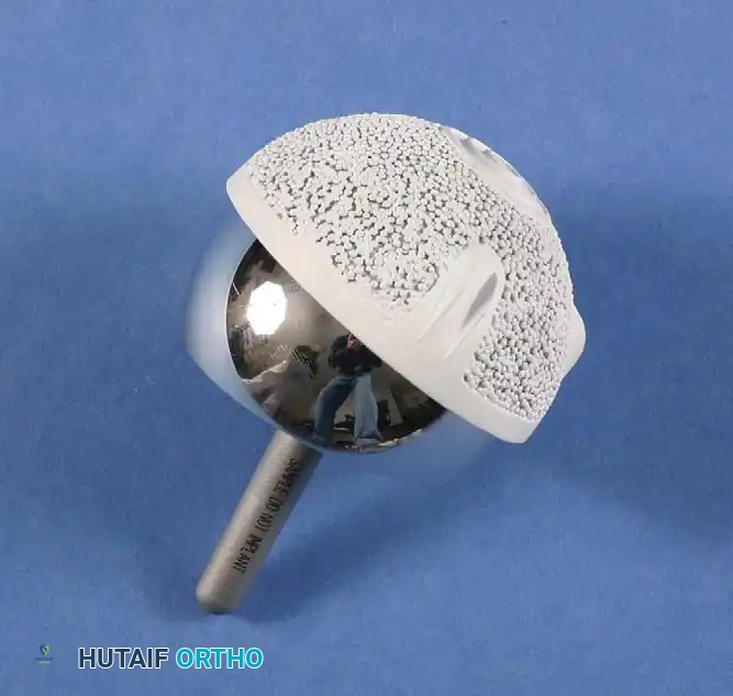
- Femoral Offset: This is the perpendicular distance from the center of rotation of the femoral head to the anatomical axis of the femur. Restoring or slightly increasing femoral offset during THA increases the abductor moment arm. This reduces the force required by the abductors to maintain a level pelvis, thereby decreasing the overall JRF and minimizing polyethylene wear.
- Center of Rotation (COR): The acetabular component should be placed at the true anatomical COR (inferior and medial). Superior and lateral placement increases the body weight lever arm, exponentially increasing the JRF and the risk of aseptic loosening.
SURGICAL APPROACHES AND POSITIONING
The choice of surgical approach depends on surgeon experience, patient anatomy, and whether the procedure is primary or revision.
1. Posterior Approach (Moore or Southern)
- Positioning: Lateral decubitus.
- Interval: Gluteus maximus is split in line with its fibers. There is no true internervous plane.
- Technique: The short external rotators (piriformis, superior gemellus, obturator internus, inferior gemellus) and the quadratus femoris are detached from the greater trochanter and reflected posteriorly to protect the sciatic nerve. The posterior capsule is incised.
- Advantages: Excellent expansile exposure for both femur and acetabulum; preserves the abductor mechanism (gluteus medius/minimus).
- Disadvantages: Historically associated with a higher rate of posterior dislocation, though meticulous posterior soft-tissue repair has mitigated this risk.
2. Direct Anterior Approach (Smith-Petersen)
- Positioning: Supine, often on a specialized traction table (e.g., Hana table).
- Interval: Internervous and intermuscular plane between the Tensor Fasciae Latae (superior gluteal nerve) and the Sartorius/Rectus Femoris (femoral nerve).
- Technique: The ascending branches of the lateral femoral circumflex artery are ligated. The anterior capsule is exposed and incised.
- Advantages: True internervous plane, no muscle detachment required, lower dislocation risk, and facilitates intraoperative fluoroscopy for component positioning and leg length assessment.
- Disadvantages: Steep learning curve, risk of lateral femoral cutaneous nerve (LFCN) neuropraxia, and difficult femoral exposure in muscular or obese patients.
3. Anterolateral Approach (Watson-Jones)
- Positioning: Supine or lateral decubitus.
- Interval: Between the Tensor Fasciae Latae (superior gluteal nerve) and the Gluteus Medius (superior gluteal nerve).
- Technique: Requires partial detachment or splitting of the anterior third of the gluteus medius and minimus to access the superior capsule.
- Advantages: Excellent acetabular exposure, very low dislocation rate.
- Disadvantages: Risk of superior gluteal nerve injury if the split extends >5 cm proximal to the greater trochanter; potential for postoperative abductor weakness and Trendelenburg gait.
STEP-BY-STEP SURGICAL TECHNIQUE
While specific techniques vary by approach and implant philosophy, the fundamental steps of a primary THA remain consistent.
Step 1: Preoperative Templating
Templating is mandatory. It determines the approximate size of the acetabular and femoral components, the level of the femoral neck cut, the required offset, and the strategy for restoring leg length.
Step 2: Exposure and Dislocation
Following the chosen surgical approach, a capsulotomy or capsulectomy is performed. The hip is dislocated (anteriorly for anterior approaches, posteriorly for posterior approaches). In cases of severe protrusio acetabuli or ankylosis, an in situ neck cut may be required before the head can be extracted.
Step 3: Femoral Neck Resection
Using the preoperative template as a guide, the distance from the lesser trochanter to the planned cut is measured. An oscillating saw is used to resect the femoral neck at the appropriate angle and version.
Step 4: Acetabular Preparation and Implantation
- Exposure: Retractors are placed anteriorly, inferiorly (under the transverse acetabular ligament), and posteriorly to achieve 360-degree visualization of the acetabulum.
- Reaming: The acetabulum is reamed sequentially. The goal is to remove all cartilage down to bleeding subchondral bone, medializing to the true floor (cotyloid fossa) without breaching the medial wall.
- Implantation: The acetabular shell is impacted into place. The target orientation is typically 40° to 45° of inclination (abduction) and 15° to 20° of anteversion. Supplemental screws may be used in the posterosuperior quadrant (safe zone) if press-fit stability is suboptimal. A trial or final liner is inserted.
Step 5: Femoral Preparation
- Canal Finding: A box osteotome or canal finder is used to enter the femoral canal laterally, avoiding varus malalignment.
- Broaching: Sequential broaches are introduced to shape the cancellous bone of the proximal femur. For cementless stems, broaching continues until rotational stability and cortical chatter are achieved. For cemented stems, the canal is prepared to allow a 2 mm uniform cement mantle.
- Calcar Planing: The calcar is planed to ensure flush seating of the collar (if a collared stem is used).
Step 6: Trialing and Kinematic Testing
Trial neck and head components are placed. The hip is reduced. The surgeon assesses:
1. Leg Length: Comparing the medial malleoli or using intraoperative pins/calipers.
2. Offset: Palpating abductor tension.
3. Stability: Taking the hip through a full range of motion. For a posterior approach, the hip is flexed to 90°, internally rotated, and adducted to check for posterior impingement and subluxation.
Step 7: Final Implantation and Closure
The trial components are removed. The final femoral stem is impacted (or cemented) into place. The final femoral head is impacted onto the trunnion. The hip is reduced, and a final stability check is performed. The capsule and soft tissues are meticulously repaired, followed by layered closure of the fascia, subcutaneous tissue, and skin.
POSTOPERATIVE PROTOCOLS AND REHABILITATION
Modern THA utilizes Enhanced Recovery After Surgery (ERAS) protocols to minimize complications and accelerate functional return.
Weight-Bearing and Mobilization
- Cemented and Press-Fit THA: The vast majority of primary THAs are allowed weight-bearing as tolerated (WBAT) immediately postoperatively.
- Physical Therapy: Mobilization begins on postoperative day 0. Patients are instructed on gait training, stair climbing, and transfers.
Dislocation Precautions
Depending on the surgical approach, specific "hip precautions" may be instituted for 6 to 12 weeks while the pseudocapsule forms:
* Posterior Approach: Avoid hip flexion past 90°, adduction across the midline, and internal rotation.
* Anterior Approach: Avoid extreme hip extension and external rotation.
Venous Thromboembolism (VTE) Prophylaxis
THA carries a high risk of deep vein thrombosis (DVT) and pulmonary embolism (PE). Prophylaxis is mandatory and typically continues for 28 to 35 days postoperatively. Options include:
* Aspirin (for low-risk patients).
* Low Molecular Weight Heparin (LMWH) (e.g., Enoxaparin).
* Direct Oral Anticoagulants (DOACs) (e.g., Rivaroxaban, Apixaban).
* Mechanical prophylaxis (Sequential Compression Devices) should be used concurrently while the patient is hospitalized.
Infection Prevention
Prophylactic intravenous antibiotics (typically a first-generation cephalosporin like Cefazolin) are administered within one hour prior to incision and discontinued within 24 hours postoperatively. Patients are educated on the need for lifelong antibiotic prophylaxis prior to high-risk dental procedures to prevent hematogenous seeding of the implant.
