Vascularized Bone Grafts in Carpal Reconstruction: The Zaidemberg Technique and Capitate Fractures
Key Takeaway
The Zaidemberg vascularized bone graft, utilizing the 1,2 intercompartmental supraretinacular artery (1,2 ICSRA), is a reliable salvage procedure for recalcitrant scaphoid nonunions with proximal pole avascular necrosis. This technique provides structural support and osteogenic potential to promote union. Meticulous surgical dissection, preservation of the vascular pedicle, and rigid internal fixation are paramount for achieving successful clinical outcomes and restoring carpal kinematics.
INTRODUCTION TO VASCULARIZED BONE GRAFTS IN THE WRIST
Scaphoid fractures account for the vast majority of carpal bone injuries. Due to the tenuous, retrograde blood supply of the scaphoid—primarily derived from the dorsal carpal branch of the radial artery entering the distal pole—fractures at the waist or proximal pole are at a disproportionately high risk of delayed union, nonunion, and avascular necrosis (AVN). When conventional non-vascularized bone grafting fails, or when magnetic resonance imaging (MRI) confirms established AVN of the proximal pole without advanced carpal collapse, a vascularized bone graft (VBG) becomes the gold standard of treatment.
The pedicled vascularized bone graft described by Zaidemberg et al. utilizes the 1,2 intercompartmental supraretinacular artery (1,2 ICSRA). This technique transposes a segment of the distal radial metaphysis, along with its robust periosteal blood supply, directly into the scaphoid defect. By delivering live, osteogenic tissue with an intact microcirculation, the VBG bypasses the creeping substitution required by non-vascularized grafts, accelerating consolidation and significantly improving union rates in recalcitrant cases.
💡 Clinical Pearl: The Biological Imperative
A vascularized bone graft does not merely provide a structural scaffold (osteoconduction); it delivers living osteocytes and osteoblasts (osteogenesis) alongside an immediate blood supply. This is critical in the setting of proximal pole sclerosis, where the recipient bed is biologically inert.
SURGICAL ANATOMY AND BIOMECHANICS
The 1,2 Intercompartmental Supraretinacular Artery (1,2 ICSRA)
The 1,2 ICSRA is a consistent ascending branch of the radial artery. It originates approximately 5 cm proximal to the radiocarpal joint, coursing distally over the extensor retinaculum between the first dorsal compartment (abductor pollicis longus [APL] and extensor pollicis brevis [EPB]) and the second dorsal compartment (extensor carpi radialis longus [ECRL] and extensor carpi radialis brevis [ECRB]). It provides rich periosteal branches to the distal radius before anastomosing with the radiocarpal arch.
Scaphoid Biomechanics and the "Humpback" Deformity
In chronic scaphoid waist nonunions, palmar comminution and the unopposed pull of the surrounding carpal ligaments lead to a flexion deformity of the distal pole and extension of the proximal pole. This results in the classic "humpback" deformity and secondary dorsal intercalated segment instability (DISI). Successful VBG surgery must not only achieve biological union but also restore the anatomical length and alignment of the scaphoid to normalize carpal kinematics and prevent Scaphoid Nonunion Advanced Collapse (SNAC).
PREOPERATIVE PLANNING AND INDICATIONS
Indications for 1,2 ICSRA Vascularized Bone Graft:
* Scaphoid waist or proximal pole nonunion with MRI-confirmed avascular necrosis of the proximal fragment.
* Failed previous non-vascularized bone grafting (e.g., Russe or Fisk-Fernandez procedures).
* Absence of advanced radiocarpal or midcarpal osteoarthritis (SNAC stages II-III are contraindications).
Preoperative Imaging:
* Plain Radiographs: PA, lateral, scaphoid views, and clenched-fist views to assess the humpback deformity, carpal height, and radiolunate angle.
* CT Scan (Sagittal/Coronal Reconstructions): Essential for quantifying the exact size of the bony defect and the degree of humpback deformity.
* MRI (Without and With Gadolinium): The definitive modality for assessing the vascularity of the proximal pole. Lack of enhancement confirms AVN.
SURGICAL TECHNIQUE: THE ZAIDEMBERG PROCEDURE (1,2 ICSRA VBG)
1. Patient Positioning and Preparation
Place the patient supine on the operating table with the affected arm extended on a radiolucent hand table. The forearm is fully pronated. Apply a well-padded pneumatic tourniquet to the proximal arm. Administer prophylactic intravenous antibiotics prior to tourniquet inflation. Following standard skin preparation and sterile draping, exsanguinate the limb with an Esmarch bandage and inflate the tourniquet to 250 mm Hg (or 100 mm Hg above systolic pressure).
2. Incision and Superficial Dissection
With the forearm pronated, design a curvilinear or oblique skin incision on the dorsoradial aspect of the wrist. The incision should be centered over the radiocarpal joint, extending from the anatomic snuffbox proximally along the course of the 1,2 ICSRA.
⚠️ Surgical Warning: The Superficial Radial Nerve
The branches of the superficial radial nerve (SRN) are highly variable and lie directly in the surgical path. Meticulous blunt dissection in the subcutaneous tissues is mandatory. Identify, mobilize, and gently retract the SRN branches using vessel loops to prevent debilitating postoperative neuromas.
3. Deep Dissection and Pedicle Identification
Incise the deep fascia to expose the extensor retinaculum. Carefully identify the longitudinal course of the ascending irrigating branch of the radial artery (the 1,2 ICSRA) on the surface of the retinaculum and distal radial periosteum.
- Incise the extensor retinaculum of the first dorsal extensor compartment.
- Retract the EPB and APL tendons in a palmar direction.
- Retract the wrist extensors (ECRL, ECRB) and finger extensors toward the ulnar side.
- Design the bone graft on the distal radial metaphysis, ensuring the longitudinal vessel remains perfectly centered over the planned harvest site.
4. Scaphoid Preparation and Debridement
Perform a dorsal capsulotomy to expose the scaphoid nonunion.
* Use sharp curettes or a high-speed power burr under continuous saline irrigation to freshen the sclerotic bone ends.
* Excise all fibrous tissue and necrotic bone until punctate bleeding is observed (the "paprika sign"), though this may be absent in the proximal pole.
* Reduce the fracture. A 1.14 mm (0.045-inch) Kirschner wire can be inserted into the proximal or distal pole to act as a "joystick" for manipulation.
💡 Clinical Pearl: The Volar Approach for Irreducible Deformities
If the humpback deformity is rigid and the fracture cannot be anatomically reduced from the dorsal approach, do not force it. Make a secondary palmar incision over the distal flexor carpi radialis (FCR). Retract the FCR tendon ulnarward, incise the volar capsule, and release the volar tethering structures to achieve reduction and restore scaphoid length.
5. Graft Harvesting
Once the scaphoid defect is measured, return to the distal radius.
* Make a 15- to 20-mm long trough in the scaphoid parallel to its long axis to receive the graft.
* Using narrow, sharp osteotomes or a small gouge, harvest the corticocancellous bone graft from the distal radius, directly beneath the periosteal vessel.
* Crucial Step: Avoid comminution of the cortical shell and strictly avoid any traction or direct injury to the delicate pedicle. Include a generous cuff of periosteum and retinaculum around the vessel to preserve venous drainage.
* The harvested graft should precisely match the dimensions of the scaphoid defect.
6. Graft Transposition and Fixation
Carefully transpose the pedicled graft distally into the scaphoid defect. Ensure the pedicle is not kinked, twisted, or under undue tension.
* Impact the graft gently into the prepared trough.
* Stabilize the bone graft and the scaphoid nonunion with multiple Kirschner wires (or a headless compression screw, depending on surgeon preference and fragment size).
* Strict Rule: Do not cross the radiocarpal joint with a Kirschner wire, as this will tether the joint and risk wire breakage.
* If small voids remain, obtain additional non-vascularized cancellous bone from the radial donor site and pack it around the VBG.
7. Hemostasis and Closure
Before closure, deflate the pneumatic tourniquet. Observe the pedicle and the edges of the bone graft for active bleeding, confirming the patency of the microvascular supply.
* Achieve meticulous hemostasis.
* Close the dorsal capsule loosely. Never close the capsule tightly over the pedicle, as this will strangulate the vessel and lead to graft thrombosis.
* Close the skin with non-absorbable sutures and apply a sterile, bulky dressing supported by a long-arm thumb spica splint or cast.
POSTOPERATIVE CARE AND REHABILITATION
The postoperative protocol must balance the need for rigid immobilization to allow bony consolidation with the prevention of profound wrist stiffness.
- Weeks 0-4: The patient is maintained in a long-arm thumb spica cast to neutralize forearm rotation, which imparts significant shear stress across the scaphoid. Sutures are removed at the 2-week mark.
- Weeks 4-8: The long-arm cast is transitioned to a short-arm thumb spica cast.
- Week 8: Clinical and radiographic evaluation is performed. Plain radiographs (PA, lateral, scaphoid views) are obtained. If union is equivocal, fine-cut computed tomography (CT) or tomograms are mandatory to assess bridging trabeculae.
- Weeks 8-16: If early union is progressing, the wrist is transitioned to a removable functional brace. Active range-of-motion (ROM) exercises for the wrist and forearm are initiated under the guidance of a certified hand therapist.
- Hardware Removal: When stable, mature bony union is radiographically confirmed (typically around 4 months postoperatively), the Kirschner wires are removed in the outpatient clinic or minor procedure room.
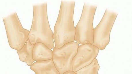
FRACTURES OF OTHER CARPAL BONES: THE CAPITATE
While the scaphoid dominates carpal fracture epidemiology, fractures of the capitate, though rare, carry significant biomechanical implications due to the capitate's role as the keystone of the distal carpal row.
Pathoanatomy and the Scaphocapitate Syndrome
Isolated fractures of the capitate are highly unusual. They most frequently occur in conjunction with scaphoid fractures, a complex injury pattern known as the scaphocapitate syndrome (often referred to as Fenton's syndrome). This typically results from a high-energy hyperextension injury. The force propagates through the scaphoid waist and strikes the neck of the capitate. As the wrist hyperextends, the proximal fragment of the capitate can rotate 180 degrees.
⚠️ Surgical Warning: Capitate Osteonecrosis
Much like the scaphoid, the blood supply to the capitate enters distally and courses proximally. Fractures through the neck or proximal pole of the capitate severely disrupt this intraosseous vascularity, placing the proximal fragment at a high risk of osteonecrosis (AVN) and subsequent midcarpal collapse.
Management of Capitate Fractures
1. Nondisplaced Fractures:
Nondisplaced fractures of the body of the capitate can be managed nonoperatively. Treatment consists of strict immobilization in a short-arm cast for 6 to 8 weeks, followed by serial radiographic evaluation to ensure no secondary displacement occurs.
2. Displaced and Complex Fractures:
Displaced fractures, particularly those involving the articular surface of the midcarpal joint, demand operative intervention.
* Open Reduction and Internal Fixation (ORIF): The joint must be anatomically reduced to prevent post-traumatic arthrosis. Fixation is typically achieved using buried Kirschner wires or headless compression screws. In scaphocapitate syndrome, both the scaphoid and the capitate must be anatomically reduced and rigidly fixed.
* Salvage Procedures: Historically, some surgeons excised the rotated proximal capitate fragment. However, modern practice dictates reduction and internal fixation. If osteonecrosis of the capitate develops and becomes highly symptomatic, salvage procedures are indicated. These include:
* Excisional-interposition arthroplasty.
* Midcarpal arthrodesis (e.g., four-corner fusion).
* Capitate-hamate arthrodesis to stabilize the central column.
CONCLUSION
The management of complex carpal fractures requires a profound understanding of carpal kinematics and vascular anatomy. The Zaidemberg 1,2 ICSRA vascularized bone graft represents a sophisticated, biologically sound solution for scaphoid nonunions complicated by proximal pole ischemia. Meticulous execution of the surgical approach, absolute protection of the vascular pedicle, and rigid internal fixation are the cornerstones of success. Similarly, vigilance in identifying and anatomically fixing associated carpal injuries, such as capitate fractures, is essential to preventing long-term degenerative collapse of the wrist.
📚 Medical References
- vascularized bone grafting for large-gap nonunion of long bones, Orthop Clin North Am 15:131, 1984.
- Ostrum RF, Chao EYS, Bassett CAL, et al: Bone injury, regeneration, and repair. In Simon SR, ed: Orthopaedic basic science, Chicago, 1994, American Academy of Orthopaedic Surgeons. Paley D, Young MC, Wiley AM, et al: Percutaneous bone marrow grafting of fractures and bony defects: an experimental study in rabbits, Clin Orthop Relat Res 208:300, 1986.
- Phemister DB: Treatment of ununited fractures by onlay bone grafts without screw or tie fi xation and without breaking down of the fi brous union, J Bone Joint Surg 29:946, 1947.
- Pho RWH, Vajara R, Satku K: Free
You Might Also Like