Posterior Glenohumeral Dislocation: Epidemiology, Anatomy, Diagnosis & Management
Key Takeaway
Posterior glenohumeral dislocation (PGHD) is an uncommon shoulder injury, often misdiagnosed, accounting for 2-5% of dislocations. It's classically linked to high-energy muscle contractions from epileptic seizures or electric shock. Key features include a reverse Hill-Sachs lesion (humeral head impaction) and potentially a reverse Bankart lesion (posterior labral tear), requiring prompt diagnosis and tailored management.
A 45-year-old male presents to the Emergency Department following a witnessed generalized tonic-clonic seizure. He is complaining of severe right shoulder pain. On examination, the arm is held in internal rotation and adduction. You note a flattening of the anterior contour. How would you investigate this patient, and what radiographic features might you look for?
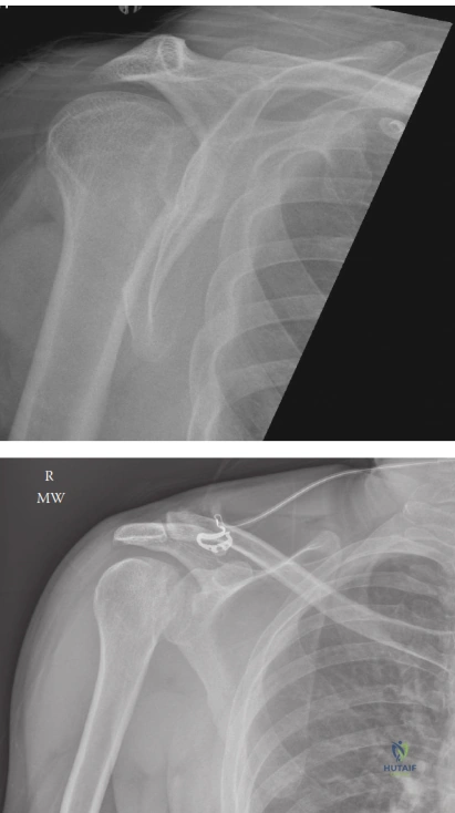
Candidate: I would start with an orthopaedic trauma series: AP, scapular Y, and an axillary lateral view. On the AP view, I would look for the 'lightbulb sign' due to fixed internal rotation and the 'trough line' sign representing an impaction fracture. The axillary view is the most critical for confirming the posterior displacement. If the diagnosis is unclear or to plan for surgery, I would order a CT scan.
Candidates often rely solely on the AP radiograph, missing the posterior dislocation because they fail to order an axillary or trans-scapular Y view. They also frequently forget to mention the 'Rim Sign' or the significance of the seizure history in the context of bilateral injuries.
Structure your response: 1. Clinical suspicion: Highlight that high-energy muscle contractions (the "3 Es") warrant a high index of suspicion. 2. Radiographic markers: Explicitly define the 'Lightbulb sign' (symmetrical humeral head), 'Trough line' (impaction), and 'Rim sign' (>6mm widening). 3. Definitive imaging: Emphasize the axillary view as the gold standard for diagnosis and CT with 3D reconstruction as mandatory for preoperative planning to quantify bone loss.
During your assessment of a chronic posterior dislocation, you obtain the following imaging. Describe the pathology and discuss the management strategy based on the defect size.
Candidate: This image displays a reverse Hill-Sachs lesion (McLaughlin lesion), an impaction fracture on the anteromedial humeral head. Management depends on the size of this defect. If <20%, closed reduction is usually sufficient. Between 20-40%, a subscapularis or lesser tuberosity transfer (McLaughlin/modified McLaughlin) is indicated. If >40-50%, I would consider structural allograft or arthroplasty.
Failure to distinguish between the standard McLaughlin procedure (tendon transfer) and the Neer modification (lesser tuberosity osteotomy). The latter is biomechanically superior because it achieves bone-to-bone healing.
Systematize by defect percentage: <20% (stable, closed reduction), 20-40% (Modified McLaughlin using lesser tuberosity transfer for bone-to-bone healing), >40% (Structural allograft), and >50% or chronic with arthritis (Arthroplasty). Mention that clinical decision-making also hinges on the patient's age and functional demands.
Post-reduction, you are planning the rehabilitation protocol. What are the specific positional considerations, and why?
Candidate: The shoulder should be immobilized in a brace that maintains external rotation and slight abduction, often called a "gunslinger" brace. We must strictly avoid internal rotation, adduction, and cross-body flexion, as these positions stress the posterior repair and risk re-dislocation by engaging the humeral head defect against the posterior glenoid rim.
Suggesting a standard sling. A standard sling keeps the arm in internal rotation, which is the exact position of instability for a posterior dislocation. This is a catastrophic error in postoperative management.
Clearly explain the biomechanical rationale: The posterior capsule is the primary restraint to posterior translation. By holding the humerus in external rotation (neutral or slightly abducted), we reduce the tension on the posterior repair and ensure the humeral head defect is directed anteriorly, away from the posterior glenoid rim (the "engaging" position).
