Greater Tuberosity Fracture: Epidemiology, Surgical Anatomy, Biomechanics, and Management
Key Takeaway
Greater tuberosity (GT) fractures are distinct shoulder injuries, comprising 15-20% of proximal humerus fractures. They often result from high-energy trauma or avulsion by the rotator cuff muscles, which insert here. Integrity is crucial for rotator cuff function, making accurate diagnosis of displacement and associated injuries like dislocations vital for effective management.
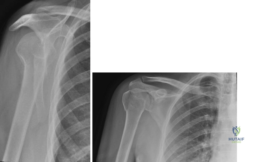
A 45-year-old active patient presents to the clinic 3 days after a fall onto their outstretched hand. They complain of severe shoulder pain and weakness in abduction. You are presented with their initial trauma radiographs, followed by this CT scan.
Describe your assessment of this injury and outline your decision-making process for management.
Candidate: This is a displaced greater tuberosity fracture. I would classify it based on the amount of displacement—looking for >3-5mm—and check for associated injuries like glenohumeral dislocation or rotator cuff tears. I'd perform a physical exam focusing on neurovascular status, specifically the axillary nerve. Given the patient's age and activity level, if displacement is significant, I would lean toward surgical fixation to restore the rotator cuff footprint.
Candidates often fail to mention the specific directions of displacement (superior vs. posterior) or omit the critical need to assess for associated anterior dislocation, which is common. They may jump immediately to "surgery" without framing it through the lens of patient-specific functional demand and the exact threshold of displacement.
A structured response is key:
1. Classification: Identify the fracture morphology and confirm displacement magnitude (superior >3-5mm is the classic indication for fixation due to subacromial impingement).
2. Associated Pathology: Highlight the 20-30% association with anterior dislocation; explicitly mention the need to exclude axillary nerve injury.
3. Decision Framework: Use a matrix of Patient Factors (age, activity level, bone quality) vs. Deformity Factors (displacement, comminution, articular involvement).
4. Management Plan: If operative, detail the goal: anatomical reduction to restore the rotator cuff footprint (supraspinatus/infraspinatus/teres minor) using either suture anchors or lag screws, depending on fragment size and bone quality.
You have decided to proceed with operative fixation for a displaced greater tuberosity fracture in a young patient. Discuss the surgical approach and the potential for iatrogenic complications.
Candidate: I would choose between an anterolateral deltoid-splitting approach or a deltopectoral approach. The deltoid-splitting approach provides direct access to the GT, but I must be careful of the axillary nerve. I would limit the split to 5cm distal to the acromion. The deltopectoral approach is better if I need to address an associated dislocation.
Forgetting the specific anatomical landmarks for the axillary nerve (5-7 cm from the lateral acromion) or failing to discuss the internervous plane (deltoid vs. pectoralis major in the DP approach) demonstrates a lack of deep anatomical knowledge.
The candidate should articulate a high-level mastery of surgical safety:
Approach: Use the deltoid-splitting approach for superior/posterior GT fragments, strictly keeping the split <5cm from the acromion to protect the axillary nerve. Use the deltopectoral approach (internervous plane: cephalic vein landmark) for associated dislocations.
Complication Management: Explicitly mention protecting the axillary nerve, managing the cephalic vein, and the risk of hardware prominence in the subacromial space. Briefly touch on the role of intraoperative fluoroscopy to ensure the hardware is buried and not penetrating the articular surface.
