
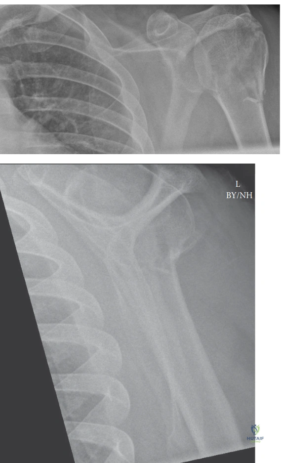
An 82-year-old female presents to the emergency department following a mechanical fall onto her outstretched hand. She is complaining of severe right shoulder pain. A radiograph is performed as shown below.
Describe your systematic approach to this radiographic assessment and define the fracture according to the Neer classification.
Candidate: I would begin by assessing the adequacy of the images, ensuring I have the trauma series: True AP, Scapular Y, and Axillary views. I am looking for the number of displaced parts: the humeral head, greater tuberosity, lesser tuberosity, and the humeral shaft. I would assess for >1cm displacement or >45 degrees of angulation. Based on this image, I see a displaced fracture of the surgical neck and likely involvement of the greater tuberosity. I would classify this as at least a two or three-part fracture and correlate this with the patient's age and bone quality.
Candidates often jump straight to the classification without describing the image systematically. Failing to mention the "Neer criteria" (1cm/45 degrees) for defining a "part" suggests a lack of fundamental knowledge. Furthermore, many neglect to comment on the glenohumeral relationship or the axillary nerve status, which must be addressed in the clinical context.
A high-scoring answer is structured: 1. Systematic Review: Confirm the trauma series (AP, Y, Axillary), assess joint congruency, and evaluate glenohumeral stability. 2. Classification: Apply the Neer criteria, defining the four segments (head, greater tuberosity, lesser tuberosity, shaft) and checking for the >1cm / >45-degree displacement threshold. 3. Clinical Integration: Specifically mention that the Neer system is a prognostic tool for vascularity (risk of AVN) and rotator cuff integrity. 4. Anatomical Landmarks: Note the status of the medial calcar, as its comminution is a critical predictor of failure in ORIF.
Considering the patient's age and the fracture pattern, what is the role of the "medial calcar" in your surgical planning, and why is its status a deal-breaker for certain fixation methods?
Candidate: The medial calcar is the dense, medial metaphyseal bone that provides the primary structural support for the humeral head. In locking plate fixation, if the calcar is comminuted or lost, the construct becomes unstable, leading to varus collapse, secondary screw cutout, and hardware failure. Therefore, if I am planning ORIF, I must ensure anatomical reduction of the medial column or use calcar-supporting screws and possibly bone grafting to restore that buttress.
Ignoring the biomechanical necessity of the medial column. Candidates who say "I will just use a locking plate" without discussing the specific need for calcar-directed screws or structural support display a "cookbook" approach rather than a mechanical understanding of the fracture.
The Gold Standard answer identifies the medial calcar as the "load-sharing" key to the proximal humerus. Mention that in the absence of an intact medial column, the locking plate acts in "load-bearing" mode, which leads to high rates of screw cutout in osteoporotic bone. The candidate should mention that if reconstruction of the medial column is impossible, they might shift the indication toward Reverse Total Shoulder Arthroplasty (RTSA) to avoid predictable mechanical failure.
Detailed Chapters & Topics
Dive deeper into specialized chapters regarding proximal-humerus-fracture