What is Pubic Symphysis? Essential Definition & Implications

Key Takeaway
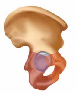
For anyone wondering about What is Pubic Symphysis? Essential Definition & Implications, The pubic symphysis definition definition definition is a fibrocartilaginous disc between the two pubic bones, an amphiarthrodial joint stabilized by superior and inferior arcuate ligaments. A diastasis of the pubic symphysis indicates a disruption of the pelvic ring and an unstable pelvis. This condition is commonly observed in anterior-posterior compression injuries, with widening over 2.5 cm defining rotational instability.
Introduction and Epidemiology
The pubic symphysis represents a critical component of the pelvic ring, a unique amphiarthrodial joint that facilitates load transfer between the axial skeleton and the lower extremities while accommodating the biomechanical demands of locomotion and parturition. As a non-synovial, fibrocartilaginous joint, it possesses limited inherent mobility, yet its integrity is paramount for maintaining pelvic stability. Disruptions of the pubic symphysis often signify severe trauma to the entire pelvic ring, necessitating a comprehensive understanding of its anatomy, biomechanics, and implications for patient management.
Epidemiologically, pelvic ring injuries are relatively uncommon, accounting for approximately 1-3% of all skeletal fractures, but they are frequently associated with high-energy trauma, often involving motor vehicle collisions, falls from height, or crush injuries. Disruption of the pubic symphysis, specifically diastasis, is a hallmark of anterior pelvic ring injury, ranging from isolated stable pubic symphysis separation to devastating "open-book" injuries (Young-Burgess classification, anteroposterior compression types II and III) or vertically unstable patterns (Tile classification, B2 and C types). These injuries carry substantial morbidity and mortality, primarily due to associated hemorrhage, genitourinary compromise, neurological deficits, and systemic inflammatory responses. The incidence of symphyseal diastasis specifically varies, but it is a consistent feature in a significant proportion of pelvic ring disruptions requiring surgical stabilization. A thorough understanding of these epidemiological patterns informs resource allocation, pre-hospital care protocols, and surgical decision-making in the acute trauma setting.
Surgical Anatomy and Biomechanics
The pubic symphysis is an intricate anatomical structure critical for pelvic ring stability. It is a secondary cartilaginous joint composed of two articular surfaces of the pubic bones, each covered by a thin layer of hyaline cartilage, separated by a fibrocartilaginous disc. This disc, often thicker anteriorly and inferiorly, contributes significantly to the joint's load-bearing capacity and limited flexibility.
Ligamentous Support of the Pubic Symphysis
The primary stabilizers of the pubic symphysis are robust ligamentous structures:
* Superior pubic ligament: Extends across the superior aspect of the pubic bones, reinforcing the superior margin of the joint.
* Inferior (arcuate) pubic ligament: A thick, dense arch of fibers spanning the inferior aspect of the symphysis, forming the superior boundary of the urogenital diaphragm. It provides significant vertical and shear stability.
* Anterior pubic ligaments: Multiple layers of interweaving fibers extending obliquely and transversely across the anterior aspect of the joint. These are the strongest and most complex of the symphyseal ligaments, intimately blended with the aponeuroses of the rectus abdominis and external oblique muscles.
* Posterior pubic ligaments: Thinner and less defined compared to their anterior counterparts, offering comparatively less structural support.
Muscular and Neurovascular Relationships
Several muscle groups have important attachments or close proximity to the pubic symphysis. The paired rectus abdominis muscles insert onto the pubic crests and symphysis, providing anterior support and contributing to core stability. The adductor muscles of the thigh originate from the inferior pubic ramus, influencing symphyseal biomechanics during lower limb movement.
Neurovascular structures in the immediate vicinity demand meticulous attention during surgical approaches. The obturator nerve and vessels pass through the obturator foramen, lateral to the symphysis. More critically, the anomalous obturator artery, historically known as the "Corona Mortis" (Crown of Death), represents a significant surgical hazard. This variant, present in approximately 20-30% of individuals, can originate from the inferior epigastric artery or external iliac artery and cross the superior pubic ramus, close to the symphysis. Injury to this vessel can result in torrential, difficult-to-control hemorrhage. The pudendal neurovascular bundle, while more posterior, is also relevant to pelvic floor integrity and potential iatrogenic injury.
Biomechanical Function of the Pelvic Ring
The pelvic ring functions as a closed kinematic chain, distributing axial loads from the spine to the lower extremities and resisting forces generated by lower limb activity. The symphysis pubis acts as the anterior articulation, complementing the stronger sacroiliac joints posteriorly. Its fibrocartilaginous nature allows for minimal rotational and translational movement, which is essential for gait and childbirth.
In the context of trauma, symphyseal disruption unhinges the anterior ring. Anteroposterior compression (APC) forces, as seen in "open-book" injuries, cause external rotation of the hemipelvis, leading to symphyseal widening. This often progresses from purely ligamentous rupture to possible avulsion fractures of the pubic rami. Vertical shear (VS) forces, indicative of highly unstable Tile C type injuries, involve not only symphyseal diastasis but also significant superior or inferior displacement of one hemipelvis relative to the other, invariably accompanied by complete disruption of the posterior sacroiliac complex. Understanding these biomechanical principles is fundamental for accurate diagnosis and appropriate surgical stabilization.
Indications and Contraindications
Surgical stabilization of the pubic symphysis is a cornerstone in the management of unstable pelvic ring injuries. The decision to intervene operatively is guided by the degree of symphyseal disruption, associated posterior instability, patient hemodynamics, and the presence of associated injuries.
Operative Indications for Pubic Symphysis Injury
Primary indications for surgical intervention typically include:
* Symphyseal Diastasis: Generally, an anterior gap exceeding 2.5 cm on static AP radiographs is considered unstable and warrants operative fixation. Some literature suggests thresholds as low as 2.0 cm, particularly if associated with posterior instability.
* Open Book Injuries (APC II and III): These involve significant external rotation of the hemipelves with symphyseal widening. APC II denotes partial disruption of the posterior sacroiliac ligaments, while APC III signifies complete posterior ligamentous disruption. Surgical fixation addresses the symphyseal component, often in conjunction with posterior stabilization.
* Vertical Shear Injuries (Tile Type C): These represent the most unstable pelvic injuries, characterized by significant vertical displacement of the hemipelvis. Symphyseal disruption is uniformly present and requires robust anterior fixation as part of a comprehensive strategy that always includes posterior stabilization.
* Unstable Mallegaigne Fractures: While primarily describing posterior instability, associated pubic rami fractures or symphyseal diastasis require anterior fixation to restore ring integrity.
* Chronic Symphyseal Instability or Non-union: Following previous trauma or in cases of chronic pain and instability from other etiologies (e.g., osteitis pubis refractory to conservative management, pregnancy-related symphyseal dysfunction with persistent diastasis).
* Open Pelvic Fractures: Any symphyseal disruption in the setting of an open fracture mandates aggressive surgical debridement and often definitive stabilization, though temporary external fixation may be preferred initially in grossly contaminated wounds.
Contraindications to Pubic Symphysis Surgery
Absolute contraindications are few and generally relate to the patient's overall medical status:
* Profound Hemodynamic Instability Unresponsive to Resuscitation: In these emergent cases, immediate external fixation or angiography with embolization takes precedence. Definitive internal fixation is deferred until the patient is physiologically stable.
* Active Infection at the Surgical Site: Local infection precludes internal fixation, necessitating debridement, antibiotic therapy, and potentially temporary external fixation.
* Severe Comorbidities: Patients with overwhelming medical conditions where the risks of anesthesia and surgery outweigh the benefits of symphyseal fixation.
Relative contraindications may include:
* Minor Symphyseal Widening (<2.0-2.5 cm) without Posterior Instability: These may be managed non-operatively with protected weight-bearing, particularly if there are no associated fractures or if the patient is medically unfit for surgery.
* Closed Head Injury or Polytrauma with Higher Priority Injuries: While pelvic stability is crucial, life-threatening intracranial hemorrhage or other injuries may temporarily delay definitive symphyseal fixation.
| Indication Category | Operative Indications | Non-Operative Indications |
|---|---|---|
| Traumatic | Symphyseal Diastasis > 2.5 cm (or 2.0 cm with posterior instability) | Symphyseal Diastasis < 2.0 cm (isolated, stable) |
| Open Book Pelvic Fractures (APC II, APC III) | Stable pubic rami fractures without symphyseal diastasis | |
| Vertical Shear Pelvic Fractures (Tile C) | Minor avulsion fractures of the symphysis | |
| Unstable Mallegaigne Fractures | ||
| Open Pelvic Fractures involving symphysis | ||
| Chronic | Chronic symphyseal instability with functional impairment | Osteitis pubis refractory to conservative therapy |
| Symphyseal non-union or symptomatic malunion | Acute osteitis pubis, pregnancy-related mild symphysiolysis |
Pre Operative Planning and Patient Positioning
Thorough preoperative planning is indispensable for achieving optimal outcomes in pubic symphysis stabilization. This process encompasses detailed imaging review, patient resuscitation, and meticulous surgical setup.
Imaging Assessment
A comprehensive imaging protocol is critical for accurate diagnosis and surgical planning:
* Standard Radiographs: Antero-posterior (AP), inlet, and outlet views of the pelvis are fundamental.
* AP View: Assesses symphyseal diastasis, pubic ramus fractures, and gross pelvic alignment.
* Inlet View: Evaluates internal or external rotation of the hemipelves and anterior-posterior displacement.
* Outlet View: Assesses vertical displacement of the hemipelves.
* Computed Tomography (CT) Scan: The gold standard for detailed assessment.
* Axial, Coronal, Sagittal Reconstructions: Crucial for defining fracture patterns, comminution, and the extent of symphyseal disruption.
* 3D Reconstructions: Provide invaluable spatial understanding of the pelvic ring deformity, aid in templating hardware, and identify occult posterior injuries.
* CT Angiography: Indicated in hemodynamically unstable patients or those with suspicion of vascular injury to identify and manage hemorrhage sources.
* Magnetic Resonance Imaging (MRI): Less commonly used in acute trauma but can be beneficial for assessing soft tissue injuries, posterior ligamentous integrity, and discerning inflammatory conditions like osteitis pubis.
Patient Resuscitation and Medical Optimization
Patients with unstable pelvic ring injuries are often polytraumatized and hemodynamically compromised. Adherence to Advanced Trauma Life Support (ATLS) protocols is paramount:
* Hemodynamic Stabilization: Aggressive fluid resuscitation, blood product transfusion, and early control of hemorrhage (pelvic binder, external fixation, angiography/embolization) are critical priorities before definitive symphyseal internal fixation.
* Associated Injuries: A thorough secondary survey to identify and manage concomitant head, thoracic, abdominal, and extremity injuries.
* Prophylactic Measures: Perioperative broad-spectrum antibiotics, deep vein thrombosis (DVT) prophylaxis (mechanical and pharmacological as appropriate), and Foley catheterization to monitor urine output and decompress the bladder are standard. Preoperative cystourethrogram should be considered if urethral injury is suspected.
Patient Positioning and Surgical Setup
- Operating Table: The patient is positioned supine on a radiolucent operating table to allow for intraoperative fluoroscopy. A dedicated fracture table is often not necessary for an isolated symphyseal fixation but can be beneficial if concomitant lower extremity fractures require traction.
- Fluoroscopy: Ensure adequate C-arm access for AP, inlet, and outlet views of the pelvis. This requires careful table placement and draping.
- Prep and Drape: Standard sterile preparation from the umbilicus to the midthighs, bilaterally, including the iliac crests if iliac fixation is contemplated. Urinary catheter should be sterilely placed.
- Surgeon and Assistant Positioning: The primary surgeon typically stands on the patient's contralateral side to the approach, or on the patient's right side for a standard lower abdominal incision, with assistants positioned for optimal retraction and visibility.
- Intraoperative Traction/Reduction Aids: Availability of a pelvic reduction clamp (e.g., Matta, Weber), pointed reduction clamps, bone hooks, and potential external fixator components is essential.
Detailed Surgical Approach and Technique
Surgical stabilization of the pubic symphysis primarily involves an anterior approach, typically via a lower abdominal incision. The goal is accurate anatomical reduction and rigid internal fixation to restore pelvic ring stability.
Surgical Approach and Exposure
The most common approach is the Pfannenstiel incision (bikini line incision). This transverse incision, 10-15 cm in length, is made approximately 2 cm superior to the pubic crests. It offers excellent cosmetic results and adequate exposure for symphyseal fixation.
Alternatively, a lower midline incision may be used, particularly in trauma settings where a laparotomy may be required for concurrent abdominal pathology or if a wider exposure is anticipated. The incision extends from the umbilicus to the symphysis.
Once the skin and subcutaneous tissues are incised, the rectus sheath is exposed. For a Pfannenstiel approach, the anterior rectus sheath is incised transversely. The rectus abdominis muscles are then separated in the midline and reflected superiorly and laterally. The inferior epigastric vessels, running on the posterior rectus sheath, should be identified and protected, or ligated if necessary for superior exposure. Dissection continues deep to the rectus abdominis muscles, exposing the preperitoneal space. The bladder, which lies immediately posterior to the symphysis, must be carefully identified and protected. A Foley catheter assists in bladder decompression and identification. The periosteum over the superior and anterior aspects of the pubic symphysis and pubic rami is incised and elevated to allow direct visualization of the diastasis and fracture fragments. Care must be taken to avoid stripping too much periosteum, which can devascularize bone fragments.

Reduction of the Symphysis Diastasis
Accurate anatomical reduction is paramount for restoring biomechanical integrity.
1. Initial Assessment: Visually inspect the symphyseal diastasis. Remove any interposed soft tissue (e.g., entrapped bladder, rectus muscle, or periosteum) from the joint space.
2. Indirect Reduction: For significant widening and external rotation (APC II/III), a pelvic clamp (e.g., Matta clamp, Weber clamp) is often employed. The clamp is applied from iliac crest to iliac crest, exerting compression to reduce the symphysis. Fluoroscopy is used to confirm reduction in AP, inlet, and outlet views, ensuring proper alignment without rotation or vertical translation.
3. Direct Reduction: In some cases, pointed reduction clamps applied directly to the pubic bones can achieve reduction. Bone hooks can also be used to manipulate fragments. The goal is to restore the normal anatomical width (typically 4-5 mm in adults) and align the superior borders of the pubic bodies. Over-compression should be avoided, as it can lead to posterior ring gapping or compromise sacroiliac joint function.
Internal Fixation of the Pubic Symphysis
Several methods exist for internal fixation, with plate osteosynthesis being the most common and robust.
Plate Osteosynthesis
- Plate Selection: A 3.5 mm reconstruction plate is frequently used, or dedicated symphysis plates (e.g., supra-acetabular plates) are becoming more common. Plates can be pre-contoured for anatomical fit. A longer plate spanning at least three cortices on each side of the symphysis is generally recommended.
- Plate Placement:
- Superior Plating: A single plate applied to the superior aspect of the pubic bones is the most common technique. It provides excellent resistance to distraction and external rotation. The plate should be contoured to fit the superior surface and allow for bicortical screw purchase into both pubic bodies. Avoid extending the plate too far anteriorly, which can irritate soft tissues or the bladder.
- Anterior Plating: Less common as a sole method due to reduced biomechanical stability against vertical shear, but can be used in conjunction with a superior plate.
- Double Plating: In highly unstable vertical shear injuries or comminuted symphyseal fractures, two plates (e.g., one superior, one anterior or two superior plates slightly offset) may be used to enhance rotational and vertical stability.
-
Screw Insertion:
- Screws should achieve bicortical purchase for maximal stability. The length of screws must be carefully chosen to avoid penetrating the bladder, urethra, or obturator neurovascular structures. Intraoperative fluoroscopy with AP, inlet, and outlet views is critical to confirm screw trajectory and length.
- Typically, 3-4 screws are placed on each side of the symphysis.
- It's imperative to recognize the anatomical proximity of the Corona Mortis. When drilling and placing screws in the superior pubic ramus near the symphysis, meticulous care must be taken to avoid this anomalous vessel. Preoperative CT angiography or careful intraoperative dissection can help identify its presence. In particular, the most medial screws on the superior aspect are at highest risk.

Suture Cerclage or Tension Band Wiring
These techniques are generally less rigid than plate fixation and are typically reserved for:
* Minor symphyseal instability without significant diastasis.
* As an adjunct to plate fixation, especially in osteoporotic bone or where soft tissue integrity is compromised.
* Temporary stabilization in specific circumstances.
* They provide good compression but are poor at resisting rotation or vertical shear.
External Fixation
- Indications: Primarily used for temporary stabilization in hemodynamically unstable patients, open pelvic fractures with gross contamination, or when definitive internal fixation is delayed. It can also be used as a definitive treatment for certain stable symphyseal diastasis patterns if internal fixation is contraindicated.
- Technique: Pins are typically placed into the anterior superior iliac spines (ASIS) or iliac crests and connected by an external frame. It provides immediate stabilization but does not offer the same anatomical reduction or rigid fixation as internal plating.
- Role in Definitive Fixation: In some cases, external fixation may be combined with limited internal fixation (e.g., for posterior ring) or used as a preliminary step to facilitate patient resuscitation before conversion to internal fixation.
Closure
After achieving satisfactory reduction and fixation, the operative field is meticulously irrigated. Hemostasis is confirmed, especially paying attention to any bleeding from the space of Retzius or from the drill holes. The rectus abdominis muscles are approximated in the midline, and the rectus sheath is closed. Subcutaneous tissues are closed, and finally, the skin is approximated. A drain may be considered if significant hemorrhage or dead space is present, though it's not universally used.
Complications and Management
Despite meticulous surgical technique, complications associated with pubic symphysis fixation can occur, ranging from minor annoyances to life-threatening events. Proactive identification and appropriate management are crucial for optimizing patient outcomes.
Common Complications
- Infection: Surgical site infection (SSI) can occur, both superficial and deep. Deep infections involving the hardware can lead to osteomyelitis or non-union.
- Hardware Failure: This includes plate breakage, screw pullout, or loss of reduction, often due to inadequate fixation, premature weight-bearing, or unrecognized persistent posterior instability.
- Non-union or Malunion: Inadequate reduction or fixation, infection, or poor bone quality can lead to failure of the symphysis to heal, resulting in persistent pain and instability (non-union) or healing in a deformed position (malunion).
- Genitourinary Injury: The close proximity of the bladder and urethra makes them susceptible to injury during dissection, drilling, or screw placement. This can manifest as bladder perforations, urethral tears, or fistula formation.
- Neurovascular Injury: Injury to the obturator nerve or vessels, or the anomalous Corona Mortis artery, can lead to paresthesia, motor weakness, or severe hemorrhage.
- Persistent Pain: Chronic pain around the symphysis, groin, or perineum is a common complaint, even after successful fixation, potentially due to soft tissue irritation, nerve entrapment, or inflammatory processes.
- Heterotopic Ossification (HO): The formation of ectopic bone in the soft tissues surrounding the symphysis is a known complication, potentially leading to pain and reduced range of motion.
- Sexual Dysfunction: Less common, but symphyseal instability or iatrogenic nerve injury can contribute to sexual dysfunction, particularly in males.
Management Strategies for Complications
| Complication | Incidence (Approximate) | Salvage Strategies |
|---|---|---|
| Surgical Site Infection | 1-5% (deep) | Superficial: Wound care, antibiotics. Deep: Surgical debridement, organism-specific antibiotics, hardware retention vs. removal (if stable union), or staged hardware removal and reimplantation. External fixation may be needed if hardware removed prior to union. |
| Hardware Failure | 5-15% | Revision surgery with stronger fixation (e.g., longer plate, double plating), bone grafting if non-union suspected. Address underlying biomechanical issues (e.g., inadequate posterior fixation). |
| Non-union/Malunion | 5-10% | Non-union: Revision internal fixation with bone grafting (autograft or allograft), potentially combined with posterior stabilization. Malunion: Corrective osteotomy and refixation. |
| Genitourinary Injury | 1-3% | Immediate intraoperative repair (cystorrhaphy, urethral repair) by urology, prolonged catheterization. Fistula: Surgical repair after inflammatory control. |
| Neurovascular Injury | <1% | Arterial: Immediate ligation or repair. Venous: Ligation. Nerve: Exploration and neurolysis or repair, symptomatic management of neuropathy. |
| Persistent Pain | Up to 20-30% | Conservative management (physical therapy, NSAIDs, injections). Hardware removal if symptomatic. Nerve blocks, neuromodulation, or revision surgery for specific identifiable causes (e.g., nerve entrapment, instability). |
| Heterotopic Ossification | 10-30% (radiographic) | Prophylaxis (NSAIDs, radiation therapy) for high-risk patients. Symptomatic HO: Surgical excision after maturation (usually >6-12 months post-injury). |
| Sexual Dysfunction | Variable (often underestimated) | Multidisciplinary approach, including urology/sexology consultation, physical therapy for pelvic floor, psychological support. |
Post Operative Rehabilitation Protocols
Postoperative rehabilitation following pubic symphysis fixation is crucial for optimizing functional recovery and ensuring the longevity of surgical outcomes. Protocols vary based on the stability of the fixation, the presence of associated posterior pelvic ring injuries, and surgeon preference, but generally follow a progressive approach.
Initial Postoperative Phase (Weeks 0-6)
- Weight-Bearing Restrictions:
- Stable Fixation (Isolated Symphysis or with adequately fixed posterior ring): Often allows for protected weight-bearing (toe-touch or partial weight-bearing) on crutches or a walker immediately postoperative. Some surgeons may allow full weight-bearing if posterior stability is unequivocally restored and anterior fixation is robust.
- Unstable Fixation/Complex Injuries: Non-weight-bearing on the affected side or bilateral non-weight-bearing may be prescribed for 6-8 weeks to allow for initial soft tissue and bony healing.
- Pain Management: Aggressive multimodal analgesia is essential to facilitate early mobilization and prevent chronic pain.
- Early Mobilization: Out-of-bed transfers and ambulation with appropriate assistive devices are initiated as soon as pain allows and weight-bearing status permits.
- Range of Motion (ROM): Gentle hip and knee active and passive range of motion exercises, avoiding extremes of abduction or external rotation that could stress the healing symphysis.
- Core Stability: Initiation of gentle isometric core muscle activation exercises (e.g., transverse abdominis contractions), avoiding intense Valsalva maneuvers or direct rectus abdominis activation.
- Deep Vein Thrombosis (DVT) Prophylaxis: Continued chemical and mechanical prophylaxis per institutional protocol.
Intermediate Rehabilitation Phase (Weeks 6-12)
- Progression of Weight-Bearing: Gradually advance weight-bearing as tolerated, based on radiographic evidence of healing and clinical stability. Full weight-bearing is typically achieved by 8-12 weeks in uncomplicated cases.
- Strengthening: Initiate progressive strengthening exercises for the hip abductors, adductors, extensors, and trunk musculature. Exercises should be performed in pain-free ranges.
- Gait Training: Focus on normalizing gait pattern and reducing reliance on assistive devices.
- Scar Management: Begin scar massage and desensitization exercises to improve pliability and reduce discomfort.
- Soft Tissue Mobilization: Address any muscle tightness or myofascial restrictions in the groin, hip, and lumbar regions.
Advanced Rehabilitation Phase (Months 3-6+)
- Functional Progression: Transition to more advanced functional exercises, including proprioception, balance training, and sport-specific drills if applicable.
- Return to Activity: Gradual return to activities of daily living, work, and recreational sports. High-impact or contact sports may be restricted for 6-12 months or until complete radiographic union and full strength return are confirmed.
- Imaging Follow-up: Regular radiographic follow-up (AP, inlet, outlet views) is critical to monitor healing, assess for loss of reduction, hardware failure, or heterotopic ossification. CT scans may be indicated to confirm bony union.
- Hardware Removal: Symptomatic hardware (e.g., plate prominence, chronic irritation, pain) may be considered for removal after adequate bony union, typically 12-18 months post-surgery. Asymptomatic hardware is often left in situ.
A multidisciplinary approach involving orthopedic surgeons, physical therapists, and pain management specialists is often beneficial for optimal patient recovery. Education on activity modification and body mechanics is essential to prevent re-injury and promote long-term stability.
Summary of Key Literature and Guidelines
The management of pubic symphysis injuries, particularly in the context of pelvic ring disruptions, has evolved significantly with advancements in surgical techniques, imaging, and understanding of biomechanics.
Classification Systems and Prognostic Indicators
The Tile classification (A, B, C based on stability) and the Young-Burgess classification (Lateral Compression - LC, Anteroposterior Compression - APC, Vertical Shear - VS) remain the foundational systems for categorizing pelvic ring injuries, directly influencing treatment algorithms. Symphyseal diastasis is a defining feature of APC and VS injuries. Literature consistently demonstrates that posterior pelvic ring integrity is the primary determinant of overall pelvic stability, and therefore, operative fixation of the symphysis must always consider the status of the posterior elements. The presence of significant symphyseal diastasis (typically >2.5 cm) is a robust indicator of an unstable pelvic ring requiring surgical stabilization, irrespective of posterior injury patterns, although co-existing posterior instability necessitates combined anterior and posterior fixation.
Evolution of Fixation Strategies
Early management often relied on external fixation or bed rest. However, numerous studies, including biomechanical analyses and clinical outcomes, have established the superiority of internal plate fixation for unstable symphyseal injuries.
* Single Plate vs. Double Plating: Biomechanical studies by groups like Routt et al. have shown that a single superiorly placed plate provides substantial stability against distraction and external rotation, but dual plating (e.g., superior and anterior) or a broad superior plate may offer enhanced resistance to vertical shear forces, particularly in Tile C type injuries or cases with poor bone quality. Clinical series suggest acceptable outcomes with single superior plating for most APC type injuries, reserving dual plating for higher energy injuries or those with persistent vertical instability.
* Screw Length and Trajectory: The importance of bicortical screw purchase for optimal fixation strength is well-established. However, careful attention to screw length and trajectory is critical to avoid iatrogenic injury to the bladder, urethra, or obturator neurovascular structures, especially the Corona Mortis. Intraoperative fluoroscopy and knowledge of regional anatomy are indispensable.
Outcomes and Complications
Contemporary literature reports favorable outcomes for surgically treated unstable pubic symphysis injuries, with union rates typically exceeding 90%. Functional outcomes, as assessed by various scores (e.g., Majeed, Iowa Pelvic Score), are generally good to excellent, though persistent pain and sexual dysfunction can occur in a significant minority of patients.
* Persistent Pain: Chronic groin pain, often related to hardware prominence, nerve irritation, or residual instability, remains a common long-term issue, contributing to the decision for elective hardware removal in a substantial number of patients (20-40%).
* Hardware Failure: Rates of hardware failure have decreased with improved plate design and surgical techniques but still occur, prompting re-operation.
* Heterotopic Ossification: While typically asymptomatic, radiographic HO is frequently observed and may contribute to stiffness or pain in some cases. Prophylaxis with NSAIDs or radiation therapy remains debated for routine use but is considered in high-risk patients.
Current Guidelines and Recommendations
Current consensus guidelines from major orthopedic trauma associations (e.g., AO Foundation, Orthopaedic Trauma Association - OTA) emphasize:
1. Early Hemodynamic Stabilization: Prioritizing resuscitation and hemorrhage control, with external fixation or pelvic binder application as initial temporizing measures.
2. Anatomic Reduction: Striving for anatomical reduction of the symphysis, especially the superior aspect, to restore the pelvic ring's load-bearing capacity.
3. Rigid Internal Fixation: Plate osteosynthesis is the preferred method for definitive stabilization of unstable symphyseal injuries.
4. Combined Anterior and Posterior Fixation: Recognizing that isolated symphyseal fixation is insufficient for most high-energy pelvic ring injuries that involve posterior instability. The posterior ring dictates overall stability and must be addressed.
5. Multidisciplinary Approach: Highlighting the importance of collaboration with trauma surgery, urology, critical care, and rehabilitation specialists for optimal patient care.
The continued emphasis on precise anatomical reduction, robust internal fixation, and a holistic approach to polytraumatized patients underlies the current paradigm for managing pubic symphysis injuries, aiming to minimize morbidity and maximize functional recovery.
You Might Also Like