Operative Management of Congenital Foot and Toe Anomalies
Key Takeaway
The operative management of congenital foot and toe anomalies requires a profound understanding of pediatric biomechanics and pathoanatomy. This comprehensive guide details evidence-based surgical interventions for conditions such as talipes equinovarus, metatarsus adductus, and complex toe deformities. Emphasizing both soft-tissue releases and osseous reconstructions, it provides orthopedic surgeons with step-by-step protocols to optimize functional outcomes and minimize recurrence in the pediatric patient.
Comprehensive Introduction and Patho-Epidemiology
The surgical management of congenital anomalies of the foot and toes represents one of the most intricate and demanding domains of pediatric orthopedics. Historically characterized by aggressive, extensive soft-tissue releases that often resulted in stiff, painful, and arthritic feet in adulthood, the modern paradigm has shifted toward a profound respect for biomechanics, tissue-sparing techniques, and the preservation of joint kinematics. Conditions ranging from isolated digital anomalies (macrodactyly, polydactyly) to complex multi-planar deformities (talipes equinovarus, metatarsus adductus) require a meticulous, stage-specific approach that balances the immediate correction of the deformity with the long-term functional demands of the growing pediatric skeleton.
Congenital talipes equinovarus (clubfoot) is among the most prevalent of these anomalies, with an incidence of approximately 1 to 2 per 1,000 live births. The patho-epidemiology of clubfoot is multifactorial, involving a complex interplay of genetic predisposition and environmental factors. Recent genomic studies have implicated variants in the PITX1-TBX4 transcriptional pathway, which is critical for early hindlimb development. While idiopathic clubfoot is the most common presentation, orthopedic surgeons must maintain a high index of suspicion for syndromic or teratologic associations, such as arthrogryposis multiplex congenita, myelomeningocele (spina bifida), or amniotic band sequence. These syndromic variants typically present with profound soft-tissue rigidity and a markedly higher resistance to conservative management, necessitating earlier and more comprehensive surgical intervention.
Metatarsus adductus, conversely, is the most common congenital foot deformity, occurring in roughly 1 in 1,000 live births, though mild cases are often underreported. The etiology is widely considered to be related to intrauterine packaging phenomena, particularly in primigravida mothers or in cases of oligohydramnios. While the vast majority of these flexible deformities resolve spontaneously or with a brief period of serial casting, a subset of rigid, structural deformities persists. The patho-epidemiology of persistent metatarsus adductus involves an abnormal osseous morphology of the medial cuneiform and a subluxation of the tarsometatarsal (Lisfranc) complex, driven by an overriding abductor hallucis contracture.
Digital anomalies, including macrodactyly (macrodystrophia lipomatosa), cleft foot (ectrodactyly), and overlapping toes, present unique epidemiological profiles. Macrodactyly is a rare, non-hereditary somatic mutation often linked to the PIK3CA gene, resulting in a localized overgrowth of mesenchymal tissues, particularly fibrofatty infiltration along the plantar nerve sheaths. Cleft foot, often termed "lobster-claw deformity," is a longitudinal deficiency characterized by the absence of central rays. It frequently demonstrates an autosomal dominant inheritance pattern with variable penetrance and is often associated with cleft hand or broader syndromic conditions like EEC (Ectrodactyly, Ectodermal dysplasia, and Cleft lip/palate) syndrome. Understanding the distinct epidemiological and pathophysiological foundations of these diverse conditions is paramount for the orthopedic surgeon, as it directly dictates the timing, extent, and prognosis of operative intervention.
Detailed Surgical Anatomy and Biomechanics
A profound mastery of the surgical anatomy and biomechanics of the pediatric foot is the cornerstone of successful operative intervention. The infant foot is not merely a miniature adult foot; it is a highly cartilaginous, dynamically evolving structure where physeal preservation is paramount. In congenital talipes equinovarus, the deformity is three-dimensional and encompasses four distinct components, universally remembered by the acronym CAVE: Cavus (midfoot), Adductus (forefoot), Varus (hindfoot), and Equinus (ankle). The biomechanical fulcrum of this deformity is the talar head. In a clubfoot, the calcaneus, navicular, and cuboid are medially rotated and inverted around the talus. The talar neck is typically shortened and deviated medially, while the body of the talus is extruded anterolaterally from the ankle mortise.
The soft-tissue anatomy in clubfoot is characterized by severe contractures of the posteromedial tether. Medially, the tibialis posterior, flexor digitorum longus (FDL), and flexor hallucis longus (FHL) tendons are profoundly shortened. The talonavicular joint capsule, the spring ligament (plantar calcaneonavicular ligament), and the superficial components of the deltoid ligament form a dense, unyielding fibrotic mass that tethers the navicular to the medial malleolus. Posteriorly, the Achilles tendon and the posterior capsules of both the tibiotalar and subtalar joints are contracted, driving the calcaneus into rigid equinus. Biomechanically, surgical release must restore the normal kinematic coupling of the subtalar joint, allowing the calcaneus to abduct, evert, and dorsiflex relative to the talus without destabilizing the midfoot.
In metatarsus adductus, the anatomical pathology is localized strictly to the midfoot and forefoot, distinguishing it from the hindfoot involvement seen in clubfoot. The hindfoot is typically in neutral or slight valgus. The primary anatomical distortion occurs at the tarsometatarsal (Lisfranc) articulation, where the metatarsals are deviated medially. Biomechanically, the medial column (first ray) is shortened and tethered by a hyperactive or contracted abductor hallucis muscle, while the lateral column (fourth and fifth rays) is relatively elongated. If left untreated, this rigid deformity disrupts the windlass mechanism of the plantar fascia during the terminal stance phase of gait, leading to abnormal weight distribution, lateral border pain, and an increased propensity for fifth metatarsal base stress fractures in adulthood.
The surgical anatomy of congenital toe deformities requires meticulous attention to the delicate neurovascular bundles and the intricate balance of the extensor and flexor mechanisms. In macrodactyly, the pathoanatomy is dominated by the proliferation of adipose tissue within the epineurium of the digital nerves, most commonly the branches of the medial plantar nerve. This fibrofatty infiltration not only causes grotesque hypertrophy of the digit but also distorts the normal vascular arborization, rendering the toe highly susceptible to ischemia during surgical debulking. In overlapping fifth toe deformities, the anatomy involves a dorsal and medial subluxation of the metatarsophalangeal joint, driven by a contracture of the extensor digitorum longus, the dorsal capsule, and an abnormal insertion of the lumbrical. Correction requires a comprehensive rebalancing of these dynamic forces to restore a plantigrade, functional ray.
Exhaustive Indications and Contraindications
The decision-making process regarding operative intervention for congenital foot and toe anomalies requires a nuanced understanding of the natural history of the specific deformity, the age of the patient, and the functional limitations imposed by the condition. The overarching goal is to achieve a painless, plantigrade, and shoeable foot, avoiding the temptation to operate solely for cosmetic perfection if it risks joint stiffness or neurovascular compromise.
| Congenital Anomaly | Primary Operative Indications | Absolute Contraindications | Relative Contraindications |
|---|---|---|---|
| Talipes Equinovarus (Clubfoot) | Failure of Ponseti casting; Relapsed deformity with dynamic supination; Rigid syndromic/teratologic clubfoot; Late-presenting neglected clubfoot (>2 years). | Active local infection; Non-ambulatory patient with painless deformity (where surgery offers no functional gain). | Severe vascular insufficiency; Extreme medical comorbidities precluding anesthesia. |
| Metatarsus Adductus | Rigid deformity persisting beyond 4-5 years of age; Painful shoe wear; Failure of serial casting; Development of a skewfoot deformity. | Flexible deformity passively correctable to neutral; Children under 3 years of age (high rate of spontaneous resolution). | Mild, asymptomatic deformity; Presence of open physes if considering non-physeal-sparing osteotomies. |
| Macrodactyly | Progressive angular deformity; Inability to wear standard footwear; Recurrent ulceration or skin breakdown; Digit length exceeding adult parent's toe. | Single-stage bilateral debulking (high risk of necrosis); Asymptomatic static enlargement. | Poor peripheral perfusion; Unrealistic parental expectations regarding cosmetic outcome. |
| Cleft Foot (Lobster-Claw) | Progressive splaying of the forefoot; Inability to accommodate standard footwear; Severe central cleft impairing weight-bearing mechanics. | Mild cleft with stable, functional forefoot; Severe syndromic involvement precluding ambulation. | Surgery before 1 year of age (anesthetic risk and delicate soft tissues). |
| Overlapping 5th Toe / Curly Toes | Persistent pain with shoe wear; Failure of conservative taping; Recurrent blistering or callus formation. | Asymptomatic curly toes in infants (high spontaneous resolution rate). | Mild overlapping without shoe-wear interference. |
In the management of clubfoot, the primary indication for extensive soft-tissue release (Comprehensive Posteromedial Release) is a rigid, syndromic clubfoot (e.g., arthrogryposis) that is entirely refractory to the Ponseti method, or a severe, neglected clubfoot in a toddler. In the modern era, idiopathic clubfeet rarely require comprehensive release; however, limited operative interventions, such as a percutaneous Achilles tenotomy, are indicated in over 90% of Ponseti-managed cases to correct residual equinus. For relapsed clubfeet presenting in early childhood (ages 3-5) with dynamic supination during the swing phase of gait, a Tibialis Anterior Tendon Transfer (TATT) is strictly indicated, provided the foot is passively correctable.
For metatarsus adductus, surgical indications are strictly reserved for rigid, structural deformities that persist into later childhood (typically over 5 years of age). Operating on flexible deformities or intervening too early exposes the child to unnecessary surgical risks, including premature physeal closure and joint stiffness, for a condition that frequently resolves spontaneously. In digital anomalies like macrodactyly, the indications for surgery are driven by the progressive nature of the disease. Epiphysiodesis is indicated to arrest longitudinal growth once the toe matches the anticipated adult length, while ray resection is indicated as a definitive, functional salvage for grotesquely enlarged, non-functional digits that preclude normal ambulation.
Pre-Operative Planning, Templating, and Patient Positioning
Meticulous pre-operative planning is non-negotiable in pediatric foot and ankle reconstruction. The foundation of this planning rests on high-quality, standardized radiographic imaging. For infants and toddlers unable to stand, simulated weight-bearing anteroposterior (AP) and lateral radiographs are obtained by applying a rigid radiolucent board to the plantar aspect of the foot with the ankle dorsiflexed to neutral. In clubfoot evaluation, the surgeon must scrutinize Kite’s angle (the AP talocalcaneal angle), which is typically reduced to less than 20 degrees (parallelism of the talus and calcaneus), and the lateral talocalcaneal angle. In metatarsus adductus, the AP radiograph is utilized to assess the metatarsal-cuneiform angles and to rule out a complex skewfoot deformity, which involves paradoxical hindfoot valgus.
Advanced imaging modalities are selectively employed. Magnetic Resonance Imaging (MRI) is the gold standard for evaluating macrodactyly, allowing the surgeon to delineate the extent of fibrofatty infiltration along the neural elements and to plan the margins of soft-tissue debulking. In complex, neglected, or relapsed clubfeet, particularly those being considered for Ilizarov external fixation or complex midfoot osteotomies, a fine-cut Computed Tomography (CT) scan with 3D reconstruction is invaluable for mapping the distorted osseous morphology and planning precise osteotomy trajectories.
Pre-operative templating is critical when planning osseous reconstructions, such as the Berman-Gartland procedure for metatarsus adductus or a lateral column shortening/medial column lengthening for relapsed clubfoot. The surgeon must calculate the exact angle of the wedge to be resected from the cuboid or the degree of opening required at the medial cuneiform to achieve a straight lateral border of the foot. Templating ensures that appropriate bone graft volumes are anticipated and that the correct size and trajectory of internal fixation (typically smooth K-wires in the pediatric population) are available.
Patient positioning and operating room setup must be optimized for the specific procedure. For a comprehensive posteromedial release (PMR) in clubfoot, the patient is ideally placed in the prone position. This allows simultaneous, unhindered access to the posterior ankle structures (Achilles tendon, posterior capsules) and the medial midfoot. A pediatric pneumatic tourniquet is applied to the proximal thigh to ensure a bloodless field, which is critical for identifying the delicate neurovascular bundles. For forefoot procedures, such as metatarsal osteotomies, cleft foot reconstruction, or toe arthroplasties, the patient is positioned supine with a small bump placed under the ipsilateral hip to internally rotate the leg, bringing the foot into a neutral, upward-facing position. Intraoperative fluoroscopy (C-arm) must be positioned to easily swing into the field for orthogonal views without compromising sterility.
Step-by-Step Surgical Approach and Fixation Technique
Comprehensive Posteromedial Release (PMR) for Clubfoot
When soft-tissue release is definitively indicated, the PMR systematically addresses the contracted medial and posterior tethers.
1. Incision and Exposure: With the patient prone, the Cincinnati incision is utilized. This transverse incision begins at the anteromedial aspect of the foot (over the navicular-cuneiform joint), curves horizontally around the posterior heel just proximal to the heel crease, and terminates at the anterolateral foot.
2. Superficial Dissection: The sural nerve and short saphenous vein are isolated laterally and protected. Medially, the neurovascular bundle (posterior tibial artery and tibial nerve) is meticulously identified proximal to the flexor retinaculum, mobilized, and protected with a vessel loop.
3. Tendon Lengthening: A Z-lengthening of the Achilles tendon is performed in the coronal plane. The tibialis posterior tendon is identified at its navicular insertion and Z-lengthened. If severe contractures persist, the FDL and FHL tendons are also lengthened, often utilizing a fractional lengthening technique at the musculotendinous junction to preserve strength.
4. Capsular Releases: A comprehensive posterior capsulotomy of the tibiotalar and subtalar joints is executed, sectioning the posterior talofibular and calcaneofibular ligaments. Moving medially, the talonavicular joint capsule is sharply opened. The spring ligament and the superficial deltoid ligament are released, allowing the navicular to physically reduce laterally and dorsally onto the talar head.
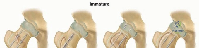
5. Reduction and Fixation: The talonavicular and subtalar joints are anatomically reduced. The reduction is provisionally held, and fluoroscopy confirms the restoration of Kite's angle. Smooth K-wires (typically 1.6mm or 2.0mm) are driven retrograde from the navicular into the talus, and from the calcaneus into the talus, locking the foot in a neutral, plantigrade position.
Osseous Reconstructions for Metatarsus Adductus (Berman-Gartland)
For rigid metatarsus adductus in a child over 5 years old, multiple metatarsal osteotomies are the gold standard to realign the forefoot without sacrificing joint mobility.
1. Approach: The patient is supine. Two or three dorsal longitudinal incisions are made: one between the 1st and 2nd metatarsals, one between the 3rd and 4th, and occasionally a third over the 5th metatarsal base.
2. Osteotomy: Subperiosteal dissection exposes the bases of all five metatarsals. Using a pediatric oscillating saw or an osteotome, dome-shaped or crescentic osteotomies are performed at the metaphyseal-diaphyseal junction. Crucial Step: The osteotomies must be strictly distal to the proximal physis of the first metatarsal and the proximal physis of the fifth metatarsal to prevent devastating growth arrest. The dome shape allows for rotational and angular correction without shortening the structural length of the rays.
3. Correction and Fixation: The forefoot is abducted as a single unit, pivoting on the dome osteotomies until the lateral border of the foot is clinically straight. Fixation is achieved by driving smooth K-wires retrogradely through the first and fifth metatarsals, crossing the osteotomy sites and anchoring into the medial cuneiform and cuboid, respectively.
Digital Reconstructions: Macrodactyly and Overlapping Toes
- Macrodactyly Debulking: The procedure must be staged. A mid-axial longitudinal incision is made on one side of the digit. The skin flaps are elevated, and the massive, fibrofatty tissue infiltrating the digital nerve is meticulously excised under loupe magnification. The digital artery must be preserved. The contralateral side is addressed no sooner than 3 to 6 months later to prevent ischemic necrosis.
- Butler Arthroplasty for Overlapping 5th Toe: A dorsal racquet-shaped incision is made, with the handle extending proximally over the 5th metatarsal. The extensor digitorum longus tendon is Z-lengthened. A radical dorsal, medial, and lateral capsulotomy of the metatarsophalangeal joint is performed. The toe is plantarflexed and derotated into proper alignment. The skin is closed in a V-Y fashion, utilizing the racquet handle to tether the toe in its corrected plantarflexed position. A K-wire is rarely needed if the soft-tissue rebalancing is adequate.
Complications, Incidence Rates, and Salvage Management
Surgical intervention in the pediatric foot carries significant risks. The delicate soft-tissue envelope, the presence of vulnerable physes, and the complex three-dimensional nature of the deformities create a landscape where technical errors can lead to profound, lifelong disability.
| Complication | Estimated Incidence | Etiology / Risk Factors | Salvage Management & Corrective Strategy |
|---|---|---|---|
| Over-correction (Planovalgus Foot) | 5% - 15% (after PMR) | Excessive release of the interosseous talocalcaneal ligament; pinning the foot in excessive valgus/dorsiflexion. | Initial conservative management (orthotics). If rigid and painful: Subtalar arthrodesis or lateral column lengthening (Evans osteotomy) in older children. |
| Under-correction / Recurrence | 10% - 30% | Incomplete release of the posteromedial tether; failure of postoperative bracing compliance. | Repeat casting (Ponseti). If dynamic: Tibialis Anterior Tendon Transfer (TATT). If rigid osseous deformity: Medial column lengthening / Lateral column shortening. |
| Skin Necrosis / Wound Dehiscence | 2% - 10% | Closing the Cincinnati incision under excessive tension; aggressive single-stage bilateral debulking in macrodactyly. | Immediate cessation of casting pressure. Local wound care, negative pressure wound therapy (VAC). Severe cases require split-thickness skin grafting or local rotational flaps. |
| Avascular Necrosis (AVN) of the Talus | 1% - 3% | Excessive stripping of the dorsal neck of the talus; damage to the artery of the tarsal canal during subtalar release. | Prolonged non-weight-bearing. If collapse occurs leading to severe arthritis: Talectomy (Astragalectomy) or definitive Triple Arthrodesis at skeletal maturity. |
| Premature Physeal Closure | 2% - 5% (Metatarsal osteotomies) | Direct thermal or mechanical injury to the physis during osteotomy; K-wire placement through the physis. | Observation if minimal growth remaining. If severe brachymetatarsia develops: Callus distraction lengthening (mini-Ilizarov) of the affected metatarsal. |
The most devastating complication of clubfoot surgery is the "over-released" or planovalgus foot. This iatrogenic deformity occurs when the surgeon aggressively sections the interosseous talocalcaneal ligament or forcefully pins the foot into excessive valgus and dorsiflexion, destroying the intrinsic stability of the subtalar complex. The resulting foot is often more painful and dysfunctional than the original uncorrected clubfoot. Salvage of a severe planovalgus foot is exceedingly difficult; it often requires complex osseous realignments, such as an Evans calcaneal lengthening osteotomy, or ultimately, a subtalar arthrodesis once the child approaches skeletal maturity.
In cases of severe, neglected clubfeet or syndromic deformities where acute surgical correction carries an unacceptably high risk of neurovascular stretch injury or skin necrosis, the Ilizarov method serves as both a primary and salvage technique. By applying a multi-planar circular external fixator (such as the Taylor Spatial Frame), the surgeon can perform gradual distraction histogenesis. This technique safely stretches the contracted neurovascular bundle and soft tissues at a rate of 1mm per day, allowing for the correction of severe osseous deformities without acute ischemic compromise. For the skeletally mature patient presenting with a rigid, painful, arthritic foot following multiple failed pediatric interventions, joint-sparing procedures are no longer viable. In these salvage scenarios, a Triple Arthrodesis (fusion of the subtalar, talonavicular, and calcaneocuboid joints) remains the gold standard, providing a stable, plantigrade, and pain-free base of support, albeit at the permanent sacrifice of hindfoot inversion and eversion.
Phased Post-Operative Rehabilitation Protocols
The technical success of any operative intervention for congenital foot and toe anomalies is inextricably linked to the rigor and compliance of the postoperative rehabilitation protocol. The pediatric foot has a profound tendency to drift back into deformity if not meticulously maintained during the healing and remodeling phases.
Phase 1: Immediate Post-Operative Immobilization (Weeks 0 to 6)
Following major soft-tissue releases (PMR) or complex osteotomies (Berman-Gartland), the foot and leg are immediately immobilized in the operating room. A well-padded, bivalved long-leg cast is applied. The knee must be flexed to 90 degrees; this serves two critical biomechanical purposes: it relaxes the gastrocnemius muscle (reducing tension on the Achilles repair and posterior capsule), and it prevents the infant or toddler from kicking off the cast. The foot is held in a neutral, plantigrade position—never forced into extreme over-correction. The patient is strictly non-weight-bearing. At the 4-to-6-week mark, the patient is brought to the clinic or operating room (depending on age and tolerance) for cast removal and extraction of the smooth K-wires.
Phase 2: Transition and Bracing (Weeks 6 to 12)
Once the K-wires are removed and clinical union of osteotomies or healing of tendons is confirmed, the patient transitions to a short-leg weight-bearing cast or a rigid walking boot for an additional 3 to 4 weeks to allow for progressive loading of the osseous structures. Following this, a strict bracing protocol is initiated. For clubfoot, this is the most vulnerable period for recurrence. A foot abduction orthosis (FAO), such as the Denis Browne splint or Mitchell brace, is prescribed. The shoes are set at 60 to 70 degrees of external rotation for the operative foot and 30 to 40 degrees for the non-operative foot, with the bar bent to provide 10 to 15 degrees of dorsiflexion.
Phase 3: Long-Term Maintenance and Physical Therapy (Months 3 to Years)
The FAO is worn for 23 hours a day for the first 3 months post-casting, and then strictly during nighttime and naps until the child is 4 to 5 years of age. Compliance with this bracing regimen is the single most critical factor in preventing relapse. Concurrently, formal physical therapy is instituted. The focus is on gait training, proprioceptive re-education, and active range-of-motion exercises. Parents are instructed on daily passive stretching routines, particularly focusing on dorsiflexion and eversion. The child is monitored clinically and radiographically at 6-month intervals until skeletal maturity to ensure the maintenance of correction and to monitor for any signs of premature physeal arrest or recurrent contractures.
Summary of Landmark Literature and Clinical Guidelines
The evolution of operative management for congenital foot anomalies is deeply rooted in landmark orthopedic literature, which has driven a massive paradigm shift over the last half-century. The most profound contribution to this field is the work of Dr. Ignacio Ponseti. His seminal publications in the mid-20th century, and subsequent long-term follow-up studies published in the Journal of Bone and Joint Surgery (JBJS), definitively proved that his method of serial manipulation and casting, based on a precise understanding of subtalar kinematics, yielded superior long-term functional outcomes compared to the aggressive comprehensive surgical releases advocated by his contemporaries. Today, the American Academy of Orthopaedic Surgeons (AAOS) and the Pediatric Orthopaedic Society of North America (POSNA) universally endorse the Ponseti method as the gold-standard first-line treatment for idiopathic clubfoot, relegating extensive surgery to relapsed, syndromic, or neglected cases.
In the realm of metatarsus adductus, the landmark paper by Berman and Gartland (1971) established the standard for operative correction in the older child. Their description of dome-shaped, non-physeal-involving osteotomies of the metatarsal bases provided a reliable, reproducible method to correct the severe skewfoot without the high rates of joint stiffness and arthritis associated with the historically popular Heyman-Herndon tarsometatarsal mobilization. Current clinical guidelines dictate that the Heyman-Herndon procedure should be largely abandoned in favor of the Berman-Gartland osteotomies for rigid, late-presenting deformities.
For congenital toe anomalies, the classification systems and surgical algorithms proposed by Blauth and Borisch for cleft foot remain the definitive framework for surgical decision-making. Their work emphasized the necessity of closing the central defect early (between 1 and 2 years of age) to prevent the progressive, biomechanically devastating splaying of the forefoot during the acquisition of mature gait. Similarly, the Butler arthroplasty for overlapping fifth toes, first described in the mid-20th century, remains the most biomechanically sound procedure for this condition, addressing all vectors of the deformity through a single, elegant soft-tissue realignment. The synthesis of this landmark literature underscores a unifying principle in modern pediatric foot surgery: respect the biology, preserve the joints, and intervene surgically only when conservative measures have been exhausted or are biomechanically destined to fail.
