ABOS Part I & AAOS OITE Orthopedic Review: Deformity Correction, Paley's Laws & FAN | Part 21913
Key Takeaway
This module offers a comprehensive review for ABOS Part I and AAOS OITE exams, focusing on advanced lower extremity deformity correction. It covers Paley's Laws, Fixator-Assisted Nailing (FAN), osteotomy principles, mechanical axis deviation, and joint orientation angles, providing essential knowledge for orthopedic board preparation.
ABOS Part I & AAOS OITE Orthopedic Review: Deformity Correction, Paley's Laws & FAN | Part 21913
A 55-year-old patient presents with a complex lower extremity deformity. Preoperative radiographs are obtained as shown below. Based on the provided case description and these images, which of the following statements best characterizes the primary deformities observed and their implications for surgical planning?
Correct Answer: C
The case explicitly states the patient presents with 'femoral diaphyseal varus, distal metaphyseal valgus, and multilevel procurvatum, compounded by tibial valgus and proximal tibial recurvatum.' Image (a), the standing AP view, visually confirms severe genu valgum, which corresponds to a lateral mechanical axis deviation. Image (b), the lateral view, clearly shows femoral procurvatum (anterior bowing) and tibial recurvatum (posterior bowing). This combination of deformities across multiple bones and planes (frontal and sagittal) necessitates identifying multiple Centers of Rotation of Angulation (CORAs) and planning separate osteotomies for each apex, as emphasized by Paley's principles for complex, multi-apical deformities. This makes option C the most accurate and comprehensive description.
Option A is incorrect because the AP view clearly shows genu valgum, not varum, and a lateral mechanical axis deviation, not medial. Option B incorrectly identifies the sagittal plane deformities; the case and image (b) show femoral procurvatum and tibial recurvatum, not the reverse. Option D is incorrect as the deformity is clearly multi-level and multi-planar, not isolated distal femoral valgus, and the lateral view shows significant sagittal plane deformities. Option E misinterprets the early postoperative image (c) and underestimates the complexity of the femoral deformity, which the case describes as profoundly complex and requiring meticulous, multi-level planning.
A surgeon is planning to correct a complex, multi-apical femoral deformity involving both diaphyseal varus and distal metaphyseal valgus, along with significant procurvatum. Due to time constraints and a desire to minimize surgical invasiveness, the surgeon decides to perform a single osteotomy at the mid-diaphysis, attempting to correct all frontal and sagittal plane deformities simultaneously by angulating the bone around this single cut. Based on Paley's Three Osteotomy Rules, what is the most likely outcome of this surgical approach?
Correct Answer: C
The scenario described is a direct violation of Paley's Osteotomy Rule Three. The case explicitly states that a multi-apical deformity (like the described femoral diaphyseal varus and distal metaphyseal valgus, plus procurvatum) has multiple distinct CORAs. Attempting to correct such a deformity with a single osteotomy performed *away* from all individual CORAs, and then angulating around an axis of correction also *away* from the true CORAs, will inevitably lead to the creation of a new, iatrogenic deformity. This results in a massive translational step-off, malalignment of the mechanical axis, and biomechanical failure, as the original angular deformity is simply traded for a translational one. This is described as a 'catastrophic surgical error' in the text.
Option A is incorrect because a multi-apical deformity does not have a single, global CORA that can perfectly correct all apices with one cut. Option B describes Paley's Rule Two, which applies when the osteotomy is away from the CORA but the axis of correction is *at* the CORA, leading to controlled translation. This is not the scenario described. Option D is incorrect; while some shortening can occur with acute corrections, the primary issue with violating Rule Three is malalignment and translation, not necessarily just shortening. Option E introduces a rotational deformity, which is not the primary consequence described for violating Paley's Rule Three in this context.
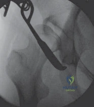
Following a multi-level femoral osteotomy for severe valgus and procurvatum deformities, an intramedullary nail (IMN) is inserted. Postoperative radiographs, as shown in image (d) below, demonstrate the use of distal blocking screws. Given the preoperative distal femoral valgus that was corrected to varus, what is the correct placement and purpose of these blocking screws?
Correct Answer: B
The case explicitly states the rule for blocking screw placement: 'Always place blocking screws on the concave side of the deformity you are correcting.' For frontal plane corrections, to maintain a varus correction (which means pushing the distal segment medially to correct a preoperative valgus deformity), the blocking screw must be placed on the **lateral** side of the nail path. This creates an artificial inner cortex, forcing the nail medially and maintaining the desired varus alignment. Image (d) clearly shows the distal blocking screws placed laterally, consistent with maintaining a varus correction of a preoperative distal femoral valgus.
Option A is incorrect because placing screws medially would maintain a valgus correction, which is the opposite of correcting a preoperative valgus to varus. Option C and D relate to sagittal plane corrections (procurvatum/recurvatum), where screws are placed anteriorly or posteriorly, respectively, and are not the primary function of the distal screws shown in the AP view for frontal plane correction. Option E is incorrect; while blocking screws contribute to overall stability, their primary role is to prevent angular loss of correction (the 'bell-clapper effect') by forcing the nail into a specific trajectory, not solely to prevent rotation.
A 30-year-old male presents with knee pain and a 'knock-kneed' appearance. Radiographic analysis reveals a Mechanical Lateral Distal Femoral Angle (mLDFA) of 80° and a Medial Proximal Tibial Angle (MPTA) of 95°. The Joint Line Convergence Angle (JLCA) is 1°. Based on these measurements and Paley's principles, what is the most accurate interpretation of this patient's frontal plane alignment?
Correct Answer: B
According to the provided table of Joint Orientation Angles:
- mLDFA: Normal range is 85° to 90° (Avg 87°). A value <87° indicates femoral valgus. This patient's mLDFA of 80° indicates significant femoral valgus.
- MPTA: Normal range is 85° to 90° (Avg 87°). A value >87° indicates tibial valgus. This patient's MPTA of 95° indicates significant tibial valgus.
- JLCA: Normal range is 0° to 2°. This patient's JLCA of 1° is within the normal range, suggesting no significant intra-articular pathology, cartilage loss, or ligamentous laxity contributing to the angular deformity.
Therefore, the patient has a combined femoral valgus and tibial valgus deformity, with a normal joint space (JLCA). This combination would result in a 'knock-kneed' (genu valgum) appearance.
Option A is incorrect as both angles indicate valgus, not varus. Option C and D are incorrect as both femur and tibia contribute to the valgus deformity, not isolated or compensatory. Option E is incorrect as both mLDFA and MPTA are outside their normal ranges, indicating bony deformity, not just soft tissue laxity.
In the management of a patient with a profoundly complex, multi-apical femoral deformity and a complex tibial deformity, the surgical team decides to stage the procedures, addressing the femur first and the tibia in a secondary operation several weeks later. Which of the following is the *most critical* advantage of this staged approach, particularly in a patient with compromised osteoporotic bone stock?
Correct Answer: B
The case highlights several critical advantages of staging procedures. While physiological tolerance and complication mitigation are important, the text specifically mentions: 'Intraoperative Adjustment: Provides the surgeon an opportunity to assess the clinical and radiographic outcome of the initial correction. If the femoral correction slightly alters the global limb length or mechanical axis unexpectedly, the subsequent tibial plan can be dynamically adjusted to compensate.' This ability to fine-tune the overall limb alignment based on the first stage's outcome is a paramount advantage in complex, multi-level corrections.
Option A is incorrect; while early mobilization is a goal, immediate full weight-bearing after multi-level osteotomies in osteoporotic bone is unlikely and not the primary reason for staging. Option C is incorrect; staging procedures often increases overall costs due to multiple hospitalizations and anesthetic events. Option D is incorrect; staging reduces the *risk* of complications but does not *eliminate* them. Option E is incorrect; functional restoration and biomechanical alignment are the primary goals, with cosmesis being a secondary benefit.
A surgeon is planning a proximal tibial osteotomy for a patient with significant tibial valgus and recurvatum, complicated by poor bone stock. The goal is to achieve simultaneous multiplanar correction with minimal limb length change and maximal bone-on-bone healing surface. Which osteotomy technique, as described in the case, is best suited for this scenario?
Correct Answer: C
The case specifically advocates for the focal dome osteotomy in complex cases like the proximal tibia with valgus and recurvatum, especially in osteoporotic bone. It states: 'A curvilinear, cylindrical cut centered exactly on the CORA. It allows for massive, multiplanar angular correction (e.g., simultaneous varus and flexion) with minimal to no change in overall limb length. Most importantly, it creates a large, highly congruent, bone-on-bone surface area for healing. This maximizes intrinsic stability and biological healing potential.'
Option A (opening wedge) is less ideal in poor bone stock due to the need for grafting and increased soft tissue tension. Option B (closing wedge) sacrifices bone stock and shortens the limb, which may not be desired, and is less versatile for multiplanar correction. Option D (transverse diaphyseal osteotomy for distraction) is primarily for lengthening, not necessarily for acute multiplanar angular correction with maximal bone contact. Option E (simple oblique osteotomy) lacks the precision, stability, and multiplanar correction capabilities of a dome osteotomy and would be unstable.
During a Lengthening Over Nail (LON) procedure for a femoral deformity, the surgeon has performed the osteotomy and is preparing to insert the intramedullary nail. A critical step to ensure successful distraction and prevent jamming of the nail during the lengthening phase is:
Correct Answer: C
The case explicitly details the step-by-step masterclass for femoral LON, stating: 'Crucially, over-ream the canal by 2 mm greater than the diameter of the nail to be inserted. This extra space prevents the nail from jamming during the distraction phase.' This is a critical technical pearl for LON procedures.
Option A is incorrect because the nail is locked proximally but *not* distally until the target length is achieved, to allow for distraction. Option B is incorrect; reaming to the exact diameter would lead to jamming during distraction. Option D is incorrect; canal reaming is essential for IMN insertion and stability, and a multiple drill hole osteotomy (corticotomy) is typically performed. Option E is incorrect; the nail is inserted and proximally locked before the external fixator is applied.
A 40-year-old patient undergoes a proximal tibial osteotomy for severe genu valgum. The surgeon plans an acute valgus-to-varus correction. Based on the surgical pearls outlined in the case, what prophylactic measure is mandatory during this procedure?
Correct Answer: B
The case specifically highlights a crucial surgical pearl: 'As noted in our radiographic review, when performing an acute valgus-to-varus correction of the proximal tibia, the common peroneal nerve is placed under sudden, extreme stretch. Prophylactic surgical decompression of the peroneal nerve at the fibular neck is mandatory to prevent devastating stretch neuropraxia or permanent foot drop.'
Option A (fasciotomy) is not routinely mandatory for this type of osteotomy unless compartment syndrome is suspected. Option C (corticosteroids) might be used for swelling but is not a mandatory prophylactic measure against nerve injury. Option D (saphenous nerve block) is for pain management, not nerve protection from stretch. Option E (prophylactic antibiotics) is standard for any orthopedic surgery but is not specific to preventing nerve injury from acute valgus-to-varus correction.
A 60-year-old patient with osteoporotic bone stock requires a complex tibial realignment using an external fixator, as depicted in the clinical image below. To maximize pin purchase and minimize the risk of pin loosening or bone failure in this compromised bone, which of the following external fixation pearls is most critical?
Correct Answer: C
The case provides specific 'Surgical Pearls: External Fixation in Osteoporotic Bone.' It states: 'Always use hydroxyapatite (HA) coated half-pins. HA coating significantly increases bone-to-pin integration, drastically improving purchase and pull-out strength in weak, osteoporotic bone.' This directly addresses the challenge of osteoporotic bone stock.
Option A is incorrect; standard stainless steel pins have less bone integration than HA-coated pins, making them less ideal in osteoporotic bone. Option B is partially correct in that tensioned fine wires are excellent for metaphyseal fixation in osteoporotic bone, but the question asks about maximizing *pin* purchase, and half-pins are also used. Relying *solely* on fine wires for all fixation might not be appropriate for all segments or constructs. Option D is incorrect; the text explicitly warns against this: 'If mounted obliquely, any subsequent distraction or adjustment will introduce iatrogenic translational forces.' Option E is incorrect; while meticulous daily pin site care is paramount, the text does not specify hydrogen peroxide, and aggressive sterilization can sometimes be detrimental to healing. The focus here is on the *hardware choice* for osteoporotic bone.
The mechanical axis is a fundamental concept in lower extremity deformity analysis. Which of the following statements accurately describes the mechanical axis and its significance in a healthy, well-aligned limb?
Correct Answer: B
The case clearly defines the mechanical axis: 'The mechanical axis is the true line of weight-bearing. In the lower extremity, it is drawn from the center of the femoral head to the center of the ankle plafond. In a perfectly aligned, healthy limb, this line bisects the knee joint, passing slightly medial to the tibial spines.' Normalizing the Mechanical Axis Deviation (MAD) is the ultimate goal of corrective osteotomy to normalize joint loading and prevent osteoarthritis progression.
Option A is incorrect; it incorrectly states the origin (greater trochanter instead of femoral head) and the normal path (lateral compartment instead of bisecting the knee). Option C describes the anatomical axis, not the mechanical axis, and misstates its primary use. Option D is incorrect; the mechanical axis is about weight-bearing and alignment, not muscle pull or primarily rotational deformities. Option E is incorrect; it misidentifies the origin and destination points of the mechanical axis.
A 32-year-old patient presents with a severe multiapical femoral deformity, as might be seen in the clinical and radiographic presentation in image (a) and (b) below. Preoperative planning identifies multiple CORAs along the femur. The surgeon opts for a Fixator-Assisted Nailing (FAN) approach. Following the initial application of an external fixator as shown in image (c), what is the *primary* immediate goal of this initial external fixator application in the context of FAN for such a complex deformity?
Correct Answer: C
The case explicitly states that the intraoperative radiograph (c) demonstrates 'the initial application of a robust external fixator to the femur—this is the crucial first step in gaining rigid control over the individual bone segments to execute a precise, mathematically planned correction.' In the FAN technique, the external fixator is applied temporarily to act as a 'powerful, multiplanar joystick' to acutely and precisely correct the deformity directly on the operating table. This allows for minute, intraoperative fine-tuning before the definitive internal fixation with an intramedullary nail.
Option A is incorrect because the external fixator in FAN is temporary and removed at the end of the case; the IM nail provides definitive, long-term stabilization.
Option B is incorrect because FAN is designed for acute intraoperative correction, not gradual correction over weeks, which is characteristic of traditional external fixation.
Option D is incorrect because the external fixator's role is to control bone segments for correction, not to guide reaming directly. Reaming occurs after the osteotomy and correction, with the fixator holding the alignment.
Option E is incorrect because while the IM nail allows for early weight-bearing, the temporary external fixator itself is not the primary means of facilitating immediate weight-bearing in the long term; it's the IM nail that provides this stability after the fixator is removed.
A 40-year-old male undergoes a FAN procedure for a severe femoral deformity. Postoperatively, a standing full-length radiograph reveals a Mechanical Lateral Distal Femoral Angle (mLDFA) of 80° and a Medial Proximal Tibial Angle (MPTA) of 95°. Based on the provided table of Joint Orientation Angles, what is the most accurate interpretation of these findings?
Correct Answer: B
The case provides a table of Joint Orientation Angles with normal ranges and target goals:
- mLDFA (Mechanical Lateral Distal Femoral Angle): Normal range 85° - 90°, Target 88°. An mLDFA of 80° (less than 85°) indicates that the distal femur is angled more medially than normal, signifying a varus deformity of the distal femur.
- MPTA (Medial Proximal Tibial Angle): Normal range 85° - 90°, Target 87° - 90°. An MPTA of 95° (greater than 90°) indicates that the proximal tibia is angled more laterally than normal, signifying a valgus deformity of the proximal tibia.
Therefore, the distal femur is in excessive varus, and the proximal tibia is in excessive valgus.
Option A is incorrect because the mLDFA of 80° indicates varus, not valgus, of the distal femur.
Option C is incorrect because the mLDFA of 80° indicates varus, not valgus, of the distal femur.
Option D is incorrect because the MPTA of 95° indicates valgus, not varus, of the proximal tibia.
Option E is incorrect because the mLDFA of 80° is outside the normal range (85°-90°) and indicates varus.
A surgeon is planning a corrective osteotomy for a patient with a single-level angular deformity of the tibia. Preoperative planning identifies the CORA. To avoid a region of poor bone quality at the CORA, the surgeon decides to perform the osteotomy 3 cm distal to the CORA. The corrective hinge of the external fixator is accurately placed at the CORA. According to Paley's Osteotomy Rules, what is the expected outcome of this correction, and what challenge might it pose for subsequent intramedullary nailing?
Correct Answer: C
This scenario directly describes Paley's Osteotomy Rule Two: 'When the corrective hinge is placed accurately at the CORA, but the actual osteotomy is performed at a different level (e.g., to avoid poor soft tissue, or for biological healing reasons), the axes will become collinear, but translation will inevitably occur at the osteotomy site.' The case further explains that 'this offset can make passing a rigid IM nail across the osteotomy site extremely difficult, or even impossible, if the translational offset is significant.'
Option A is incorrect because translation will occur if the osteotomy is not at the CORA, even if the hinge is. Pure angulation without translation only occurs when both osteotomy and hinge are at the CORA (Rule One).
Option B is incorrect because Rule Two states the axes will become collinear, not parallel but non-collinear. A secondary iatrogenic deformity with parallel but non-collinear axes occurs under Rule Three.
Option D is incorrect because the rule describes translation, not overcorrection, and the difficulty for IM nailing is a direct consequence.
Option E is incorrect because the rule describes the geometric outcome and challenge for IMN, not a failure of osteotomy healing due to CORA separation.
The diagram below illustrates different approaches for intramedullary nail insertion in the femur. In the context of Fixator-Assisted Nailing (FAN), which statement best describes the flexibility and rationale for choosing an antegrade versus a retrograde approach for femoral nailing?
Correct Answer: C
The case states: 'As clearly illustrated in the diagram above, intramedullary nails can be inserted in an antegrade fashion (starting from the hip/piriformis fossa or greater trochanter) or a retrograde fashion (starting from the intercondylar notch of the knee) for the femur. The choice depends entirely on the specific location of the deformity, the presence of retained hardware, and the surgeon's clinical preference. The versatile FAN technique is fully compatible with both approaches.'
Option A is incorrect because the choice is not always antegrade; it depends on several factors, and FAN is compatible with both.
Option B is incorrect because retrograde nailing is not exclusively used, and the choice is multifactorial.
Option D is incorrect because the case explicitly states that FAN is 'fully compatible with both approaches.'
Option E is incorrect because while reaming is part of the nailing process, the diagram and accompanying text specifically discuss the antegrade and retrograde *insertion approaches* for IM nails, and their compatibility with FAN.
A 28-year-old patient undergoes a FAN procedure for a complex multiapical tibial deformity. During the procedure, the external fixator is used to acutely correct the deformity. Intraoperative fluoroscopy confirms perfect alignment, with the mechanical axis passing directly through the center of the knee and ankle joints, and all joint orientation angles within normal limits. An intramedullary nail is then inserted. What is the *most significant* patient-centric advantage of this FAN approach compared to a traditional long-term external fixator for definitive correction?
Correct Answer: C
The case highlights 'Reduced Patient Morbidity' as a key advantage: 'Patients are completely freed from the physical restrictions, pain, and psychological burden of a long-term external frame, dramatically improving their overall recovery experience.' This is a direct patient-centric benefit of removing the external fixator immediately after the IM nail is placed.
Option A is incorrect because while FAN offers 'Unmatched Precision,' the question asks for a *patient-centric* advantage compared to a traditional external fixator. Traditional external fixators are also capable of precise angular correction, albeit over a longer period and with higher patient morbidity.
Option B is incorrect because while the risk of *long-term* pin-site infections is eliminated, pin-site infections can still occur during the temporary period the fixator is in place. The primary benefit is the *removal* of the frame, not the complete elimination of infection risk during the temporary application.
Option D is incorrect because while FAN can manage translation, this is a technical advantage, not the *most significant patient-centric* advantage over a traditional fixator, which can also correct translation.
Option E is incorrect because FAN often involves the cost of both an external fixator and an IM nail, potentially making it more expensive than a traditional external fixator alone, though the overall cost-benefit of faster recovery and reduced complications might be favorable.
A 55-year-old patient with a history of Paget's disease presents with a complex, multiapical femoral deformity. Preoperative planning is initiated. According to the foundational principles of deformity planning outlined in the case, what are the two overarching, non-negotiable goals of surgical correction for this patient?
Correct Answer: A
The case explicitly states: 'The overarching goal of deformity correction is twofold: 1. To normalize the overall limb alignment, which is mathematically represented by the Mechanical Axis Deviation (MAD). 2. To ensure that the weight-bearing joints are perfectly parallel to the ground during the stance phase of gait, represented by the Joint Orientation Angles.'
Option B is incorrect because while identifying the CORA and using an IM nail are crucial *methods* in modern correction, they are not the overarching *goals* themselves. The CORA dictates osteotomy placement to achieve the goals of alignment.
Option C is incorrect because minimizing surgical time and blood loss are general surgical principles, not the specific, primary biomechanical goals of deformity correction.
Option D is incorrect because while restoring length and preventing rotational malalignment are important aspects of a complete correction, they are sub-goals that contribute to the primary goals of MAD and joint orientation, which encompass overall limb alignment.
Option E is incorrect because while pain relief and improved range of motion are desired clinical outcomes, they are not the direct, measurable biomechanical goals of deformity correction as defined in the text.
A 60-year-old patient presents with a severe varus knee deformity. Preoperative planning reveals a Mechanical Axis Deviation (MAD) of 20mm medial to the center of the knee. What is the clinical significance of this finding, and what is the ultimate goal for this measurement after correction?
Correct Answer: B
The case defines MAD: 'Medial MAD: Indicates a varus (bowleg) deformity. The mechanical axis passes medial to the center of the knee, overloading the medial compartment.' It further states: 'The Ultimate Goal: A MAD of exactly zero millimeters. At this neutral point, the mechanical axis passes directly through the center of the knee joint, ensuring optimal, symmetric load distribution across the articular cartilage and preventing the rapid progression of unilateral compartment osteoarthritis.'
Therefore, a MAD of 20mm medial indicates a varus deformity overloading the medial compartment, and the ultimate goal is a MAD of exactly zero millimeters.
Option A is incorrect because medial MAD indicates varus, not valgus, and the goal is zero, not 20mm lateral.
Option C is incorrect because MAD primarily measures angular alignment in the coronal plane, not rotational deformity directly.
Option D is incorrect because MAD measures alignment, not leg length discrepancy, although severe deformities can be associated with length issues.
Option E is incorrect because MAD is an extra-articular measurement of overall limb alignment, not an indicator for intra-articular deformity or arthroplasty, although it can contribute to joint degeneration.
A surgeon is performing a complex femoral deformity correction. Due to an error in preoperative planning and intraoperative execution, both the osteotomy and the corrective hinge of the external fixator are inadvertently placed away from the identified CORA. According to Paley's Osteotomy Rules, what is the most likely geometric outcome of this surgical error?
Correct Answer: C
This scenario directly describes Paley's Osteotomy Rule Three: 'When both the osteotomy and the corrective hinge are located away from the CORA, a secondary iatrogenic deformity is created. The mechanical axes will become parallel but will *not* be collinear. This results in a persistent, unsightly translational deformity.'
Option A is incorrect because pure angulation without translation occurs only when both osteotomy and hinge are at the CORA (Rule One).
Option B is incorrect because collinear axes with translation occur when the hinge is at the CORA but the osteotomy is away (Rule Two).
Option D is incorrect because Rule Three describes an angular and translational deformity, not primarily a lengthening error, although length can be affected.
Option E is incorrect because Rule Three describes the geometric outcome of the correction, not necessarily a failure of union, although malalignment can contribute to nonunion.
A patient has undergone a Fixator-Assisted Nailing (FAN) procedure for a complex lower extremity deformity. The images below represent typical post-operative radiographs following such a procedure. What do these images primarily confirm regarding the success of the FAN technique?
Correct Answer: B
The images provided (ch_142_fig_fbe55b.webp and ch_142_fig_fa46a4.webp) show an intramedullary nail in place, with the bone segments appearing well-aligned. The case describes FAN as a technique where 'The correction is meticulously confirmed with intraoperative fluoroscopy to ensure the MAD and all joint orientation angles are absolutely perfect. While the fixator rigidly holds the bone segments in this idealized position, an IM nail is passed across the osteotomy site, permanently locking in the correction internally. The external fixator is immediately removed at the end of the surgical case.' Therefore, these post-operative images confirm the successful acute correction and internal fixation of the deformity.
Option A is incorrect because the external fixator is removed at the end of the FAN procedure; these images show internal fixation only.
Option C is incorrect because the goal of FAN is definitive correction in one stage, not to require further immediate intervention.
Option D is incorrect because the images show a well-aligned bone with an IM nail, which is the desired outcome, not an iatrogenic deformity.
Option E is incorrect because FAN is a hybrid technique that uses a *temporary* external fixator, not a traditional Ilizarov frame for long-term stabilization.
The case emphasizes that rigorous, exhaustive geometric analysis is paramount before surgery. Which of the following statements best encapsulates the fundamental reason why this meticulous preoperative planning is considered 'non-negotiable' for complex lower extremity deformities?
Correct Answer: C
The case states: 'Before a single scalpel is lifted or a single incision is made, a rigorous, exhaustive geometric analysis of the patient's deformity is absolutely paramount. This is not merely an academic exercise to be glossed over; it is the definitive blueprint for surgical success.' It further emphasizes: 'Identifying the CORA is non-negotiable. It dictates the ideal biomechanical location for the osteotomy and defines the exact pivot point around which the bone segments must be manipulated to restore a straight, collinear axis.'
Option A is incorrect because insurance eligibility is an administrative concern, not a surgical planning principle.
Option B is incorrect because while good planning can contribute to a smoother recovery, minimizing hospital stay is a secondary outcome, not the fundamental reason for geometric analysis.
Option D is incorrect because antibiotic prophylaxis is a standard perioperative measure, unrelated to geometric deformity analysis.
Option E is incorrect because patient education, including cosmetic benefits, is important, but it is not the fundamental reason for the detailed geometric analysis that dictates surgical execution.
The case describes Fixator-Assisted Nailing (FAN) as a 'revolutionary, hybrid surgical technique.' Which of the following statements most accurately describes how FAN resolves the classic orthopedic dilemma of choosing between the precision of external fixation and the comfort/rehabilitation of internal fixation?
Correct Answer: C
The case explicitly states: 'Fixator-Assisted Nailing (FAN) is a revolutionary, hybrid surgical technique that masterfully harnesses the unique power of two distinct fixation methods to achieve a vastly superior clinical result. It elegantly resolves the classic orthopedic dilemma: having to choose between the ultimate precision of an external fixator (at the cost of high patient morbidity) versus the comfort and rapid rehabilitation of an IM nail (at the risk of an inaccurate, malaligned correction).' It then details the process: 'A temporary, highly rigid external fixator is strategically applied... The external fixator is utilized as a powerful, multiplanar 'joystick' to acutely and precisely correct the deformity... While the fixator rigidly holds the bone segments in this idealized position, an IM nail is passed across the osteotomy site, permanently locking in the correction internally. The external fixator is immediately removed...' This perfectly describes the combination of acute precision and immediate internal stability.
Option A is incorrect because FAN is a hybrid technique that *combines* both, not replaces external fixation entirely.
Option B is incorrect because FAN involves a *temporary* external fixator for *acute* correction, not long-term gradual correction.
Option D is incorrect because FAN is specifically highlighted for 'complex multiplanar and multiapical deformities,' not simple ones.
Option E is incorrect because FAN involves performing a planned osteotomy to correct the bone deformity.
A 55-year-old male presents with chronic right knee pain and a progressive valgus deformity. A weight-bearing long-leg anteroposterior radiograph is obtained, as depicted in the foundational diagram below, illustrating key alignment parameters. The mechanical axis is measured to pass 15 mm lateral to the center of the knee joint. The mLDFA is measured at 78°, and the MPTA is 88°.
Based on these findings and the principles of deformity correction, which of the following statements is TRUE?
Correct Answer: C
The mechanical axis deviation (MAD) is the perpendicular distance from the center of the knee to the mechanical axis line. A negative MAD indicates the mechanical axis falls lateral to the center of the knee, which represents a valgus deformity. A MAD of -15 mm (15 mm lateral) is outside the neutral zone (0 ± 8 mm) and confirms a significant valgus deformity. The normal mLDFA is 85° to 90° (average 87°). An mLDFA of 78° is less than 85°, indicating a valgus deformity of the distal femur. The normal MPTA is 85° to 90° (average 87°). An MPTA of 88° is within the normal range, suggesting the proximal tibia is not the primary source of the frontal plane deformity.
Option A is incorrect: A negative MAD indicates valgus, not physiologic varus, and -15 mm is outside the neutral zone.
Option B is incorrect: The MPTA is normal, ruling out a primary deformity in the proximal tibia. A varus malalignment would be indicated by a positive MAD, not a negative one.
Option D is incorrect: An mLDFA of 78° indicates a valgus deformity of the distal femur (less than 85°), not a varus deformity. Correction would require a varus-producing osteotomy (e.g., lateral closing wedge or medial opening wedge) to increase the mLDFA towards normal.
Option E is incorrect: An MPTA of 88° is within the normal range (85°-90°), indicating no significant valgus deformity of the proximal tibia. Therefore, a medial opening wedge osteotomy of the tibia is not indicated based on this angle.
A 30-year-old active duty military personnel presents with a chronic distal femoral deformity following a combat injury. Preoperative planning reveals a significant procurvatum deformity of the distal femur, with a Posterior Distal Femoral Angle (PDFA) measured at 70°. The surgeon plans a Fixator-Assisted Nailing (FAN) procedure to correct this deformity. According to Paley's principles, what is the most appropriate interpretation of this PDFA measurement and the primary goal for its correction?
Correct Answer: B
The Posterior Distal Femoral Angle (PDFA) evaluates the sagittal plane alignment of the distal femur. The normal value range is 79° to 87°, with an average of 83°. An angle less than 79° indicates a procurvatum (flexion deformity), while an angle greater than 87° indicates recurvatum (hyperextension). Therefore, a PDFA of 70° is significantly less than the normal range, confirming a procurvatum deformity. The primary goal of correction would be to increase this angle to the average normal value of 83° to restore proper sagittal alignment.
Option A is incorrect: A PDFA of 70° is less than 79°, which indicates procurvatum, not recurvatum.
Option C is incorrect: A PDFA of 70° indicates procurvatum, not recurvatum. The goal is to increase the angle, not decrease it.
Option D is incorrect: While a PDFA of 70° indicates procurvatum, the goal is to increase the angle to 83°, not decrease it.
Option E is incorrect: A PDFA of 70° is well outside the normal range of 79° to 87°, indicating a significant deformity requiring correction.
A surgeon is planning a distal femoral osteotomy for a patient with a valgus deformity. Preoperative planning identifies the Center of Rotation of Angulation (CORA) at the junction of the distal metaphysis and diaphysis. The surgeon decides to perform a focal dome osteotomy precisely at the CORA and uses an external fixator as a temporary reduction tool, with its hinge axis also passing through the CORA. During the procedure, the deformity is acutely corrected. According to Paley's Three Laws of Osteotomy, what is the expected outcome of this surgical approach?
Correct Answer: B
This scenario perfectly describes Paley's Osteotomy Rule One: 'When the osteotomy and the hinge axis both pass directly through the CORA, pure angular correction is achieved without any translation.' This is the surgical ideal, maximizing bone-to-bone contact and promoting robust healing. A focal dome osteotomy centered at the CORA is a classic example of achieving pure angular correction.
Option A is incorrect: Angulation with intentional translation occurs when the hinge axis passes through the CORA, but the osteotomy is performed at a different level (Rule Two).
Option C is incorrect: Iatrogenic translation deformity (Rule Three) occurs when both the hinge axis and the osteotomy are separate from the CORA, leading to misaligned mechanical axes.
Options D and E are incorrect: A focal dome osteotomy centered at the CORA, performing pure angular correction, does not typically result in significant limb shortening or lengthening. These are more associated with large opening or closing wedge osteotomies or specific lengthening/shortening procedures.
A 40-year-old patient undergoes Fixator-Assisted Nailing (FAN) for a distal femoral valgus deformity. During the procedure, after the osteotomy is completed and the external fixator is applied, the surgeon proceeds with reaming and nail insertion, as depicted in the FAN workflow diagram. Despite achieving excellent frontal plane correction, post-operative radiographs reveal an iatrogenic procurvatum deformity. Which of the following is the most likely cause of this complication?
Correct Answer: C
The case explicitly states that the most common and critical error in distal femoral Fixator-Assisted Nailing is the failure to adequately control the sagittal plane during a frontal plane correction, almost invariably leading to iatrogenic procurvatum. The text highlights that a simple, single-bar, two-pin external fixator is biomechanically incompetent in the sagittal plane, acting as a simple hinge and offering zero resistance to flexion or extension forces. This allows the powerful gastrocnemius muscle pull and the posterior trajectory of the intramedullary nail in the wide distal metaphysis to drive the distal fragment into procurvatum.
Option A is incorrect: While inadequate reaming can cause issues, it's not the primary mechanism described for iatrogenic procurvatum in the context of sagittal plane control failure.
Option B is incorrect: Osteotomy placement relative to the CORA primarily affects translation (Paley's Rules Two and Three), not directly the iatrogenic procurvatum in the sagittal plane during nail insertion, assuming the correction is held.
Option D is incorrect: Over-correction of the frontal plane is a different type of malalignment and not the direct cause of procurvatum as described.
Option E is incorrect: Premature removal of the fixator would lead to loss of the *corrected* alignment, but the procurvatum occurs *during* the reaming and nailing process when the fixator is still in place but inadequate for sagittal control.
A surgeon is performing a distal femoral osteotomy with Fixator-Assisted Nailing (FAN) to correct a valgus deformity and prevent iatrogenic procurvatum. To achieve robust multi-planar stability and counteract the forces leading to procurvatum, which advanced hardware strategy is most appropriate for sagittal plane control?
Correct Answer: C
The case explicitly states that to prevent procurvatum, the surgeon must abandon simplistic fixation and proactively build a construct that provides true, rigid multi-planar stability. The most direct and effective solution is to supplement the standard two lateral Schanz pins with at least one anterior pin placed in the distal segment (and ideally one in the proximal segment as well). This anterior pin acts as a rigid buttress, directly opposing the flexion force exerted by the gastrocnemius and the posteriorly directed force of the advancing intramedullary nail, transforming an unstable hinge into a locked, immovable block.
Option A is incorrect: The text specifically warns against the single-bar, two-pin construct, stating it is biomechanically incompetent in the sagittal plane, regardless of pin diameter.
Option B is incorrect: To prevent procurvatum, a blocking screw must be placed in the *posterior half* of the distal segment, just anterior to the nail's desired final posterior cortex position. Placing it anteriorly would exacerbate procurvatum by pushing the nail even further posteriorly.
Option D is incorrect: The type of osteotomy (closing wedge vs. dome) primarily relates to the frontal plane correction and bone contact, not directly to the intraoperative sagittal plane stability during nail insertion in the context of external fixation.
Option E is incorrect: While nail entry point is important, the primary method for *controlling* the distal fragment's sagittal plane position during nail insertion, especially against gastrocnemius pull and nail trajectory, is through robust external fixation or blocking screws, not solely by adjusting the entry point.
A 60-year-old female presents with a chronic distal femoral varus deformity. Preoperative planning indicates a need for a valgus-producing osteotomy and Fixator-Assisted Nailing (FAN). To prevent recurrent varus deformity and ensure the intramedullary nail maintains the corrected alignment, where should a blocking (Poller) screw be strategically placed in the distal fragment?
Correct Answer: B
The 'Golden Rule' for blocking screw placement is to 'Always place the blocking screw on the concave side of the deformity you are trying to prevent.' For a varus deformity, the concave side is lateral. Therefore, to prevent recurrent varus (i.e., to hold the bone in the corrected valgus alignment), a blocking screw should be placed laterally in the distal fragment. This narrows the lateral canal, forcing the nail medially and preventing the distal fragment from sliding back into varus.
Option A is incorrect: Placing a screw medially would be done to prevent recurrent valgus, not varus.
Option C is incorrect: Placing a screw anteriorly is not the primary strategy for frontal plane varus/valgus control; it would be used to prevent recurvatum.
Option D is incorrect: Placing a screw posteriorly is used to prevent procurvatum, not recurrent varus.
Option E is incorrect: While blocking screws contribute to stability, their primary role is to guide the nail's trajectory and prevent specific malalignments, not just general stabilization at the osteotomy site.
A surgeon is performing a distal femoral osteotomy for a patient with a complex multi-planar deformity. The surgeon chooses a focal dome osteotomy, as it is considered biomechanically and biologically superior. Post-osteotomy, the segments are held with a temporary external fixator, as shown in the image. Which of the following is a key advantage of the focal dome osteotomy over a simple transverse cut or wedge osteotomy?
Correct Answer: B
The case explicitly highlights the advantages of a focal dome osteotomy. One of its profound benefits is that, unlike a wedge osteotomy which often creates gaps, a dome osteotomy creates a massive, highly congruent surface area of cancellous bone contact. This promotes rapid, robust endosteal and periosteal healing, which is crucial for successful deformity correction.
Option A is incorrect: While deformity correction can be combined with lengthening, the dome osteotomy itself is not primarily designed for easier lengthening compared to other osteotomy types; its advantage lies in angular correction and stability.
Option C is incorrect: When the center of the dome's radius is placed precisely at the CORA, it perfectly fulfills Paley's Osteotomy Rule One, allowing for *pure angular correction without translation*. It does not inherently create controlled translation; that is a feature of Rule Two when the osteotomy is separate from the CORA.
Option D is incorrect: A focal dome osteotomy requires meticulous drill-hole placement along a pre-marked arc and careful connection with an osteotome, making it arguably more technically demanding and not necessarily simpler or faster than a straightforward transverse or wedge cut.
Option E is incorrect: The dome osteotomy provides inherent rotational and translational stability, which *reduces* stress on the internal hardware, but it does not minimize the need for definitive internal fixation (like an IM nail) in a FAN procedure. The internal hardware is still essential for load sharing and long-term stability.
A 48-year-old patient is undergoing a distal femoral osteotomy with Fixator-Assisted Nailing (FAN) for a complex valgus deformity. The surgical team is meticulously following the comprehensive workflow. At which specific step in the surgical sequence should the blocking (Poller) screws be inserted?
Correct Answer: C
The comprehensive surgical workflow explicitly states: 'Step 3: Poller Screw Insertion. Based on the preoperative template, the blocking screws are inserted *before* the osteotomy is performed. This ensures they are perfectly positioned relative to the native anatomy to guide the reamer and nail later in the case.'
Option A is incorrect: Blocking screws are used to *guide* the nail; inserting them after the nail is locked would defeat their purpose.
Option B is incorrect: While the fixator holds the correction, the blocking screws need to be in place *before* the osteotomy to ensure their precise placement relative to the original bone anatomy and to guide the reamer and nail through the osteotomy site.
Option D is incorrect: Blocking screws are in place *before* reaming to create the desired trajectory for the reamer and subsequent nail.
Option E is incorrect: The external fixator is removed *after* the nail is fully locked, long after the blocking screws have served their purpose.
A 25-year-old patient requires a distal femoral osteotomy for a severe valgus deformity. Preoperative planning indicates that the Center of Rotation of Angulation (CORA) is located very close to the knee joint line, making an osteotomy at this precise location technically challenging and potentially compromising joint integrity. The surgeon decides to perform the osteotomy slightly proximal to the CORA, while still ensuring the hinge axis of the temporary external fixator passes through the CORA. According to Paley's Three Laws of Osteotomy, what is the expected outcome of this approach?
Correct Answer: B
This scenario describes Paley's Osteotomy Rule Two: 'When the hinge axis passes through the CORA, but the osteotomy is performed at a different level (proximal or distal to the CORA), the correction results in angulation plus translation.' The mechanical axes will still align correctly at the end of the procedure, but the bone ends at the actual osteotomy site will be offset (displaced). This rule is a powerful tool when the CORA is in an undesirable location, allowing the surgeon to achieve overall mechanical alignment while accepting an intentional offset at the osteotomy site.
Option A is incorrect: Pure angular correction (Rule One) occurs only when both the osteotomy and the hinge axis pass directly through the CORA.
Option C is incorrect: Iatrogenic translation deformity (Rule Three) occurs when both the hinge axis and the osteotomy are separate from the CORA, leading to misaligned mechanical axes. In this case, the hinge axis still passes through the CORA, ensuring mechanical axis alignment.
Options D and E are incorrect: While some minor length changes can occur, the primary outcome described by Rule Two is angulation with translation, not significant lengthening or shortening as the main feature.
A 35-year-old patient is undergoing a distal femoral osteotomy for a varus deformity. The surgeon has completed the focal dome osteotomy and is now applying the temporary external fixator. To ensure optimal control of the deformity during the subsequent reaming and nailing, which of the following statements accurately describes the ideal placement of the external fixator pins?
Correct Answer: C
The comprehensive surgical workflow explicitly states: 'Step 2: Patient Positioning and Multi-Planar Pin Placement... Crucially, these pins must be placed *out of the path* of the future intramedullary nail. The surgeon must place at least two pins proximally and two pins distally, ensuring a multi-planar construct (e.g., lateral and anterior pins) to control both the frontal and sagittal planes.'
Option A is incorrect: The text warns against single-plane fixation (e.g., two lateral pins) as it is biomechanically incompetent in the sagittal plane, leading to procurvatum.
Option B is incorrect: While proximity to the osteotomy can increase stability, the paramount concern is that the pins do not obstruct the path of the intramedullary nail. Placing them too close without considering the nail trajectory would be a critical error.
Option D is incorrect: The text specifically highlights the 'Two-Pin Fixator Dilemma,' stating it is a 'recipe for failure' due to its inability to control the sagittal plane. A multi-planar construct with at least two pins per segment is required.
Option E is incorrect: Pins are placed *before* the osteotomy and certainly before reaming, as they are used to hold the correction during reaming and nailing. Placing them after reaming would mean the correction is not held during these critical steps.
A 50-year-old male presents with a long-standing distal femoral valgus deformity and associated lateral compartment knee pain. Preoperative planning indicates an mLDFA of 75° and a negative MAD of 20 mm. The surgeon plans a distal femoral osteotomy using Fixator-Assisted Nailing (FAN). To achieve optimal correction and prevent the most common iatrogenic complications, which combination of strategies is most critical?
Correct Answer: B
The patient has a valgus deformity (mLDFA 75°, negative MAD 20 mm). To correct this, the nail needs to be guided to create a varus-producing effect. According to the 'Golden Rule' for blocking screws, to prevent recurrent valgus (the original deformity), a blocking screw should be placed on the concave side, which is medially in the distal fragment. This forces the nail laterally, correcting the valgus. Furthermore, the case emphasizes that the most common and critical error in distal femoral FAN is iatrogenic procurvatum due to inadequate sagittal plane control. This is prevented by using a multi-planar external fixator, specifically by supplementing lateral pins with at least one anterior pin in the distal segment to counteract gastrocnemius pull and nail trajectory.
Option A is incorrect: A simple transverse osteotomy is inferior to a dome osteotomy biomechanically, and a two-pin external fixator is biomechanically incompetent in the sagittal plane, leading to procurvatum.
Option C is incorrect: Placing a blocking screw laterally would be for a varus deformity to prevent recurrent varus, not for a valgus deformity. A single-bar fixator is inadequate for sagittal control.
Option D is incorrect: An opening wedge osteotomy is a valid technique, but avoiding blocking screws for a valgus deformity would risk recurrence. Limb lengthening is not the primary goal here, and the choice of osteotomy type (opening wedge vs. dome) is a separate consideration from the critical hardware strategies for stability.
Option E is incorrect: The workflow clearly states that the external fixator is applied and correction achieved *before* reaming and nail insertion. Inserting the nail first would make acute correction impossible and risk malalignment.
None
