Orthopaedic Oncology: Examination Question Biopsy Decoded
Key Takeaway
Learn more about Orthopaedic Oncology: Examination Question Biopsy Decoded and how to manage it. An examination question biopsy addresses obtaining tissue diagnosis via excisional, incisional, or percutaneous methods. Excisional biopsies are for benign or small lesions. Incisional biopsies require careful planning to allow en bloc removal of the tract. Percutaneous biopsies, often image-guided with a Tru-Cut needle, are safe, efficient, and performed in clinic, though necrosis assessment may be less reliable.
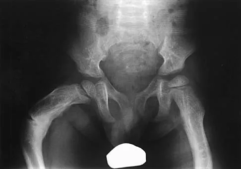
A 14-year-old male presents with a painful, enlarging mass in the distal femoral metaphysis. Plain radiographs show a permeative, moth-eaten lytic lesion with cortical breakthrough and aggressive periosteal reaction. You are the registrar tasked with performing the biopsy. The consultant emphasizes the need to plan the biopsy to allow for subsequent limb salvage.

Describe your systematic approach to the biopsy of this lesion, focusing on site selection and technique.
Candidate: I would discuss this at the MDT first. I'd avoid the joint and major neurovascular bundles. I'd do a longitudinal incision and use a core needle under image guidance to make sure I don't contaminate too much tissue. I’d make sure to mark the tract so it can be resected later.
Failing to emphasize the "Single Compartment" rule, opting for a transverse incision (which contaminates multiple planes), choosing the wrong biopsy path (e.g., crossing the epiphysis), or forgetting to mention that the biopsy tract itself is considered "tumor" and must be excised en bloc.
A structured approach is required: 1. Pre-operative Planning: Must be discussed at an Orthopaedic Oncology MDT. Use MRI to identify the most viable area of the tumor and avoid areas of necrosis. 2. Incision: Must be a longitudinal incision. Transverse incisions contaminate wider tissue planes, making definitive excision difficult. 3. Compartmental Control: The biopsy must stay within a single fascial compartment. It should ideally be placed in an area that can be easily sacrificed during definitive surgery (e.g., the lateral aspect for a distal femur). 4. Technique: Use image-guided (CT or Ultrasound) core needle biopsy to minimize the soft tissue tract. 5. Oncologic Integrity: Meticulous hemostasis is vital to prevent hematoma formation, which spreads tumor cells. The biopsy tract is considered a "contaminated" structure and must be resected en bloc with the primary tumor during the definitive procedure.
Following the biopsy, the pathologist reports "inconclusive, suggestive of reactive bone formation" despite the radiological appearance being highly concerning for osteosarcoma. How do you manage this discordance?
Candidate: I would tell the patient it's likely benign and repeat the scan in a few weeks to see if it changes.
Assuming the biopsy is the "gold standard" truth. If the biopsy is negative but the imaging is "screaming" malignancy, the biopsy is a False Negative. Waiting to watch it grow is a catastrophic delay in treatment.
This is a Clinicopathologic Discordance. I would never reassure the patient or wait. The steps are: 1. Review: Review the imaging and pathology with a specialist sarcoma radiologist and pathologist. 2. Re-evaluate: Ensure the biopsy didn't hit a necrotic area or normal reactive bone rather than the tumor. 3. Repeat: The biopsy must be repeated, likely as an open incisional biopsy, as this provides a larger specimen and allows for frozen section verification of tissue viability. 4. Proceed: Only once a tissue diagnosis is reached should definitive oncological treatment (chemotherapy or surgery) commence.
