Pediatric Femur Fracture Management in Cerebral Palsy with Osteopenia: Case 37
Key Takeaway
Managing femur fractures in children with GMFCS IV cerebral palsy and osteopenia presents unique challenges. Orthopedic specialists must address weakened, osteopenic bone, pre-existing spasticity, and contractures that complicate alignment and implant stability. Detailed imaging like CT is crucial for surgical planning, ensuring adequate fixation and considering long-term bone health strategies for these complex pediatric patients.
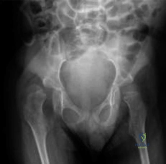
You are reviewing a patient in the emergency department: an 8-year-old male, GMFCS Level IV cerebral palsy, presenting with an acutely deformed, painful right thigh following a routine transfer. Here are the initial radiographs. How would you categorize this injury and what are the specific clinical implications of this patient's underlying physiology on your management strategy?
Candidate: This is a pathologic fracture of the femur in a patient with severe CP. The radiographs show a comminuted mid-diaphyseal fracture with significant varus angulation. Because the patient has CP, he has severe osteopenia from disuse and potentially from his medications like valproic acid. I would be worried about the spasticity causing displacement and would likely recommend surgery rather than a cast because a cast is hard to keep clean and won't hold the reduction.
The candidate focuses only on the "pathologic" label without classifying the fracture (AO Pediatric) or understanding the pathophysiology. They fail to mention the specific mechanism (insufficient bone due to disuse and metabolic bone disease) and offer a vague management plan without acknowledging why traditional pediatric methods (like ESIN or spica casting) are contraindicated in this specific high-spasticity, osteopenic patient.
The candidate classifies this as an AO Pediatric 32-D/5.1 femur fracture. They demonstrate a high-level understanding by framing this as a fragility fracture rather than a 'tumor-like' pathology. They structure the answer by highlighting: 1) Physiological risk: Valproic acid-induced hypovitaminosis D and chronic immobility causing severe osteopenia. 2) Mechanical risk: Spastic deforming forces (adductors/hamstrings) that make non-operative (spica) or flexible (ESIN) management failure-prone. 3) Solution: Submuscular plating with a locking construct to act as an internal fixator, emphasizing that biological fixation and bone health optimization (bisphosphonates) are as crucial as the hardware itself.
You have decided to proceed with surgery. Describe your considerations for patient positioning and choice of implant, specifically addressing why you would avoid certain traditional techniques used in pediatric trauma.
Candidate: I would place the patient on a standard radiolucent table and avoid a traction table. I would use a long locking plate, likely a 4.5mm LCP, inserted submuscularly. I would avoid ESIN because the bone is too soft to hold the nails, and the spasticity would cause the fracture to telescope.
The candidate identifies the correct implant but fails to explain why the traction table is dangerous in this patient (risk of physeal injury or fracture through osteopenic bone due to existing hip/knee contractures). They also fail to mention the importance of preserving the fracture hematoma/periosteum, which is vital for healing in a metabolic bone disease context.
The candidate systematically addresses: 1) Positioning: Avoidance of traction tables to prevent iatrogenic fractures or nerve palsies in the setting of severe hip/knee contractures; instead, use manual traction and bumps. 2) Implant choice: Locking plates are mandatory because they function as internal fixators, providing angular stability in cortical bone that lacks the density for standard screw purchase. 3) Technique: MIPO (Minimally Invasive Plate Osteosynthesis) to respect the biological envelope. 4) Rationale: ESIN is contraindicated due to the 'telescoping' effect caused by the patient's severe spasticity, and locking plates mitigate the risk of loss of reduction.
