ABOS Part I & AAOS OITE Orthopaedic Review: Forearm & Distal Humerus Fractures | Part 22149
Key Takeaway
This module provides a comprehensive review for the ABOS Part I and AAOS OITE examinations. It features 21 advanced multiple-choice questions covering critical topics in orthopaedic trauma, including diaphyseal radius/ulna fractures and complex distal humerus fractures. Key areas include surgical approaches, fixation principles, biomechanics, nerve protection, and post-operative rehabilitation strategies.
ABOS Part I & AAOS OITE Orthopaedic Review: Forearm & Distal Humerus Fractures | Part 22149
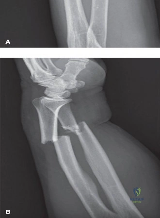
A 32-year-old male sustains a displaced diaphyseal fracture of the radius and ulna after a high-energy motor vehicle accident. During preoperative planning, the orthopedic surgeon reviews the patient's contralateral uninjured forearm radiographs to determine the optimal plate contouring for the radius. According to the seminal biomechanical studies on radial anatomy, what are the average magnitude and location of the maximum radial bow that the surgeon aims to restore?
Correct Answer: C
The teaching case explicitly states, 'Classic biomechanical studies by Schemitsch and Richards defined the normal parameters of the radial bow. The maximum radial bow averages 15.3 millimeters (range, 10 to 22 mm), and the location of this maximum bow is situated at approximately 60% of the total radial length, measured from the bicipital tuberosity to the radiocarpal joint.' This precise anatomical restoration is critical for optimal forearm rotation.
Options A, B, D, and E present incorrect values for either the magnitude or the location of the maximum radial bow, which would lead to suboptimal functional outcomes if used as a surgical target. Failure to restore these specific parameters directly correlates with a proportional loss of forearm rotation, as highlighted in the case.
A 28-year-old construction worker presents with a malunited diaphyseal radius fracture, sustained 6 months prior, resulting in a significant loss of pronation and supination. Clinical examination reveals a fixed deformity. Based on the biomechanical principles discussed in the case, which of the following angular deformities is most likely to cause a severe and clinically significant loss of forearm rotation?
Correct Answer: D
The teaching case, referencing Matthews et al., states: 'Matthews et al. demonstrated that angular deformities of less than 10 degrees in any plane do not significantly restrict forearm rotation. However, deformities exceeding 20 degrees, particularly those involving a loss of the radial bow, result in a severe and clinically significant loss of pronation and supination.' An angular deformity of 25 degrees clearly exceeds this 20-degree threshold, making it the most likely cause of severe rotational loss.
Options A, B, C, and E describe angular deformities that are either below or at the threshold of 10 degrees, which the literature suggests do not significantly restrict forearm rotation. While a 15-degree deformity (Option C) is greater than 10 degrees, the case specifically highlights deformities exceeding 20 degrees as leading to severe and clinically significant loss. Therefore, 25 degrees is the most accurate answer for a severe loss of function.
A 40-year-old female undergoes open reduction and internal fixation of a comminuted mid-diaphyseal radial fracture using the volar (Henry) approach. During the deep dissection to expose the proximal and middle thirds of the radius, the surgeon must be particularly vigilant about protecting a specific nerve. Which of the following describes the most critical nerve to protect and its anatomical relationship during this approach?
Correct Answer: C
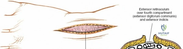
The teaching case details the Volar Henry approach: 'To expose the proximal third of the radius, the recurrent radial artery (the 'leash of Henry') must be identified, ligated, and divided. This allows the brachioradialis to be retracted laterally, exposing the supinator muscle. The supinator is sharply detached from its ulnar origin and reflected laterally. This maneuver protects the posterior interosseous nerve (PIN), which courses within the substance of the supinator.' This is the most critical nerve to protect during proximal exposure via the Henry approach.
Option A is incorrect; the ulnar nerve is on the ulnar side of the forearm and not typically encountered in the primary dissection field of the Henry approach to the radius. Option B is incorrect; the median nerve is retracted ulnarly with the FCR, but the PIN is the nerve most at risk during the deeper dissection of the proximal radius. Option D is incorrect; the radial nerve innervates the brachioradialis, but the PIN is the branch of the radial nerve that is specifically vulnerable within the supinator. Option E is incorrect; while the anterior interosseous nerve (AIN) is a branch of the median nerve and can be at risk, the PIN is the primary nerve of concern when detaching and reflecting the supinator for proximal radial exposure in the Henry approach.
A 55-year-old male presents with a comminuted fracture of the proximal third of the radial diaphysis. The surgeon opts for a dorsal (Thompson) approach for open reduction and internal fixation. During the deep dissection, after incising the fascia and developing the interval between the ECRB and EDC, the supinator muscle is exposed. What is the most crucial step to prevent iatrogenic nerve injury during the subsequent exposure of the radial shaft?
Correct Answer: C
The teaching case describes the Dorsal Thompson approach: 'The supinator muscle is exposed. The critical step in this approach is the identification and protection of the PIN. The nerve emerges from the supinator approximately 1 cm proximal to the distal edge of the muscle. The supinator must be carefully split along the course of the nerve, or elevated off the radius from ulnar to radial, ensuring the nerve remains protected within the muscle belly during retraction.' This maneuver is paramount to avoid injury to the PIN.
Options A, B, D, and E describe steps or anatomical structures relevant to other approaches or different parts of the forearm, or incorrect nerve relationships for the dorsal Thompson approach. Retracting the brachioradialis and exposing the radial artery (A) is part of the Henry approach. Elevating FPL and pronator quadratus (B) is for distal radial exposure, typically volar. Ligating the leash of Henry (D) is specific to the proximal Henry approach. Identifying the median nerve deep to pronator teres (E) is relevant to the Henry approach but not the primary concern for the PIN in the Thompson approach.
A 38-year-old male undergoes ORIF for a displaced mid-diaphyseal radial fracture. The surgeon uses a standard straight 3.5 mm LC-DCP. Postoperatively, the patient experiences significant restriction in forearm pronation and supination. Intraoperative fluoroscopy confirmed length and axial alignment. What is the most likely cause of the restricted rotation in this scenario?
Correct Answer: C
The teaching case explicitly warns: 'Applying a straight plate to the curved radial diaphysis will inevitably flatten the radial bow, narrowing the interosseous space and restricting rotation.' This is a classic cause of malunion and loss of forearm rotation after radial shaft fixation, especially when standard straight plates are used without meticulous contouring.
Option A is incorrect because over-contouring would lead to excessive bowing, not flattening, and while it can also cause impingement, the scenario describes using a straight plate. Option B is incorrect; while inadequate stability can lead to nonunion, it's less directly linked to immediate mechanical restriction of rotation compared to a flattened bow. Option D is a risk factor for hardware failure or nonunion, but not the primary cause of mechanical rotational restriction due to incorrect radial bow. Option E describes a potential rehabilitation complication, but the question implies a mechanical issue related to the surgical technique and plate application, not a rehabilitation error.
A 22-year-old male sustains an open, comminuted diaphyseal fracture of both the radius and ulna after a fall from a height. He undergoes emergent irrigation and debridement followed by ORIF of both bones through separate volar (Henry) and dorsal ulnar incisions. Despite meticulous surgical technique, the patient develops progressive loss of forearm rotation over several months. Which of the following complications is most likely, and what is a key risk factor for its development that the surgeon attempted to mitigate?
Correct Answer: C
The teaching case lists 'Radioulnar Synostosis' as a complication with an incidence of 2-8%. Key risk factors include 'High-energy trauma, closed head injury, single-incision approach for both bones, severe soft tissue stripping, delayed surgery.' The case also states, 'Furthermore, the routine use of dual incisions (separate volar Henry and dorsal ulnar approaches) is universally recommended over single-incision approaches to minimize the devastating complication of radioulnar synostosis.' The patient's high-energy trauma is a risk factor, and the surgeon's use of dual incisions was a direct attempt to mitigate synostosis.
Options A, B, D, and E are all potential complications, and the listed mitigation strategies are correct. However, the question specifically asks for the complication that leads to 'progressive loss of forearm rotation over several months' and a risk factor that the surgeon 'attempted to mitigate' by using dual incisions. Radioulnar synostosis directly causes loss of rotation and is specifically mitigated by dual incisions, making it the best answer in this context.
A 60-year-old female presents with a chronic malunion of her radial shaft fracture, sustained 18 months prior, leading to severe restriction of pronation and supination. Imaging reveals a significant loss of the radial bow. The orthopedic surgeon plans a corrective osteotomy. What is the most recommended advanced imaging and planning tool to ensure precise restoration of the radial bow in this complex salvage setting?
Correct Answer: C
The teaching case emphasizes the importance of advanced imaging for complex deformities: 'In complex comminuted fractures or established malunions, computed tomography (CT) with three-dimensional reconstructions is highly recommended. Advanced planning software can mirror the contralateral intact radius, allowing for precise calculation of the required osteotomy angles or the degree of plate contouring necessary to restore the native anatomy.' This approach is crucial for accurate restoration of the radial bow in malunions.
Option A (standard radiographs) is mandatory for initial assessment but insufficient for precise 3D planning of a corrective osteotomy for a malunion. Option B (stress radiographs) is used for instability assessment, not for planning bone correction. Option D (MRI) is excellent for soft tissue but less precise for bony morphology and 3D planning of osteotomies. Option E (EMG/NCS) is for nerve function assessment, not for anatomical bone reconstruction planning.
A 42-year-old male sustains an isolated, non-displaced ulnar shaft fracture (nightstick fracture) after a direct blow. He has no associated injuries, and his PRUJ and DRUJ are stable. Based on the provided case, what is the most appropriate initial management strategy for this patient?
Correct Answer: C
The teaching case outlines operative decision-making: 'Non-operative management is reserved for a highly select, narrow subset of injuries or for patients with prohibitive surgical risks.' The table further specifies 'Non Operative Indications' including 'Truly non-displaced, isolated ulnar shaft fractures (Nightstick)' and 'Intact radial bow parameters.' Given the patient has a non-displaced, isolated ulnar shaft fracture with stable PRUJ and DRUJ, non-operative management is the most appropriate initial strategy.
Option A (immediate ORIF) is the gold standard for most displaced adult forearm fractures but not for truly non-displaced isolated ulnar shaft fractures. Option B (external fixation) is typically reserved for severe open fractures or medically unstable patients. Option D (urgent CT) is not indicated for a non-displaced isolated fracture where standard radiographs are sufficient. Option E (diagnostic arthroscopy) is overly aggressive and not indicated for this specific injury pattern.
A 30-year-old athlete undergoes ORIF for a displaced radial shaft fracture. Postoperatively, the surgeon initiates a rehabilitation protocol. In the early mobilization phase (Weeks 0-2), assuming rigid internal fixation, which of the following activities is the primary focus and which is strictly prohibited?
Correct Answer: C
The teaching case describes Phase I (Early Mobilization, Weeks 0-2) of the rehabilitation protocol: 'Assuming rigid internal fixation has been achieved, early active and active-assisted range of motion (ROM) is initiated within the first few days postoperatively. The primary focus is on digital ROM to prevent tendon adhesions and reduce edema. Gentle, pain-free pronation and supination exercises are commenced, along with elbow flexion and extension. Weight-bearing and lifting are strictly prohibited.'
Options A, B, D, and E describe activities that are either incorrect for the early phase (e.g., progressive strengthening, aggressive manipulation, full weight-bearing) or incorrectly prohibited (e.g., digital ROM, elbow flexion/extension).
A 25-year-old male sustains a complex, comminuted diaphyseal fracture of the radius. During surgical planning, the surgeon notes significant fragmentation. According to the principles of fixation for such complex fractures, what is the primary objective and the recommended plating technique?
Correct Answer: B
The teaching case differentiates fixation principles based on fracture complexity: 'Fixation principles dictate the achievement of absolute stability for simple fracture patterns (AO/OTA Type A and B) utilizing lag screws and neutralization plates, or compression plating techniques. For complex, comminuted fractures (AO/OTA Type C), bridge plating techniques are employed to preserve the soft tissue envelope and vascularity of the fracture fragments, focusing on the restoration of length, alignment, and rotation rather than anatomical reduction of every butterfly fragment.'
Option A is incorrect because anatomical reduction of every fragment is not the primary goal for comminuted fractures; bridge plating focuses on overall length, alignment, and rotation. Option C describes techniques for simple fractures, not complex comminuted ones. Option D is incorrect; external fixation is typically temporary, and early weight-bearing is not the primary objective for complex fractures. Option E is incorrect; while intramedullary nailing can be used for some long bone fractures, plating is the standard for diaphyseal forearm fractures, and minimizing exposure is a goal of bridge plating, but not the primary objective over restoration of function.
A 58-year-old female presents with a complex, comminuted intra-articular distal humerus fracture (AO/OTA Type C3) after a fall from standing height. Pre-operative CT scans confirm significant articular involvement and disruption of both medial and lateral columns. During surgical planning for a posterior approach, the surgeon decides to perform an olecranon osteotomy. Which of the following statements regarding the ulnar nerve and its management during this procedure is MOST accurate?
Correct Answer: C
The case explicitly states, 'The ulnar nerve is identified proximal to the cubital tunnel, typically lying anterior to the medial head of the triceps. Trace the nerve distally through the cubital tunnel (between the medial epicondyle and olecranon). Perform an extensive neurolysis of the ulnar nerve... While not always strictly necessary in every case, anterior transposition of the ulnar nerve is generally recommended during open reduction internal fixation (ORIF) of DHFs via a posterior approach. This protects the nerve from direct injury during drilling, plating, and screw insertion, and prevents post-operative compression from hardware or scar tissue.' The 'Summary of Key Literature / Guidelines' section further reinforces this: 'Prophylactic anterior transposition of the ulnar nerve during posterior approaches for DHF ORIF is widely recommended. Studies have shown a significant reduction in post-operative ulnar neuropathy rates with routine transposition compared to in situ decompression or no specific management.'
Option A is incorrect because prophylactic transposition is recommended regardless of pre-operative symptoms due to the high risk of iatrogenic injury or post-operative compression.
Option B is incorrect because the ulnar nerve is located posteriorly to the medial epicondyle, within the cubital tunnel, not anteriorly, and the brachialis muscle is anterior to the humerus, not directly protecting the ulnar nerve in the cubital tunnel.
Option D is incorrect because while the ulnar nerve can be at risk from hardware, its primary risk during a posterior approach is from direct injury during dissection, retraction, or compression from hardware/scar tissue in the cubital tunnel, not specifically from lateral column screw placement. Lateral column screws are more likely to endanger the radial nerve if excessively long or misplaced.
Option E is incorrect because the cubital tunnel is formed by the medial epicondyle and the olecranon, with the arcuate ligament forming the roof, not the radial head.
A 32-year-old male sustains a high-energy distal humerus fracture (AO/OTA Type C2) involving both columns and the articular surface. Surgical fixation is planned via a posterior approach with an olecranon osteotomy. The surgeon opts for an orthogonal plating strategy. Which of the following biomechanical principles BEST describes the advantage of this construct for distal humerus fractures?
Correct Answer: C
The 'Summary of Key Literature / Guidelines' section states: 'Biomechanically, both orthogonal (medial and posterior/posterolateral) and parallel (medial and lateral) plating constructs provide sufficient stability. Several studies, including cadaveric biomechanical analyses and clinical series, suggest that orthogonal plating may offer superior stiffness, particularly in torsion and bending, especially when the posterior plate is positioned to capture fragments of the lateral column and bridge the olecranon fossa effectively.' This directly supports the advantage described in option C.
Option A is incorrect because while compression is important, the primary biomechanical advantage highlighted for orthogonal plating is its stiffness in torsion and bending, not exclusively axial compression. Lag screws provide interfragmentary compression.
Option B is incorrect because the ability to place interfragmentary lag screws is dependent on the fracture pattern and surgical technique, not inherently easier or harder with orthogonal versus parallel plating. Both strategies aim to incorporate lag screws.
Option D is incorrect because both orthogonal and parallel plating strategies for complex DHFs via a posterior approach typically involve significant soft tissue dissection to expose the fracture and apply plates, so one does not inherently minimize stripping more than the other.
Option E is incorrect because orthogonal plating is widely used and often preferred for complex intra-articular fractures (AO/OTA Type C), as described in the case, due to its robust fixation and ability to capture articular fragments. It does not limit articular visualization, especially when combined with an olecranon osteotomy.
A 72-year-old osteoporotic female presents with a highly comminuted, intra-articular distal humerus fracture (AO/OTA Type C3) after a low-energy fall. Given her bone quality and the complexity of the fracture, the surgeon is considering the optimal triceps management strategy for a posterior approach. Which of the following statements regarding triceps management is MOST appropriate for this patient?
Correct Answer: B
The 'Detailed Surgical Approach / Technique' section, under 'Triceps Management', states: 'Olecranon Osteotomy: Indications: Gold standard for complex intra-articular fractures (AO/OTA C-type) requiring maximal visualization and direct access to the articular surface.' It further notes its advantage: 'Unrivaled exposure of the articular surface and both medial and lateral columns. Allows direct visualization of the fracture pattern and facilitates anatomical reduction.' While it 'Adds a second fracture to manage, potential for nonunion, symptomatic hardware, or pain at the osteotomy site,' for a highly comminuted intra-articular fracture, the benefit of optimal visualization for anatomical reduction often outweighs these risks, especially with modern osteotomy fixation techniques.
Option A is incorrect because while a triceps split avoids an osteotomy, it provides more limited exposure, which can compromise anatomical reduction of a highly comminuted intra-articular fracture. The case states that olecranon osteotomy is the gold standard for such complex fractures.
Option C is incorrect because the paratricipital approach is a triceps-sparing technique that offers more limited exposure compared to an olecranon osteotomy, especially for the entire articular surface and both columns. It is not superior for highly comminuted patterns.
Option D is incorrect because triceps-sparing approaches are suitable for 'extra-articular (AO/OTA A-type) or less comminuted intra-articular fractures where full articular exposure is not mandatory,' not generally for all DHFs, especially complex ones.
Option E is incorrect because the triceps reflecta (Kocher) is a more extensile triceps-sparing approach that provides good access to both columns, not primarily limited to lateral column fractures. However, it still does not offer the 'unrivaled exposure' of an olecranon osteotomy for complex articular fractures.
A 48-year-old male undergoes ORIF of a distal humerus fracture via a posterior approach with olecranon osteotomy. Post-operatively, he develops new onset paresthesia and weakness in the small finger and ulnar half of the ring finger, along with intrinsic muscle weakness. This complication is most consistent with ulnar neuropathy. Based on the case, what is the MOST appropriate initial management strategy for this patient?
Correct Answer: B
The 'Complications & Management' section, under 'Management Considerations for Specific Complications', states: 'Ulnar Neuropathy: Most often presents as paresthesia or weakness in the ulnar nerve distribution. Many cases are transient neurapraxias. If symptoms persist beyond 3-6 months, worsen, or present as a new deficit, EMG/NCS studies are warranted. Surgical exploration, neurolysis, and re-transposition may be indicated.'
Option A is incorrect because immediate surgical exploration is generally not the first step unless there is clear evidence of acute, severe nerve transection or entrapment. Most post-operative neuropathies are transient and resolve with observation.
Option C is incorrect because while corticosteroids can reduce inflammation, there is no specific evidence presented in the case or general guidelines recommending high-dose corticosteroids as the primary initial management for post-operative ulnar neuropathy.
Option D is incorrect because revision ORIF for hardware removal is premature. Hardware removal is considered if symptoms persist and are clearly attributable to hardware irritation after fracture union, or if the nerve was not transposed and is now compressed by hardware. Initial management is observation.
Option E is incorrect because aggressive physical therapy, especially focusing on stretching the nerve, could potentially exacerbate an irritated nerve. Rehabilitation should be guided by the nerve's status and the fracture's healing, with caution regarding nerve symptoms.
A 65-year-old male presents to the emergency department after a motor vehicle accident with a severely comminuted distal humerus fracture. Clinical examination reveals significant swelling and tenderness around the elbow. Initial radiographs are difficult to interpret due to the comminution. Which of the following pre-operative imaging modalities is MOST essential for surgical planning in this case?
Correct Answer: C
The 'Pre-Operative Planning & Patient Positioning' section, under 'Radiographic Evaluation', states: 'Computed Tomography (CT) Scan: Essential for nearly all complex DHFs. Axial, sagittal, and coronal reconstructions, along with 3D reconstructions, are invaluable for understanding fracture morphology, articular involvement, degree of comminution, and fragment orientation. This guides implant selection and surgical strategy.'
Option A is incorrect because while MRI can assess soft tissues, it is not the primary or most essential imaging for detailed bone fracture morphology in complex DHFs. CT is superior for bony detail.
Option B is incorrect because while standard radiographs are the initial assessment, the case states they are 'difficult to interpret due to the comminution,' highlighting the need for more advanced imaging like CT.
Option D is incorrect because arteriography is indicated 'if vascular injury is suspected (e.g., absent pulses, expanding hematoma),' which is not explicitly stated as the primary concern in the vignette, although it might be considered if vascular compromise were present. CT is essential for fracture planning regardless.
Option E is incorrect because ultrasound is not the primary imaging modality for detailed fracture assessment or surgical planning for complex distal humerus fractures.
A 40-year-old construction worker undergoes ORIF for a complex intra-articular distal humerus fracture. The surgeon achieves stable bicondylar fixation with an orthogonal plating construct. Post-operatively, the patient is placed in a posterior splint. Which of the following principles is MOST critical for the immediate post-operative rehabilitation protocol in this patient?
Correct Answer: B
The 'Post-Operative Rehabilitation Protocols' section, under 'General Principles', states: 'Early, Controlled Motion: The overarching goal is to prevent stiffness and heterotopic ossification by initiating controlled active and passive range of motion (ROM) as soon as safely possible.' Under 'Phase 1: Immediate Post-Operative / Early Protection (Weeks 0-3)', it specifies: 'Passive Range of Motion (PROM): Gentle, gravity-assisted flexion and extension within a pain-free arc, typically starting the first post-operative day if fixation is stable. Avoid forceful manipulation. Active-Assisted Range of Motion (AAROM): Patient uses the unaffected arm to assist the injured arm. Active Range of Motion (AROM): Gentle active flexion/extension, pronation/supination within comfort limits.'
Option A is incorrect because the case explicitly states that 'prolonged immobilization is detrimental' and that the goal is 'early, controlled motion' to prevent stiffness.
Option C is incorrect because the protocol specifies 'Strictly non-weight-bearing through the upper extremity' in Phase 1, and 'Still no significant weight-bearing or heavy lifting' in Phase 2.
Option D is incorrect because strengthening exercises are initiated gradually, with 'gentle isometric exercises' starting in Phase 2 (Weeks 3-6/8), not aggressive exercises on day 1.
Option E is incorrect because while CPM can be used, the case emphasizes active and passive ROM exercises. The specific duration and necessity of 24-hour CPM are not highlighted as the MOST critical principle, and its routine use is debated.
A 28-year-old male presents with an open, displaced intra-articular distal humerus fracture (AO/OTA Type C1) after a motorcycle accident. He has no significant comorbidities. Which of the following factors is the MOST compelling indication for operative management in this patient?
Correct Answer: B
The 'Indications for Operative Management' section lists: 'Open Fractures: Require urgent debridement and stabilization.' While all other options are also valid indications or contributing factors, an open fracture is an urgent surgical indication due to the high risk of infection and the need for immediate debridement and stabilization to prevent further contamination and facilitate healing.
Option A is incorrect because while young age and high activity level are factors favoring operative management to restore function, they are not as immediately compelling as an open fracture.
Option C is incorrect because the intra-articular nature is a strong indication for ORIF to restore joint congruity, but an open fracture adds an element of urgency and necessity for immediate intervention beyond just the fracture pattern.
Option D is incorrect because displacement is a general indication for operative management in many fractures, but an open fracture carries additional, more immediate risks that mandate surgery.
Option E is incorrect because being a good surgical candidate (absence of comorbidities) facilitates surgery but is not an indication for surgery itself; rather, it allows for the treatment of existing indications.
During definitive fixation of a complex distal humerus fracture via a posterior approach, the surgeon is applying an orthogonal plating construct. A medial plate is applied to the medial column, and a posterior/posterolateral plate is applied to the lateral column. To maximize bone purchase and construct stability, what is the MOST effective screw trajectory strategy for the distal fragments?
Correct Answer: C
The 'Detailed Surgical Approach / Technique' section, under 'Definitive Fixation' and 'Screw Trajectories', states: 'The key is to direct screws from each plate to avoid collision and to maximize bone purchase, ideally interlocking each column distally. Screws from the medial plate are directed laterally, and screws from the posterior/posterolateral plate are directed medially. Bicortical purchase is desirable where anatomically safe.' This describes the 'omega' configuration mentioned in the 'Summary of Key Literature / Guidelines' section, which maximizes interfragmentary purchase.
Option A is incorrect because directing all screws parallel to the humeral shaft axis would not effectively capture the distal articular fragments in a cross-columnar fashion, which is crucial for bicondylar fixation.
Option B is incorrect because directing screws medially from the medial plate and posteriorly from the posterior plate would not achieve the desired interlocking and cross-columnar fixation, potentially leading to inadequate stability.
Option D is incorrect because the case states 'Bicortical purchase is desirable where anatomically safe' to maximize stability, not unicortical purchase to avoid neurovascular injury. While neurovascular protection is paramount, bicortical purchase is preferred when safe.
Option E is incorrect because while some screws may be perpendicular, the primary strategy for distal humerus fixation is to direct screws to maximize purchase and interlock the columns, which often involves varying angles, not strictly perpendicular to the plate.
A 55-year-old male undergoes ORIF of a complex distal humerus fracture. Despite stable fixation and a diligent rehabilitation protocol, he develops significant elbow stiffness with a limited range of motion (30-90 degrees) and radiographic evidence of heterotopic ossification (HO) 4 months post-operatively. Which of the following is the MOST appropriate next step in managing his elbow stiffness and HO?
Correct Answer: B
The 'Complications & Management' section, under 'Management Considerations for Specific Complications' for 'Stiffness', states: 'If stiffness develops, a stepwise approach is taken: intensive physical therapy, static progressive or dynamic splinting, and if conservative measures fail, manipulation under anesthesia (MUA) or open capsular release (often combined with hardware removal).' For HO, it states: 'surgical excision after maturation (usually 6-12 months post-injury).'
At 4 months, the HO is likely not fully mature, and a conservative, stepwise approach to stiffness is indicated before considering aggressive surgical excision of HO. Intensified therapy and splinting are the next logical steps, with MUA as a potential escalation if conservative measures fail.
Option A is incorrect because surgical excision of HO is generally recommended 'after maturation (usually 6-12 months post-injury)' to reduce recurrence risk. At 4 months, it is likely too early for surgical excision of HO, though capsular release might be considered later if conservative measures fail.
Option C is incorrect because prolonged immobilization is a known cause of stiffness and would worsen the current situation, directly contradicting the principle of early motion.
Option D is incorrect because there is no mention of signs of infection in the vignette. Stiffness and HO are common complications of DHF, and infection is not the primary assumption without other clinical signs.
Option E is incorrect because while NSAIDs are used for HO prophylaxis, indefinite high-dose NSAID use is not a primary treatment for established HO and carries significant side effects. Prophylaxis is typically initiated perioperatively, not indefinitely post-HO formation.
A 68-year-old female presents with a non-displaced extra-articular distal humerus fracture (AO/OTA Type A1) after a fall. She has multiple comorbidities, including severe cardiac disease and poorly controlled diabetes. Which of the following is the MOST appropriate initial management strategy for this patient?
Correct Answer: B
The 'Indications for Non-Operative Management' section states: 'Non-displaced or Minimally Displaced Extra-Articular Fractures (AO/OTA Type A): Especially in elderly or low-demand patients.' It also lists 'Non-displaced or Minimally Displaced Intra-Articular Fractures (rare): In elderly, frail, low-demand patients with significant comorbidities where surgical risks outweigh potential benefits...' The patient in the vignette has a non-displaced extra-articular fracture and significant comorbidities, making non-operative management the most appropriate initial strategy.
Option A is incorrect because ORIF is typically indicated for displaced fractures, especially intra-articular ones. For a non-displaced extra-articular fracture, especially in a patient with severe comorbidities, the risks of surgery likely outweigh the benefits.
Option C is incorrect because external fixation is generally reserved for open fractures with severe soft tissue compromise or as a temporary measure in polytrauma, not typically for a non-displaced extra-articular fracture.
Option D is incorrect because total elbow arthroplasty is a salvage procedure for severe comminuted fractures in elderly, low-demand patients, or for post-traumatic arthritis, not for a non-displaced extra-articular fracture.
Option E is incorrect because while a second opinion is always an option, the case provides clear guidelines that support non-operative management for this specific fracture type and patient profile, making it the most appropriate initial strategy.
A 35-year-old male sustains a distal humerus fracture. During the physical examination, the orthopedic resident assesses the stability of the elbow joint. Which of the following combinations of structures provides the primary static stability to the elbow joint?
Correct Answer: C
The 'Surgical Anatomy & Biomechanics' section, under 'Biomechanics' and 'Elbow Joint Stability', states: 'The elbow derives its stability from a combination of osseous congruence (trochlear notch of ulna with trochlea of humerus), static ligamentous restraints (MCL, LCL complex), and dynamic muscular contributions.' Osseous congruence and static ligamentous restraints are the primary static stabilizers.
Option A is incorrect because the triceps and anconeus are dynamic muscular stabilizers, not primary static stabilizers.
Option B is incorrect because the radial and ulnar nerves are neurovascular structures, not stabilizers of the joint.
Option D is incorrect because the brachialis muscle and biceps tendon are dynamic muscular stabilizers, not primary static stabilizers.
Option E is incorrect because the median nerve and brachial artery are neurovascular structures, not stabilizers of the joint.
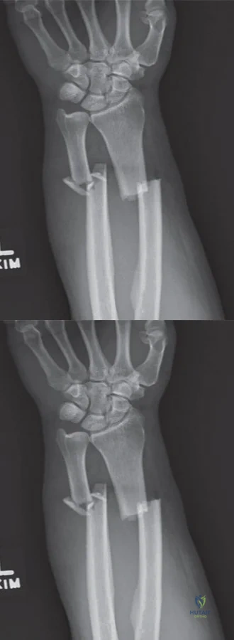
A 45-year-old mechanic sustains a Galeazzi fracture-dislocation. Following rigid plate fixation of the radial shaft, the distal radioulnar joint (DRUJ) is noted to be grossly unstable in supination. What is the recommended acute management for the DRUJ?
When performing a paratricipital (Alonso-Llames) approach for a distal humerus extra-articular fracture, which of the following best describes the management of the triceps mechanism?
None
