FRCS Exam Success: Why Orthopedic Moment of Inertia Matters
Key Takeaway
Here are the crucial details you must know about FRCS Exam Success: Why Orthopedic Moment of Inertia Matters. In orthopedics, the moment of inertia (or second moment area) quantifies an object's material distribution relative to applied load. For solid nails, it varies with the radius's fourth power. For hollow nails, it's proportional to the outer radius's fourth power minus the inner radius's fourth power. A higher moment of inertia indicates greater rigidity against torsional and bending forces, making hollow designs efficient for resisting stress.
A 72-year-old female presents with symptomatic medial compartment osteoarthritis of the knee. She has failed conservative management and is being considered for a Unicompartmental Knee Arthroplasty (UKA). You are evaluating the preoperative radiographs to ensure patient suitability.
Describe your systematic assessment of these radiographs and list the contraindications for this procedure based on the imaging findings.
Candidate: I would assess the radiographs to confirm isolated medial compartment disease. Contraindications I look for include patellofemoral arthritis, lateral compartment disease, fixed flexion deformity, or an ACL deficiency. I also check for the degree of bone loss and overall limb alignment.
The candidate focuses only on the compartment space narrowing. They fail to mention the "Stress Radiograph" requirement or specific contraindications like inflammatory arthritis (RA), chondrocalcinosis, or the status of the ACL. A "pass" requires a structured, comprehensive approach including joint status, ligamentous integrity, and deformity.
A structured approach is required: 1. **Compartmental Analysis:** Confirm isolated medial compartment disease; look for lateral or patellofemoral arthritis (clear contraindications). 2. **Ligamentous Status:** Mention the mandatory status of the ACL (it must be intact). 3. **Deformity:** Assess for fixed deformity (>10-15 degrees flexion contracture or >5 degrees varus correction requirement). 4. **Bone Quality/Stock:** Look for erosion or cyst formation >5mm. 5. **Advanced Imaging:** If questionable, mention the role of stress radiographs to confirm the lateral compartment remains reducible (open) during varus-valgus stress.
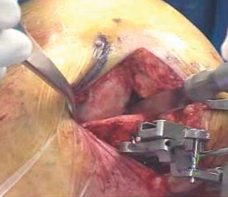
You have decided to proceed with the UKA. Intraoperatively, you notice that the medial meniscus is not as degenerated as anticipated, and there is some questionable cartilage on the lateral aspect of the medial plateau. How do you re-evaluate the joint, and what clinical decision would you make?
Candidate: I would perform a thorough arthroscopic or open inspection of all three compartments. If the cartilage is poor, I would proceed with a Total Knee Arthroplasty (TKA) instead of a UKA.
Candidates often suggest switching to a TKA without explicitly explaining the "low threshold for conversion." Failing to mention that the surgeon must be prepared to switch if the intraoperative findings are worse than preoperative imaging is a major procedural safety concern.
The candidate must emphasize the "conversion strategy." 1. **Thorough Examination:** Visualize the entire joint (lateral and patellofemoral compartments). 2. **Judgment:** If the cartilage loss is beyond the medial compartment or if the ACL is found to be incompetent intraoperatively, convert to TKA. 3. **Patient Consent:** A critical high-scoring point is stating that the patient was consented for both procedures preoperatively, acknowledging the possibility of conversion if findings are not suitable for UKA.
