Ace Your FRCS: Key Questions in Trauma & Orthopaedics
Key Takeaway
We review everything you need to understand about Ace Your FRCS: Key Questions in Trauma & Orthopaedics. In trauma and orthopaedics, managing open tibial fractures involves immediate ATLS review, wound care, analgesia, splinting, and antibiotics. Theatre planning includes thorough debridement and fracture stabilization. Key post-operative concerns are compartment syndrome, monitored clinically or invasively, often necessitating fasciotomy. Definitive soft tissue cover is typically aimed for within five days to optimize outcomes.
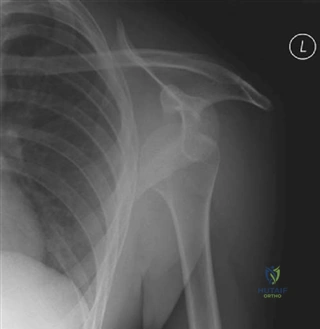
A 27-year-old patient presents following a high-energy road traffic accident. You are presented with the following radiograph. Describe your findings and outline your immediate management plan.
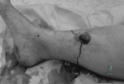
Candidate: "This is an open tibial shaft fracture. I would perform an ATLS primary survey, assess the wound, dress it with saline-soaked gauze, splint the limb, and initiate intravenous antibiotics and tetanus prophylaxis. I would then request AP and lateral radiographs."
Candidates often forget to mention the immediate involvement of the Plastic Surgical team. Relying solely on Orthopaedic management for an open fracture in the modern FRCS era is seen as suboptimal. Additionally, failing to mention the neurovascular status of the limb in the initial 'assessment' sentence is a common 'fail' point.
Structure your answer: 1. Safety & Systemic: ATLS survey and stabilization. 2. Local: Neurovascular assessment (documenting pulses/nerve status), assessment of wound (Gustilo-Anderson grade). 3. First Aid: Photograph, saline-soaked dressing, splinting. 4. Systemic Therapy: Early antibiotics and tetanus. 5. Multidisciplinary: Immediate communication with the combined Orthoplastic team, as this is a high-energy injury requiring coordinated care.
This patient was the driver in a high-speed RTA. Please describe these radiographs and identify the specific concerns associated with this fracture pattern.
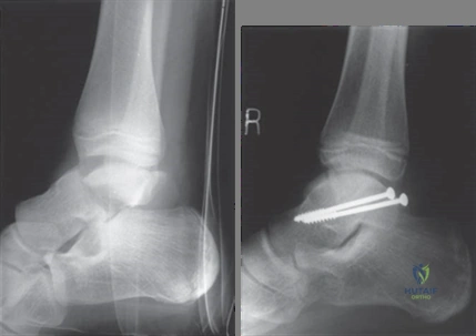
Candidate: "This is a displaced talar neck fracture. I would classify this as Hawkins Type II. It occurs due to axial loading, often in a hyper-dorsiflexed foot. I would check the neurovascular status and ensure the skin is viable."
Candidates often forget to mention the 'Hawkins Sign' and its significance. They also frequently miss the potential for foot compartment syndrome, which is a critical, limb-threatening complication in talar fractures.
A structured response: Identification: Hawkins classification. Pathophysiology: Axial load, high-energy. Assessment: Focus on skin viability (talar neck fractures are often open/tenting) and the 9 compartments of the foot. Prognosticating: Mention AVN risk (approx. 25-50% for Type II) and the Hawkins sign (subchondral lucency at 6-8 weeks indicating reperfusion). Vascularity: Describe the supply via the anastomosis of the artery of the tarsal canal, sinus tarsi, and dorsalis pedis.
This 20-year-old roofer fell from a ladder. Describe the radiograph and your approach to managing this patient.
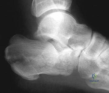
Candidate: "This is a calcaneal fracture. Bohler's angle is reduced, suggesting posterior facet involvement. I would request a CT scan to classify it using the Sanders system."
Failing to mention the high association with lumbar spine fractures (Don Juan Syndrome) and failing to address the soft tissue status (swelling/blistering), which is the limiting factor for surgery.
Address: Imaging: Bohler’s angle (20-40 deg) and Gissane’s angle. Classification: Sanders (based on CT). Associations: Screen the lumbar spine. Timing: Emphasize "waiting for the wrinkles" (soft tissue recovery). Evidence: Mention the Buckley et al. (2002) RCT showing no significant difference between operative and non-operative management for most patients, but noted better outcomes in specific subgroups (younger, anatomical reduction).
