Comprehensive Guide to the Direct Lateral Approach (DLA) for Hip Arthroplasty
Key Takeaway
The Direct Lateral Approach (DLA) is a foundational hip surgery technique, prioritizing adequate exposure while minimizing abductor damage. It's often used for total hip arthroplasty, aiming for early post-operative mobility and reduced dislocation risk. A thorough understanding of its anatomy, biomechanics, and indications is critical for successful patient outcomes and preserving crucial neurovascular structures.
Introduction and Epidemiology
The direct lateral approach (DLA), often referred to as the modified Hardinge or transgluteal approach, represents a foundational surgical pathway to the hip joint. Its development was driven by the imperative to balance adequate surgical exposure with minimizing iatrogenic soft tissue damage, particularly to the abductor mechanism, thereby facilitating early post-operative mobilization and reducing dislocation risk. Historically, approaches to the hip have evolved from extensive and often morbid exposures, such as the Ollier or Kocher-Langenbeck for trauma, to more nuanced pathways optimized for total hip arthroplasty (THA).

The DLA gained prominence due to its perceived advantages in reducing posterior dislocation rates compared to the posterior approach, and its ability to preserve the anterior abductor musculature, differentiating it from true transgluteal approaches that involve a more significant split of the gluteus medius. Epidemiologically, the choice of surgical approach for primary THA varies geographically and by surgeon preference. While the posterior approach remains highly popular globally, and the direct anterior approach has seen a recent surge in adoption, the DLA maintains a significant role in many practices. Its utility is particularly recognized in patients where robust abductor function is paramount for early rehabilitation and stability, and in cases where a direct and controlled view of the acetabulum and proximal femur is desired without the need for extensive anterior soft tissue release or complex patient positioning. National joint registries consistently demonstrate that the lateral approach yields excellent long-term survivorship, with dislocation rates historically lower than those of un-repaired posterior approaches, making it a reliable workhorse in both primary and revision arthroplasty settings.
Surgical Anatomy and Biomechanics
A thorough understanding of the regional anatomy is critical for minimizing complications and optimizing outcomes with the direct lateral approach. The approach fundamentally relies on exploiting the anatomic continuity between the gluteus medius and the vastus lateralis over the greater trochanter.
Musculature of the Lateral Hip
The muscular envelope of the lateral hip dictates the surgical windows available during the DLA.
- Gluteus Maximus: This large, superficial muscle is typically retracted posteriorly or split longitudinally in line with its fibers, without significant detachment. It acts as a primary extensor and external rotator of the hip.
- Tensor Fascia Lata: Located anterior to the gluteus medius, the TFL originates from the anterior iliac crest and inserts into the iliotibial band. The IT band must be incised longitudinally to access deeper structures. The TFL assists in hip flexion, abduction, and internal rotation.
- Gluteus Medius: The cornerstone of the hip abductor mechanism, originating from the external surface of the ilium and inserting onto the greater trochanter. In the modified DLA, the anterior portion of the gluteus medius is preserved, while the anterior one-third to one-half is split longitudinally. The posterior portion remains attached to the greater trochanter.
- Gluteus Minimus: Lying deep to the gluteus medius, it also originates from the ilium and inserts onto the anterior facet of the greater trochanter, sharing the abductor function. Careful management is essential to avoid denervation or irreparable damage.
- Vastus Lateralis: This muscle originates from the greater trochanter and linea aspera, forming part of the quadriceps femoris. It is split longitudinally from its origin on the vastus ridge of the femur to expose the femoral shaft, maintaining continuity with the anterior gluteus medius flap.

Neurovascular Structures and Safe Zones
The most critical neurovascular structure at risk during the direct lateral approach is the superior gluteal nerve.
- Superior Gluteal Nerve and Artery: These structures supply the gluteus medius, gluteus minimus, and tensor fascia lata. The nerve exits the pelvis through the greater sciatic foramen, above the piriformis, and courses anteriorly between the gluteus medius and minimus. It typically enters the deep surface of the gluteus medius approximately 3 to 5 cm proximal to the tip of the greater trochanter. Preserving the integrity of this neurovascular bundle is paramount to preventing post-operative abductor weakness and a catastrophic Trendelenburg gait. The proximal extent of the gluteus medius split must strictly adhere to this "safe zone" (no more than 3 to 5 cm proximal to the trochanteric tip).
- Ascending Branch of the Lateral Circumflex Femoral Artery: Encountered during the deep dissection between the tensor fascia lata and the rectus femoris, and during the capsulotomy. It requires meticulous ligation or electrocautery to prevent significant post-operative hematoma.

Biomechanical Considerations
The direct lateral approach directly impacts the abductor lever arm. The hip joint operates as a class I lever. The center of rotation of the femoral head acts as the fulcrum, the body weight acts as the load, and the abductor musculature provides the effort. By elevating the anterior portion of the gluteus medius and minimus, the surgeon temporarily compromises this effort arm. Precise anatomic repair of the conjoined tendon of the gluteus medius and vastus lateralis is biomechanically requisite to restore the abductor moment, stabilize the pelvis during the single-leg stance phase of gait, and prevent superior migration of the femur.
Indications and Contraindications
The direct lateral approach is highly versatile, offering excellent exposure for a variety of hip pathologies. It is particularly favored when posterior stability is a primary concern.

| Clinical Scenario | Operative Indications via Direct Lateral Approach | Non Operative Management Alternatives |
|---|---|---|
| Primary Osteoarthritis | Severe, recalcitrant pain with radiographic joint space narrowing, subchondral sclerosis, and osteophyte formation failing conservative measures. | Activity modification, NSAIDs, physical therapy, intra-articular corticosteroid injections, weight loss. |
| Femoral Neck Fracture | Displaced intracapsular fractures (Garden III/IV) in physiologically older patients requiring hemiarthroplasty or THA. High risk of posterior dislocation. | Non-operative management is strictly reserved for non-ambulatory, moribund patients with unacceptable surgical risk. |
| Avascular Necrosis | Ficat Stage III or IV with femoral head collapse and secondary degenerative changes. | Protected weight-bearing, bisphosphonates (limited efficacy), core decompression (for early, pre-collapse stages only). |
| Developmental Dysplasia | Crowe Types I-III requiring extensive acetabular reconstruction where direct visualization of the true acetabulum is beneficial. | Observation in asymptomatic cases; bracing in pediatric populations (not applicable for adult arthroplasty candidates). |
| Revision Arthroplasty | Acetabular or femoral component loosening, particularly when anterior or superior structural allografting is anticipated. | Observation for asymptomatic, non-progressive radiolucencies without impending catastrophic failure. |
Contraindications
Absolute contraindications are rare but include active local infection or severe, pre-existing abductor deficiency (e.g., poliomyelitis, severe L5 radiculopathy) where further iatrogenic trauma to the abductors would render the hip permanently unstable. Relative contraindications include the need for extensive posterior column exposure or the presence of massive posterior heterotopic ossification requiring excision.
Pre Operative Planning and Patient Positioning
Thorough pre-operative planning is the cornerstone of successful total hip arthroplasty via the direct lateral approach.
Digital Templating
Standardized anteroposterior (AP) pelvis and lateral hip radiographs are mandatory. Digital templating ensures the restoration of the center of rotation, femoral offset, and leg length.
1. Acetabular Templating: The teardrop and the ilioischial line serve as primary landmarks. The component should be positioned at approximately 40 degrees of inclination and 15 to 20 degrees of anteversion.
2. Femoral Templating: The surgeon must assess the medullary canal geometry (Dorr classification) to select the appropriate stem philosophy. Restoring femoral offset is critical in the DLA to adequately tension the repaired abductor mechanism, thereby reducing the risk of post-operative Trendelenburg gait.

Patient Positioning
The DLA is almost universally performed with the patient in the lateral decubitus position.
* The patient is placed on the operative table with the operative hip facing upwards.
* Rigid pelvic fixation is achieved using a pegboard system, specialized pelvic positioners, or a vacuum bean bag.
* The anterior superior iliac spine (ASIS) and the pubic symphysis must be meticulously aligned perpendicular to the floor. Any unrecognized pelvic tilt or rotation will directly translate to malpositioning of the acetabular component, leading to impingement, accelerated wear, or dislocation.
* The operative leg is draped free to allow for full range of motion testing during the procedure. Bony prominences of the contralateral down-leg (e.g., fibular head, lateral malleolus) must be heavily padded to prevent compressive neuropathies or decubitus ulcers.

Detailed Surgical Approach and Technique
The surgical execution of the direct lateral approach requires meticulous soft tissue handling and respect for internervous planes to ensure optimal recovery.
Superficial Dissection and Fascial Incision
A longitudinal incision, approximately 10 to 15 cm in length, is centered over the greater trochanter. The incision extends proximally toward the iliac crest and distally along the axis of the femoral shaft. Subcutaneous tissues are divided in line with the skin incision down to the fascia lata.
The fascia lata is incised longitudinally directly over the center of the greater trochanter. Proximally, this incision splits the gluteus maximus in line with its fibers. Distally, it splits the iliotibial band. Charnley or self-retaining retractors are placed deep to the fascia to expose the underlying trochanteric bursa, which is excised to reveal the gluteus medius insertion and the vastus lateralis origin.

Deep Dissection and Internervous Planes
The defining step of the modified Hardinge approach is the creation of the anterior musculotendinous flap. Technically, there is no true internervous plane in the proximal aspect of this approach, as the gluteus medius, minimus, and TFL are all innervated by the superior gluteal nerve.
- The Split: An incision is made through the conjoined tendon of the gluteus medius and vastus lateralis. This incision starts on the vastus lateralis, approximately 2 to 3 cm distal to the vastus ridge, and extends proximally over the anterior third of the greater trochanter.
- Proximal Extension: The split continues proximally into the substance of the gluteus medius. Crucially, this proximal split must not extend beyond 3 to 5 cm from the tip of the greater trochanter to avoid transecting the superior gluteal nerve.
- Elevation: The anterior portion of the gluteus medius and the underlying gluteus minimus are sharply elevated off the anterior capsule. The vastus lateralis is elevated anteriorly off the intertrochanteric line. This creates a continuous anterior sleeve of tissue.

Articular Exposure and Preparation
With the anterior musculature retracted, the anterior hip capsule is exposed. A T-shaped or H-shaped capsulotomy is performed. The hip is then dislocated anteriorly by applying external rotation, extension, and adduction to the operative leg.
The femoral neck is osteotomized according to pre-operative templating. Retractors are systematically placed around the acetabulum: an anterior retractor over the anterior wall, an inferior retractor beneath the transverse acetabular ligament, and a posterior retractor to protect the sciatic nerve and posterior soft tissues.
Acetabular reaming is performed sequentially to achieve a hemispherical bed, followed by component impaction. Femoral preparation involves elevating the proximal femur, broaching the canal, and inserting the trial components. After trial reduction, stability is assessed in all planes, paying particular attention to anterior stability in external rotation and extension.
Abductor Mechanism Repair
The success of the direct lateral approach hinges entirely on the robust repair of the abductor mechanism. Once the final components are implanted and the hip is reduced, the anterior flap (gluteus medius, minimus, and vastus lateralis) must be meticulously reapproximated to its posterior counterpart and the greater trochanter.
Heavy, non-absorbable sutures (e.g., #2 or #5 FiberWire or Ethibond) are utilized. Many surgeons employ a transosseous equivalent technique or utilize a Krackow or Mason-Allen stitch configuration to ensure a biomechanically sound repair that can withstand the forces of early mobilization. The vastus lateralis fascia is closed continuously, followed by the closure of the iliotibial band and fascia lata to re-tension the lateral column.

Complications and Management
While the direct lateral approach is generally safe and reproducible, it carries specific risks related to the surgical anatomy and the required soft tissue dissection.

| Complication | Estimated Incidence | Etiology and Pathomechanism | Salvage and Management Strategies |
|---|---|---|---|
| Trendelenburg Gait / Abductor Weakness | 4% - 20% | Denervation via superior gluteal nerve injury (split >5cm proximal), failure of the abductor tendon repair, or inadequate restoration of femoral offset. | Initial management includes prolonged physical therapy and assistive devices (cane in contralateral hand). Refractory cases may require bracing or, rarely, gluteus maximus muscle transfer procedures. |
| Heterotopic Ossification (HO) | 15% - 40% | Extensive muscle |
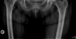
Clinical & Radiographic Imaging


You Might Also Like